

Community-acquired pneumonia is defined as pneumonia that is acquired outside the hospital. The most commonly identified pathogens are Streptococcus pneumoniae, Haemophilus influenzae, atypical bacteria (ie, Chlamydia pneumoniae, Mycoplasma pneumoniae, Legionella species), and viruses. Staphylococcus aureus (including methicillin-resistant S. aureus) and Pseudomonas aeruginosa can cause community-acquired pneumonia in patients with specific risk factors for these pathogens. Symptoms and signs are fever, cough, sputum production, pleuritic chest pain, dyspnea, tachypnea, and tachycardia. Diagnosis is based on clinical presentation and chest x-ray. Treatment is with empirically chosen antibiotics. Prognosis is excellent for relatively young or healthy patients, but many pneumonias, are serious or even fatal in older or sicker patients.
(See also Overview of Pneumonia.)
Etiology of Community-Acquired Pneumonia
Many organisms cause community-acquired pneumonia, including bacteria, viruses, and fungi. Pathogens vary by patient age and other factors, but the relative importance of each as a cause of community-acquired pneumonia is uncertain because most patients do not undergo thorough testing, and because even with testing, specific agents are identified in < 50% of cases.
Bacterial causes
The most common bacterial causes are
Pneumonias caused by chlamydia and mycoplasma are often clinically indistinguishable from other pneumonias.
C. pneumoniae is the second most common cause of lung infections in healthy people aged 5 to 35 years. C. pneumoniae is commonly responsible for outbreaks of respiratory infection within families, in college dormitories, and in military training camps. It causes a relatively benign form of pneumonia that infrequently requires hospitalization. Chlamydia psittaci pneumonia (psittacosis) is rare and occurs in patients who own or are often exposed to psittacine birds (ie, parrots, parakeets, macaws).
Since the year 2000, the incidence of community-acquired methicillin-resistant S. aureus (CA-MRSA) skin infections has increased markedly. This pathogen can rarely cause severe, cavitating pneumonia and tends to affect young adults.
S. pneumoniae and S. aureus (including MRSA) can cause necrotizing pneumonia.
Identification of possible MRSA pneumonia is based on an assessment of risk factors that include known MRSA colonization (nasal), prior MRSA infection, compatible gram stain, hospitalization or antibiotic use in the previous 3 months, immunosuppression, necrotizing or cavitary pneumonia, recent influenza, empyema.
Identification of possible P. aeruginosa pneumonia is based on assessment of risk factors that include known Pseudomonas colonization, prior Pseudomonas infection, hospitalization or antibiotic use in the previous 3 months, severe chronic obstructive pulmonary disease (COPD) or other structural lung disease, and immunosuppression.
Q fever, tularemia, anthrax, and plague are uncommon bacterial syndromes related to exposure to infected animals or animal products in which pneumonia may be a prominent feature. Tularemia, anthrax, and plague should raise the suspicion of bioterrorism.
Viral causes
Bacterial superinfection can make distinguishing viral from bacterial infection difficult.
Common viral causes include
Coronaviruses (since 2020, primarily SARS-CoV-2)
Epstein-Barr virus and coxsackievirus are common viruses that rarely cause pneumonia. Seasonal influenza can rarely cause a direct viral pneumonia but often predisposes to the development of a serious secondary bacterial pneumonia. Varicella virus and hantavirus cause lung infection as part of adult chickenpox and hantavirus pulmonary syndrome. Coronaviruses cause severe acute respiratory syndrome (SARS), the Middle East respiratory syndrome (MERS), and COVID-19.
Other causes
Common fungal pathogens include Histoplasma capsulatum (histoplasmosis) and Coccidioides immitis (coccidioidomycosis). Less common fungal pathogens include Blastomyces dermatitidis (blastomycosis) and Paracoccidioides braziliensis (paracoccidioidomycosis). Pneumocystis jirovecii commonly causes pneumonia in patients who have human immunodeficiency virus (HIV) infection or are immunosuppressed (see Pneumonia in Immunocompromised Patients).
Parasites causing lung infection in higher income countries include Toxocara canis or T. catis (toxocariasis), Dirofilaria immitis (dirofilariasis), and Paragonimus westermani (paragonimiasis).
Pulmonary tuberculosis and nontuberculous mycobacterial infections are discussed elsewhere.
Pneumonia in children
In children, the most common causes of pneumonia depend on age:
< 5 years: Most often viruses; among bacteria, S. pneumoniae, S. aureus, and S. pyogenes, are common
≥ 5 years: Most often the bacteria S. pneumoniae, M. pneumoniae, or Chlamydia pneumoniae
Pneumonia in neonates is discussed elsewhere.
Symptoms and Signs of Community-Acquired Pneumonia
Symptoms include malaise, chills, rigor, fever, cough, dyspnea, and chest pain. Cough typically is productive in older children and adults and dry in infants, young children, and older adults. Dyspnea usually is mild and exertional and is rarely present at rest. Chest pain is pleuritic and is adjacent to the infected area. Pneumonia may manifest as upper abdominal pain when lower lobe infection irritates the diaphragm. Gastrointestinal symptoms (nausea, vomiting, diarrhea) are also common.
Symptoms are variable at the extremes of age. Infection in infants may manifest as nonspecific irritability and restlessness. In older adults, infection may manifest as confusion and obtundation.
Signs include fever, tachypnea, tachycardia, crackles, bronchial breath sounds, egophony (E to A change— when, during auscultation, a patient says the letter “E” and through the stethoscope the examiner hears the letter “A”), and dullness to percussion. Signs of pleural effusion may also be present. Nasal flaring, use of accessory muscles, and cyanosis are common among infants. Fever is frequently absent in older patients.
Symptoms and signs were previously thought to differ by type of pathogen. However, manifestations in patients with typical and atypical pathogens overlap considerably. In addition, no single symptom or sign is sensitive or specific enough to predict the organism. Symptoms and signs are even similar for other noninfective inflammatory lung diseases such as acute hypersensitivity pneumonitis and cryptogenic organizing pneumonia.
Diagnosis of Community-Acquired Pneumonia
Chest x-ray
Consideration of alternative diagnoses (eg, heart failure, pulmonary embolism, inflammatory lung conditions)
Sometimes identification of pathogen
Evaluation of severity and risk stratification
Diagnosis of pneumonia is suspected on the basis of clinical presentation and infiltrate seen on chest x-ray. When there is high clinical suspicion of pneumonia and the chest x-ray does not reveal an infiltrate, doing computed tomography (CT) or repeating the chest x-ray in 24 to 48 hours is recommended.
Severity of the pneumonia is estimated using a variety of clinical and laboratory factors (see Risk Stratification), which are sometimes organized using quantitative scoring systems.
Typically, testing includes oxygen saturation, complete blood count, and a basic or complete metabolic profile.
Differential diagnosis in patients presenting with pneumonia-like symptoms includes acute bronchitis and exacerbation of chronic obstructive pulmonary disease (COPD), which can be distinguished from pneumonia by the absence of infiltrates on chest x-ray. Other disorders such as heart failure, organizing pneumonia, and hypersensitivity pneumonitis should be considered, particularly when findings are inconsistent. The most serious common misdiagnosis is pulmonary embolism, which may be more likely in patients with acute onset of dyspnea, minimal sputum production, no accompanying upper respiratory infection or systemic symptoms, and risk factors for thromboembolism (see table ); thus, testing for pulmonary embolism should be considered in patients with such symptoms and risk factors.
Quantitative cultures of bronchoscopic or suctioned specimens, if they are obtained before antibiotic administration, can help distinguish between bacterial colonization (ie, presence of microorganisms at levels that provoke neither symptoms nor an inflammatory response) and infection. However, bronchoscopy is usually done only in patients receiving mechanical ventilation or for those with other risk factors for unusual microorganisms or complicated pneumonia (eg, immunocompromise, failure of empiric therapy).
Distinguishing between bacterial and viral pneumonias is challenging. Many studies have investigated the utility of clinical, imaging, and routine blood tests, but no test is reliable enough to make this differentiation. Even identification of a virus does not preclude concomitant infection with a bacteria; therefore, antibiotics are indicated in almost all patients with a community-acquired pneumonia.
In outpatients with mild pneumonia, with the exception of COVID-19 testing and influenza testing during influenza season, no further diagnostic testing is needed (see table ). In patients with moderate or severe pneumonia, a white blood cell count and measurement of electrolytes, blood urea nitrogen (BUN), and creatinine are useful to classify risk and hydration status. Pulse oximetry or arterial blood gas (ABG) testing should also be done to assess oxygenation. For patients with moderate or severe pneumonia who require hospitalization, 2 sets of blood cultures are obtained to assess for bacteremia and sepsis. Additional tests indicated in these patients include sputum Gram stain and culture, urine pneumococcal and Legionella antigen testing, respiratory viral panel by multiplex polymerase chain reaction (PCR), and HIV screening. In selected severely ill patients, especially if they are immunocompromised, bronchoscopy to obtain lower airway specimens is indicated (1).
Pathogen identification
Diagnosis of etiology can be difficult. A thorough history of exposures, travel, pets, hobbies, and other exposures is essential to raise suspicion of less common organisms. For example, exposure to farm animals may suggest Q fever, and a recent hotel or cruise ship stay may suggest Legionella infection.
Identification of the pathogen can be useful to direct therapy and verify bacterial susceptibilities to antibiotics. However, because of the limitations of current diagnostic tests and the success of empiric antibiotic treatment, experts recommend limiting attempts at microbiologic identification (eg, cultures, specific antigen testing) unless patients are at high risk or have complications (eg, severe pneumonia, immunocompromise, asplenia, failure to respond to empiric therapy). In general, the milder the pneumonia, the less such diagnostic testing is required. Critically ill patients, patients in whom an antibiotic-resistant or unusual organism (eg, Mycobacterium tuberculosis, P. jirovecii) is suspected, and patients whose condition is deteriorating or who are not responding to treatment within 72 hours require the most intensive testing.
Chest x-ray findings generally cannot distinguish one type of infection from another, although the following findings are suggestive:
Multilobar infiltrates suggest S. pneumoniae or Legionella pneumophila infection.
Interstitial pneumonia (on chest x-ray, appearing as increased interstitial markings and subpleural reticular opacities that increase from the apex to the bases of the lungs) suggests viral or mycoplasmal etiology.
Cavitating pneumonia suggests S. aureus or a fungal or mycobacterial etiology.

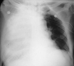
This chest x-ray shows an infiltrate that appears to blend with the right heart border (silhouette sign). The silhouette sign indicates contiguous positioning of the 2 structures that have similar radiodensity; the part of the lung contiguous with the right heart border is the right middle lobe, so that is the part with the infiltrate and pneumonia.
This chest x-ray shows an infiltrate that appears to blend with the right heart border (silhouette sign). The silhouett
SCIENCE PHOTO LIBRARY

This chest x-ray shows an infiltrate that does not obscure the right heart border (ie, there is no silhouette sign). Because the silhouette sign develops when 2 contiguous structures have a similar radiodensity, the part of the lung affected by this infiltrate is the part not contiguous with the right heart border; that part is the right lower lobe.
This chest x-ray shows an infiltrate that does not obscure the right heart border (ie, there is no silhouette sign). Be
LIVING ART ENTERPRISES, LLC/SCIENCE PHOTO LIBRARY

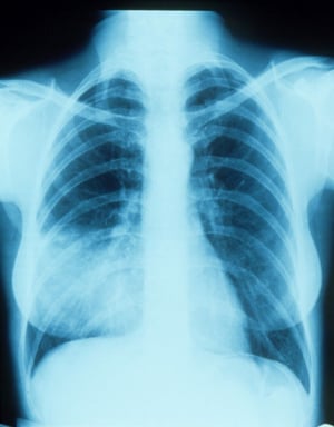
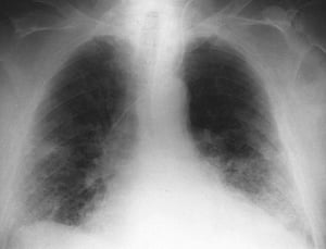
Alveolar infiltrate of the left lower lobe in a male with bacterial pneumonia.
Alveolar infiltrate of the left lower lobe in a male with bacterial pneumonia.
By permission of the publisher. From Roberts R. In Atlas of Infectious Diseases: Pleuropulmonary and Bronchial Infections. Edited by GL Mandell (series editor) and MS Simberkoff. Philadelphia, Current Medicine, 1996.

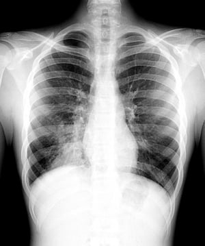
Consolidation of the right upper, middle, and lower lobes in a 64-year-old male with pneumococcal pneumonia.
Consolidation of the right upper, middle, and lower lobes in a 64-year-old male with pneumococcal pneumonia.
By permission of the publisher. From Roberts R. In Atlas of Infectious Diseases: Pleuropulmonary and Bronchial Infections. Edited by GL Mandell (series editor) and MS Simberkoff. Philadelphia, Current Medicine, 1996.

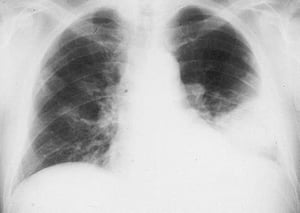
Bilateral interstitial opacities in an older male with respiratory syncytial virus pneumonia.
Bilateral interstitial opacities in an older male with respiratory syncytial virus pneumonia.
By permission of the publisher. From Betts R, Falsey A, Hall C, et al. In Atlas of Infectious Diseases: Pleuropulmonary and Bronchial Infections. Edited by GL Mandell (series editor) and MS Simberkoff. Philadelphia, Current Medicine, 1996.

This intubated patient has multiple bilateral infiltrates, most prominently in the right upper lobe. Arrow indicates the right horizontal fissure.
This intubated patient has multiple bilateral infiltrates, most prominently in the right upper lobe. Arrow indicates th
Photo courtesy of Thomas M. File, Jr., MD MSc MACP FIDSA FCCP.
This chest x-ray shows an infiltrate that appears to blend with the right heart border (silhouette sign). The silhouette sign indicates contiguous positioning of the 2 structures that have similar radiodensity; the part of the lung contiguous with the right heart border is the right middle lobe, so that is the part with the infiltrate and pneumonia.
This chest x-ray shows an infiltrate that appears to blend with the right heart border (silhouette sign). The silhouett
SCIENCE PHOTO LIBRARY
This chest x-ray shows an infiltrate that does not obscure the right heart border (ie, there is no silhouette sign). Because the silhouette sign develops when 2 contiguous structures have a similar radiodensity, the part of the lung affected by this infiltrate is the part not contiguous with the right heart border; that part is the right lower lobe.
This chest x-ray shows an infiltrate that does not obscure the right heart border (ie, there is no silhouette sign). Be
LIVING ART ENTERPRISES, LLC/SCIENCE PHOTO LIBRARY
Alveolar infiltrate of the left lower lobe in a male with bacterial pneumonia.
Alveolar infiltrate of the left lower lobe in a male with bacterial pneumonia.
By permission of the publisher. From Roberts R. In Atlas of Infectious Diseases: Pleuropulmonary and Bronchial Infections. Edited by GL Mandell (series editor) and MS Simberkoff. Philadelphia, Current Medicine, 1996.
Consolidation of the right upper, middle, and lower lobes in a 64-year-old male with pneumococcal pneumonia.
Consolidation of the right upper, middle, and lower lobes in a 64-year-old male with pneumococcal pneumonia.
By permission of the publisher. From Roberts R. In Atlas of Infectious Diseases: Pleuropulmonary and Bronchial Infections. Edited by GL Mandell (series editor) and MS Simberkoff. Philadelphia, Current Medicine, 1996.
Bilateral interstitial opacities in an older male with respiratory syncytial virus pneumonia.
Bilateral interstitial opacities in an older male with respiratory syncytial virus pneumonia.
By permission of the publisher. From Betts R, Falsey A, Hall C, et al. In Atlas of Infectious Diseases: Pleuropulmonary and Bronchial Infections. Edited by GL Mandell (series editor) and MS Simberkoff. Philadelphia, Current Medicine, 1996.
This intubated patient has multiple bilateral infiltrates, most prominently in the right upper lobe. Arrow indicates the right horizontal fissure.
This intubated patient has multiple bilateral infiltrates, most prominently in the right upper lobe. Arrow indicates th
Photo courtesy of Thomas M. File, Jr., MD MSc MACP FIDSA FCCP.
Blood cultures, which are often obtained in patients hospitalized for pneumonia, can identify causative bacterial pathogens if bacteremia is present. Approximately 7 to 12% of all patients hospitalized with pneumonia have bacteremia; S. pneumoniae is the most frequent pathogen in these cases (2).
Sputum testing can include Gram stain and culture for identification of the pathogen, but the value of these tests is uncertain because specimens often are contaminated with oral flora and overall diagnostic yield is low. Regardless, typical Gram stain findings can raise suspicion for a MRSA or P. aeruginosa, and identification of a bacterial pathogen in sputum cultures allows for susceptibility testing. Obtaining sputum samples also allows for testing for viral pathogens via direct fluorescence antibody testing or PCR, but caution needs to be exercised in interpretation because 15% of healthy adults carry a respiratory virus or potential bacterial pathogen. In patients whose condition is deteriorating and in those unresponsive to broad-spectrum antibiotics, sputum should be tested with mycobacterial and fungal stains and cultures.
Sputum samples can be obtained noninvasively by simple expectoration or, for patients unable to produce sputum, after hypertonic saline nebulization (induced sputum). Alternatively, patients can undergo bronchoscopy or endotracheal suctioning. Either of these procedures can be easily done through an endotracheal tube in mechanically ventilated patients. Otherwise, bronchoscopic sampling is usually done only for patients with other risk factors (eg, immunocompromise, failure of empiric therapy).
Urine testing for Legionella antigen and pneumococcal antigen is widely available. These tests are simple and rapid and have higher sensitivity and specificity than sputum Gram stain and culture for these pathogens. Urinary Legionella antigen remains present long after treatment is initiated, and the test detects only L. pneumophila serogroup 1 (70% of cases).
COVID-19 test using reverse transcriptase-polymerase chain reaction (RT-PCR) testing of respiratory secretions (nasopharyngeal specimen is preferred) is recommended in patients presenting with pneumonia .
Nasopharyngeal swabs, sputum, and bronchoscopic samples can also be tested in multiplex PCR for other viruses, including influenza, RSV, and other respiratory viruses, atypical pathogens, and bacteria. These results should be interpreted with caution because of baseline colonization and incomplete validation with lower respiratory tract samples.
Serum procalcitonin can help distinguish bacterial infection from other causes of infection or inflammation. However, use of the serum procalcitonin level as a criterion to initiate antibiotic therapy in community-acquired pneumonia is not recommended. It can be used, along with clinical judgement, to guide early discontinuation of antibiotics in lower respiratory tract infections.
Diagnosis references
1. Metlay JP, Waterer GW, Long AC, et al. Diagnosis and Treatment of Adults with Community-acquired Pneumonia. An Official Clinical Practice Guideline of the American Thoracic Society and Infectious Diseases Society of America. Am J Respir Crit Care Med 2019;200(7):e45-e67. doi:10.1164/rccm.201908-1581ST
2. Metersky ML, Ma A, Bratzler DW, Houck PM. Predicting bacteremia in patients with community-acquired pneumonia. Am J Respir Crit Care Med 2004;169(3):342-347. doi:10.1164/rccm.200309-1248OC
Treatment of Community-Acquired Pneumonia
Risk stratification for determination of site of care
Antibiotics
Antivirals for influenza or varicella
Systemic corticosteroids in selected critically ill patients
Supportive measures
Risk stratification
Risk stratification via risk prediction rules may be used to estimate mortality risk and thus help guide decisions regarding hospitalization (1). These rules have been used to identify patients who can be safely treated as outpatients and those who require hospitalization because of high risk of complications (see table ). However, these prediction rules should supplement, not replace, clinical judgment because many unrepresented factors, such as likelihood of adherence, ability to care for self, ability to maintain oral intake, should also influence triage decisions.
Intensive care unit (ICU) admission is required for patients who
Need mechanical ventilation
Have hypotension (systolic blood pressure ≤ 90 mm Hg) that is unresponsive to volume resuscitation
Other criteria, especially if ≥ 3 are present, that should lead to consideration of ICU admission include
Hypotension requiring fluid support
Respiratory rate > 30/minute
Partial pressure of arterial oxygen (PaO2)/fraction of inspired oxygen (FIO2) < 250
Multilobar pneumonia
Confusion
Blood urea nitrogen (BUN) > 19.6 mg/dL (> 7 mmol/L)
Leukocyte count < 4000 cells/microL (< 4 × 10⁹/L)
Platelet count < 100,000/microL (< 100 × 109/L)
Temperature < 36° C
The Pneumonia Severity Index (PSI) is the preferred validated prediction rule (1). However, because the PSI is complex and requires several laboratory assessments, simpler rules such as CURB-65 are a useful alternative. Use of these prediction rules has led to a reduction in unnecessary hospitalizations for patients who have milder illness.

In CURB-65, 1 point is allotted for each of the following risk factors:
Confusion
Uremia (BUN ≥ 19 mg/dL [6.8 mmol/L])
Respiratory rate > 30 breaths/minute
Systolic Blood pressure < 90 mm Hg or diastolic blood pressure ≤ 60 mm Hg
Age ≥ 65 years
Scores can be used as follows:
0 or 1 points: Risk of death is < 3%. Outpatient therapy is usually appropriate, especially if the only point is related to age.
2 points: Risk of death is 9%. Hospitalization should be considered.
≥ 3 points: Risk of death is 15 to 40%. Hospitalization is indicated, and, particularly with 4 or 5 points, ICU admission should be considered.
In clinical settings where access to a BUN measurement is not readily available, a CRB-65 score can be used instead. Use of CRB-65 scores is similar to that for CURB-65, with 0 points: appropriate for outpatient therapy; 1 to 2 points: consider hospitalization; ≥ 3 points: consider ICU admission.
Risk Stratification for Community-Acquired Pneumonia (the Pneumonia Severity Index)
Factor | Points | ||
|---|---|---|---|
Patient demographics | |||
| Age (in years) | ||
| Age (in years) − 10 | ||
Nursing home resident | 10 | ||
Coexisting illness | |||
Cancer | 30 | ||
Liver disease | 20 | ||
Heart failure | 10 | ||
Cerebrovascular disease | 10 | ||
Renal disease | 10 | ||
Physical examination | |||
Altered mental status | 20 | ||
Respiratory rate ≥ 30 breaths/minute | 20 | ||
Systolic blood pressure < 90 mm Hg | 20 | ||
Temperature ≥ 40° C or < 35°C | 15 | ||
Heart rate ≥ 125 beats/minute | 10 | ||
Test results | |||
Arterial pH < 7.35 | 30 | ||
Blood urea nitrogen ≥ 30 mg/dL (11 mmol/L) | 20 | ||
Sodium < 130 mEq/L (130 mmol/L) | 20 | ||
Glucose ≥ 250 mg/dL (14 mmol/L) | 10 | ||
Hematocrit < 30% | 10 | ||
PaO2< 60 mm Hg or Oxygen saturation < 90%* | 10 | ||
Pleural effusion | 10 | ||
Points | Mortality | Recommendation | |
≤ 70 | < 1% | Outpatient treatment† | |
71−90 | < 5% | Outpatient treatment† | |
91−130 | 5−15% | Admit | |
> 130 | > 15% | Admit | |
* Many clinicians consider hypoxemia an absolute indication for admission. | |||
† Acute care admission, subacute care admission, observation period, home IV antibiotics, or home nursing visits should be considered for patients who are frail, isolated, or living in unstable environments. | |||
PaO2 = partial pressure of arterial oxygen. | |||
Adapted from Fine MJ, Auble TE, Yealy DM, et al. A prediction rule to identify low-risk patients with community-acquired pneumonia. N Engl J Med 1997;336(4):243-250. doi:10.1056/NEJM199701233360402. | |||
Antimicrobials
Antibiotic therapy is the mainstay of treatment for community-acquired pneumonia. Appropriate treatment involves starting empiric antibiotics as soon as possible, preferably ≤ 4 hours after presentation. Because pathogen identification is difficult and takes time, the empiric antibiotic regimen is selected based on likely pathogens and severity of illness. Consensus guidelines have been developed by many professional organizations; one widely used set is detailed in the guideline Diagnosis and Treatment of Adults with Community-acquired Pneumonia. Guidelines should be adapted to local susceptibility patterns, drug formularies, and individual patient circumstances (eg, specific exposures). If a pathogen is subsequently identified, the results of antibiotic susceptibility testing can help guide any changes in antibiotic therapy. Omadacycline and lefamulin can be considered, especially in situations in which the usually recommended choices are not appropriate. . Guidelines should be adapted to local susceptibility patterns, drug formularies, and individual patient circumstances (eg, specific exposures). If a pathogen is subsequently identified, the results of antibiotic susceptibility testing can help guide any changes in antibiotic therapy. Omadacycline and lefamulin can be considered, especially in situations in which the usually recommended choices are not appropriate.
For children, treatment depends on age, previous vaccinations, and whether treatment is outpatient or inpatient.
For children treated as outpatients, treatments are dictated by age:
< 5 years: Amoxicillin or 5 years: Amoxicillin oramoxicillin/clavulanate is usually the medication of choice. If epidemiology suggests an atypical pathogen as the cause and clinical findings are compatible, a macrolide (eg, azithromycin, clarithromycin) can be used instead. Some experts suggest not using antibiotics if clinical features strongly suggest viral pneumonia.is usually the medication of choice. If epidemiology suggests an atypical pathogen as the cause and clinical findings are compatible, a macrolide (eg, azithromycin, clarithromycin) can be used instead. Some experts suggest not using antibiotics if clinical features strongly suggest viral pneumonia.
≥ 5 years: Amoxicillin or (particularly if an atypical pathogen cannot be excluded) amoxicillin plus a macrolide. 5 years: Amoxicillin or (particularly if an atypical pathogen cannot be excluded) amoxicillin plus a macrolide.Amoxicillin/clavulanate is an alternative. If the cause appears to be an atypical pathogen, a macrolide alone can be used.
For children treated as inpatients, antibiotic therapy tends to be more broad-spectrum and depends on the child's previous vaccinations:
Fully immunized (against S. pneumoniae and H. influenzae type b): Ampicillin or penicillin G (alternatives are ceftriaxone or cefotaxime). If MRSA is suspected, vancomycin or clindamycin is added. If an atypical pathogen cannot be excluded, a macrolide is added. type b): Ampicillin or penicillin G (alternatives are ceftriaxone or cefotaxime). If MRSA is suspected, vancomycin or clindamycin is added. If an atypical pathogen cannot be excluded, a macrolide is added.
Not fully immunized: Ceftriaxone or cefotaxime (alternative is levofloxacin). If MRSA is suspected, vancomycin or clindamycin is added. If an atypical pathogen cannot be excluded, a macrolide is added. Not fully immunized: Ceftriaxone or cefotaxime (alternative is levofloxacin). If MRSA is suspected, vancomycin or clindamycin is added. If an atypical pathogen cannot be excluded, a macrolide is added.
Full details are described in the Clinical Practice Guidelines by the Pediatric Infectious Diseases Society and the Infectious Diseases Society of America.
With empiric treatment, 90% of patients with bacterial pneumonia improve. Improvement is manifested by decreased cough and dyspnea, defervescence, relief of chest pain, and decline in white blood cell count. Failure to improve should trigger suspicion of
An unusual organism
Resistance to the antimicrobial used for treatment
Empyema
Coinfection or superinfection with a 2nd infectious agent
An obstructive endobronchial lesion
Immunosuppression
Metastatic focus of infection with reseeding (in the case of pneumococcal infection)
Nonadherence to treatment (in the case of outpatients)
Wrong diagnosis (ie, a noninfectious cause of the illness such as acute hypersensitivity pneumonitis)
When usual therapy has failed, consultation with a pulmonary and/or infectious disease specialist is indicated.
Antiviral therapy may be indicated for select viral pneumonias. Ribavirin is not used routinely for respiratory syncytial virus pneumonia in children or adults but may be used occasionally in high-risk children age may be indicated for select viral pneumonias. Ribavirin is not used routinely for respiratory syncytial virus pneumonia in children or adults but may be used occasionally in high-risk children age< 24 months.
For influenza, oseltamivir or zanamivir started within 48 hours of symptom onset and given for 5 days reduce the duration and severity of symptoms in patients who develop influenza infection. Alternatively, baloxavir started within 48 hours of symptom onset can be given in a single dose. Patients hospitalized with confirmed influenza infection may benefit even 48 hours after symptom onset. For influenza, oseltamivir or zanamivir started within 48 hours of symptom onset and given for 5 days reduce the duration and severity of symptoms in patients who develop influenza infection. Alternatively, baloxavir started within 48 hours of symptom onset can be given in a single dose. Patients hospitalized with confirmed influenza infection may benefit even 48 hours after symptom onset.
Acyclovir is recommended for children and adults with varicella lung infections. Acyclovir is recommended for children and adults with varicella lung infections.
Though pure viral pneumonia does occur, superimposed bacterial infections are common and require antibiotics directed against S. pneumoniae, H. influenzae, and S. aureus.
Follow-up x-rays are generally not recommended in patients whose pneumonia resolves clinically as expected. Resolution of radiographic abnormalities can lag behind clinical resolution by several weeks. Chest x-ray should be considered in patients with pneumonia symptoms that do not resolve or that worsen over time.
Community-Acquired Pneumonia in Adults
Group | Likely Organisms | Empiric Treatment |
|---|---|---|
I. Outpatients—no comorbidities or risk factors for MRSA and Pseudomonas aeruginosa present* | Streptococcus pneumoniae, Mycoplasma pneumoniae, Chlamydia pneumoniae, Haemophilus influenzae, respiratory viruses, miscellaneous organisms (eg, Legionella, Mycobacterium tuberculosis, endemic fungi) | AmoxicillinAmoxicillin plus either Macrolide (azithromycin or clarithromycin)Macrolide (azithromycin or clarithromycin) or Doxycycline (if allergic to macrolides)Doxycycline (if allergic to macrolides) |
II. Outpatients—comorbidities present* | S. pneumoniae, including antibiotic-resistant forms; M. pneumoniae; C. pneumoniae; mixed infection (bacteria + atypical pathogen or virus); H. influenzae; enteric gram-negative organisms; respiratory viruses; miscellaneous organisms (eg, Moraxella catarrhalis, Legionella, anaerobes [aspiration], M. tuberculosis, endemic fungi) | Beta-lactam (cefpodoxime, cefuroxime, amoxicillin, or amoxicillin/clavulanate) Beta-lactam (cefpodoxime, cefuroxime, amoxicillin, or amoxicillin/clavulanate) plus either Macrolide orally or Antipneumococcal fluoroquinolone orally or IV (eg, moxifloxacin, levofloxacin )Antipneumococcal fluoroquinolone orally or IV (eg, moxifloxacin, levofloxacin ) |
III. Inpatient—not in intensive care unit (ICU), no risk factors for MRSA or P. aeruginosa | S. pneumoniae, H. influenzae, M. pneumoniae, C. pneumoniae, mixed infection (bacteria + atypical pathogen or virus), respiratory viruses, Legionella, miscellaneous organisms (eg, M. tuberculosis, endemic fungi, Pneumocystis jirovecii) | AzithromycinAzithromycin plus Beta-lactam IV (cefotaxime or ceftriaxone)Beta-lactam IV (cefotaxime or ceftriaxone) or Antipneumococcal fluoroquinolone orally or intravenously (alone) |
IVA. ICU patient—no risk factors for MRSA or P. aeruginosa | S. pneumoniae, including antibiotic-resistant forms; Legionella; H. influenzae; enteric gram-negative organisms; Staphylococcus aureus; M. pneumoniae; respiratory viruses; miscellaneous organisms (eg, C. pneumoniae, M. tuberculosis, endemic fungi) | Beta-lactam IV (cefotaxime or ceftriaxone)Beta-lactam IV (cefotaxime or ceftriaxone) plus either Antipneumococcal fluoroquinolone IV or AzithromycinAzithromycin |
IVB. Inpatient or ICU patient—MRSA or Pseudomonas risk factors present (if prior isolation is the only risk factor, add initial coverage for that pathogen only and obtain cultures) | Same as those for category IVA (above) plus MRSA or Pseudomonas | Linezolid or vancomycinLinezolid or vancomycin or/plus Antipseudomonal beta-lactam (cefepime, cetazidime, imipenem, meropenem, piperacillin/tazobactam) or aztreonam (if allergic to or intolerant of beta-lactams) Antipseudomonal beta-lactam (cefepime, cetazidime, imipenem, meropenem, piperacillin/tazobactam) or aztreonam (if allergic to or intolerant of beta-lactams) |
* Modifying factors:
| ||
MRSA = methicillin-resistant Staphylococcus aureus | ||
Data from Metlay JP, Waterer GW, Long AC, et al. Diagnosis and Treatment of Adults with Community-acquired Pneumonia. An Official Clinical Practice Guideline of the American Thoracic Society and Infectious Diseases Society of America. Am J Respir Crit Care Med 2019;200(7):e45-e67. doi:10.1164/rccm.201908-1581ST | ||
Supportive care
Supportive care includes fluids, antipyretics (as needed for high grade fever), analgesics, and, for patients with hypoxemia, oxygen. Prophylaxis against thromboembolic disease and early mobilization improve outcomes for patients hospitalized with pneumonia. Cessation counseling should also be done for smokers.
Adjunctive corticosteroids may be used in immunocompetent patients with severe disease. Randomized studies have demonstrated reduced mortality and morbidity with early initiation of intravenous hydrocortisone 200 mg daily by continuous infusion for 4 to 7 days in immunocompetent patients with severe pneumonia (defined by need for ventilatory support or high-flow oxygen) (Adjunctive corticosteroids may be used in immunocompetent patients with severe disease. Randomized studies have demonstrated reduced mortality and morbidity with early initiation of intravenous hydrocortisone 200 mg daily by continuous infusion for 4 to 7 days in immunocompetent patients with severe pneumonia (defined by need for ventilatory support or high-flow oxygen) (2). Such treatment is not advised for less severe pneumonia, in immunocompromised patients, or when viral or fungal pneumonia is suspected.
Treatment references
1. Metlay JP, Waterer GW, Long AC, et al. Diagnosis and Treatment of Adults with Community-acquired Pneumonia. An Official Clinical Practice Guideline of the American Thoracic Society and Infectious Diseases Society of America. Am J Respir Crit Care Med 2019;200(7):e45-e67. doi:10.1164/rccm.201908-1581ST
2. Dequin PF, Meziani F, Quenot JP, et al. Hydrocortisone in Severe Community-Acquired Pneumonia. N Engl J Med 2023;388(21):1931-1941. doi:10.1056/NEJMoa2215145
Health Care-Associated Pneumonia
The category of health care-associated pneumonia was removed as a separate category of pneumonia in the 2016 Infectious Diseases Society of America (IDSA) American Thoracic Society guidelines for hospital-acquired pneumonia (1). Health care-associated pneumonia includes community-based patients who have had recent contact with the health care system, such as those who reside in nursing homes or other long-term care facilities or visit dialysis centers or infusion centers. This category was created to help identify patients at increased risk for antibiotic-resistant bacteria. However, the 2016 IDSA guidelines found increasing evidence that many patients with health care-associated pneumonia were not infected with antibiotic-resistant bacteria. Rather, the risk for antibiotic-resistant bacteria in these patients can be based on validated risk factors described for patients with community-acquired pneumonia.
Health care-associated pneumonia reference
1. Kalil AC, Metersky ML, Klompas M, et al. Management of Adults With Hospital-acquired and Ventilator-associated Pneumonia: 2016 Clinical Practice Guidelines by the Infectious Diseases Society of America and the American Thoracic Society [published correction appears in Clin Infect Dis. 2017 May 1;64(9):1298] [published correction appears in Clin Infect Dis. 2017 Oct 15;65(8):1435] [published correction appears in Clin Infect Dis. 2017 Nov 29;65(12):2161]. Clin Infect Dis 2016;63(5):e61-e111. doi:10.1093/cid/ciw353
Prognosis for Community-Acquired Pneumonia
Short-term mortality is related to severity of illness. Mortality is < 1% in patients who are candidates for outpatient treatment. Mortality in hospitalized patients is 8%. Death may be caused by pneumonia itself, progression to sepsis syndrome, or exacerbation of coexisting conditions. In patients hospitalized for pneumonia, risk of death is increased during the year after hospital discharge.
Mortality varies to some extent by pathogen. Mortality rates are highest with gram-negative bacteria and CA-MRSA. However, because these pathogens are relatively infrequent causes of community-acquired pneumonia, S. pneumoniae remains the most common cause of death in patients with community-acquired pneumonia. Atypical pathogens such as Mycoplasma have a good prognosis. Mortality is higher in patients who do not respond to initial empiric antibiotics and in those whose treatment regimen does not conform with guidelines.
Prevention of Community-Acquired Pneumonia
Some forms of community-acquired pneumonia are preventable with vaccination. Available pneumococcal vaccines and dosing schedules are discussed separately. (See Pneumococcal Vaccine.)
Recommendations for other vaccines, such as H. influenzae type b (Hib) vaccine (for patients < 2 years), varicella vaccine (for patients < 18 months and a later booster vaccine), and influenza vaccine (annually for everyone ≥ 6 months and especially for those at higher risk of developing serious flu-related complications), can also be found at the CDC website. This higher risk group includes people ≥ 65 years and people of any age with certain chronic medical conditions (such as diabetes, asthma, or heart disease), pregnant women, and young children.
In high-risk patients who are not vaccinated against influenza and household contacts of patients with influenza, oseltamivir 75 mg orally once a/ day or zanamivir 10 mg orally mg once a /day can be given as prophylaxis for 2 weeks, which differs from the dosing regimens for treatment. If started within 48 hours of exposure, these antivirals may prevent influenza (although resistance has been described for oseltamivir). In high-risk patients who are not vaccinated against influenza and household contacts of patients with influenza, oseltamivir 75 mg orally once a/ day or zanamivir 10 mg orally mg once a /day can be given as prophylaxis for 2 weeks, which differs from the dosing regimens for treatment. If started within 48 hours of exposure, these antivirals may prevent influenza (although resistance has been described for oseltamivir).
Smoking cessation can reduce the risk of developing pneumonia.
Key Points
Community-acquired pneumonia is a leading cause of death in the United States and around the world.
Common symptoms and signs include cough, fever, chills, fatigue, dyspnea, rigors, sputum production, and pleuritic chest pain.
Treat patients with mild or moderate risk pneumonia with empiric antibiotics without testing designed to identify the underlying pathogen.
Hospitalize patients at high risk, as delineated by risk assessment tools.
Consider alternate diagnoses, including pulmonary embolism, particularly if pneumonia-like signs and symptoms are not typical.
More Information
The following English-language resources may be useful. Please note that THE MANUAL is not responsible for the content of these resources.
Centers for Disease Control and Prevention: Child and Adolescent Immunization Schedule by Age. Reviewed April 27, 2023. Accessed October 19, 2023.
Kalil AC, Metersky ML, Klompas M, et al. Management of Adults With Hospital-acquired and Ventilator-associated Pneumonia: 2016 Clinical Practice Guidelines by the Infectious Diseases Society of America and the American Thoracic Society [published correction appears in Clin Infect Dis. 2017 May 1;64(9):1298] [published correction appears in Clin Infect Dis. 2017 Oct 15;65(8):1435] [published correction appears in Clin Infect Dis. 2017 Nov 29;65(12):2161]. Clin Infect Dis 2016;63(5):e61-e111. doi:10.1093/cid/ciw353
Drug Information for the Topic


