An extensive language has been developed to standardize the description of skin lesions, including:
Lesion type (sometimes called primary morphology)
Lesion configuration (sometimes called secondary morphology)
Rash is a general term for a temporary skin eruption.
(See also Evaluation of the Dermatologic Patient and Diagnostic Tests for Skin Disorders.)
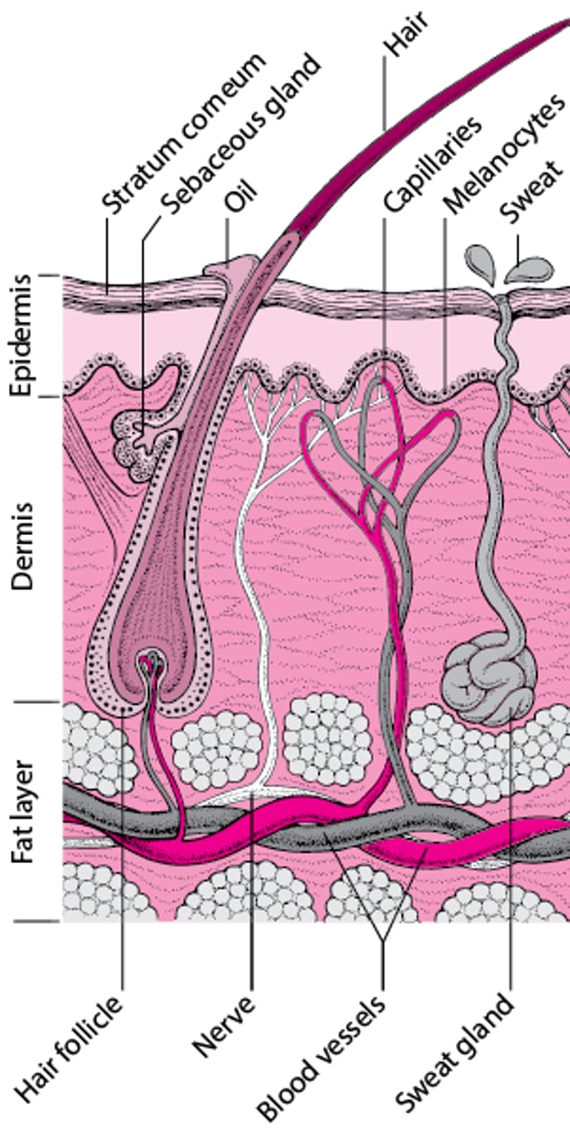
Cross-Section of the Skin and Skin Structures

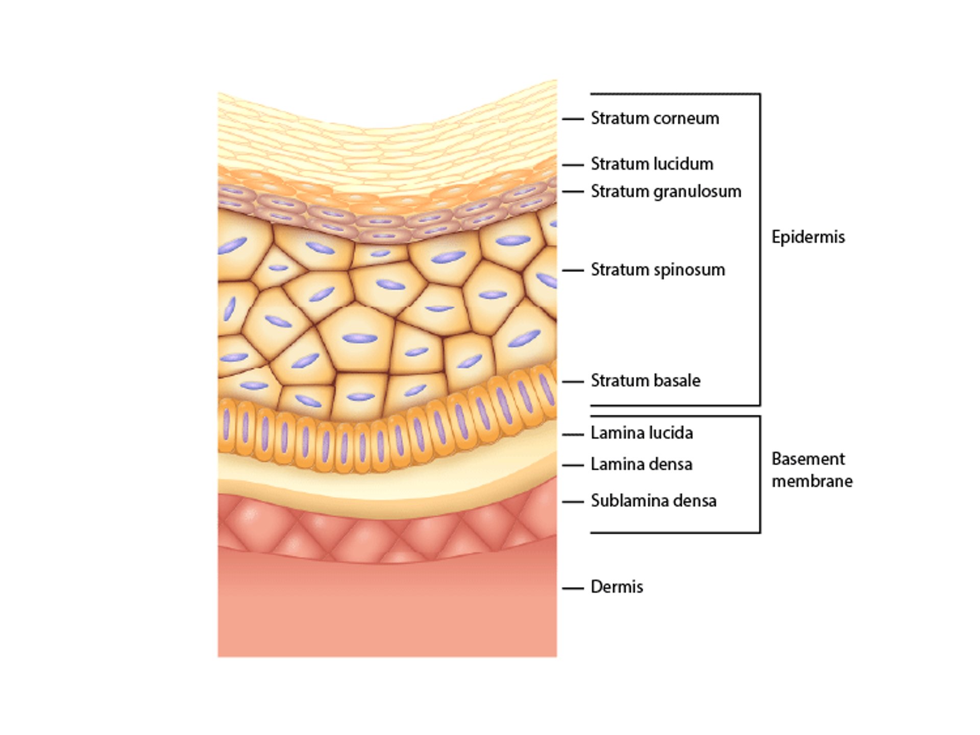
Layers of the Skin
In this figure, the 5 layers of the epidermis along with its basement membrane and the dermis can be seen. Note that the stratum lucidum is present over the palms and soles only. The basement membrane is disproportionately enlarged to display its layers. The sublamina densa anchors the dermis to the upper layers of the basement membrane. |

Lesion Type (Primary Morphology)
Macules are flat, nonpalpable lesions usually < 10 mm in diameter. Macules represent a change in color and are not raised or depressed compared to the skin surface. Examples of macules include freckles and flat moles.

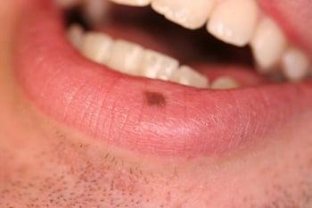
The lesion in this photo is a macule because it is flat, nonpalpable, and of small diameter.
Patches are large macules usually > 10 mm in diameter. They are not raised or depressed compared to the skin surface. Examples of conditions that may present with patches include vitiligo, pityriasis alba, and port-wine stains.
Examples of lesions that can be classified as either macules or patches, depending on size, include tattoos and the rashes of rickettsial infections, rubella, measles (can also have papules and plaques), and some allergic drug eruptions.


© Springer Science+Business Media
Papules are elevated lesions usually < 10 mm in diameter that can be felt or palpated. Examples include nevi, warts, lichen planus, insect bites, seborrheic keratoses, actinic keratoses, some lesions of acne, and skin cancers. The term maculopapular is often loosely and improperly used to describe many erythematous rashes; because this term is nonspecific and easily misused, it should be avoided.

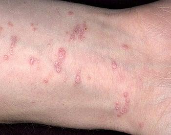
Papules are elevated, usually palpable lesions < 10 mm in diameter.
Lichen planus (pictured) may manifest as a papular rash.
Plaques are palpable lesions > 10 mm in diameter that are elevated or depressed compared to the skin surface. Plaques may be flat topped or rounded or may be comprised of multiple groups of small papules. Lesions of psoriasis and granuloma annulare commonly form plaques.

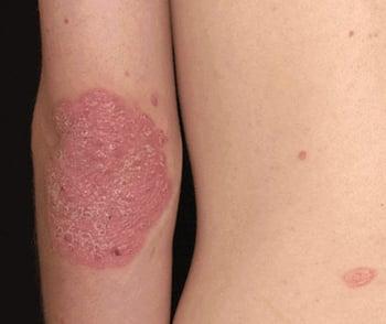
Psoriasis plaques are elevated or depressed, palpable lesions > 10 mm in diameter.
Psoriasis typically manifests as plaques covered with thick, silvery, shiny scales.


This photo shows well-demarcated psoriatic plaques in a patient with more deeply pigmented skin.
Nodules are firm papules or lesions that extend into the dermis or subcutaneous tissue. Examples include cysts, lipomas, fibromas, and infectious conditions (eg, parapoxvirus lesions, sporotrichosis).


This photo shows cutaneous nodules in a lymphangitic (sporotrichoid) distribution over the dorsum of the right hand.
Image courtesy of Karen McKoy, MD.
Vesicles are small, fluid-filled blisters < 10 mm in diameter. Vesicles are characteristic of herpes infections, acute allergic contact dermatitis, and some autoimmune bullous disorders (eg, dermatitis herpetiformis).

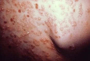
Vesicles are small fluid-filled blisters < 10 mm in diameter.
Dermatitis herpetiformis (pictured) typically manifests as crops of vesicles.
Bullae are fluid-filled blisters > 10 mm in diameter. These may be caused by burns, bites, irritant contact dermatitis or allergic contact dermatitis, and drug reactions. Classic autoimmune bullous diseases include pemphigus vulgaris and bullous pemphigoid. Bullae also may occur in inherited disorders of skin fragility (eg, epidermolysis bullosa).

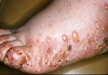
Bullae are fluid-filled blisters > 10 mm in diameter.
Bullous pemphigoid (pictured) is characterized by eruptions of tense bullae on normal-appearing skin or on skin with inflammatory color changes, which may appear erythematous or hyperpigmented depending on skin tone, in older adults.


Bullae are fluid-filled blisters > 10 mm in diameter.
Bullous fixed drug eruption (pictured) is characterized by tense bullae on inflamed skin. Color varies depending on skin tone, eg, violaceous, gray-brown, or hyperpigmented in patients with more deeply pigmented skin (shown here).
Pustules are vesicles that contain pus. Pustules are common in bacterial infections and folliculitis and may arise in some inflammatory disorders including pustular psoriasis.

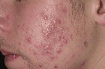
Pustules are elevated, usually yellow-topped lesions that contain pus. Scattered pustules appear on the face of this person with acne.
Urticaria (wheals or hives) is characterized by elevated lesions caused by localized edema. Wheals may appear as localized lesions or as a more generalized eruption. Wheals are typically pruritic and erythematous; however, in patients with a dark skin tone, erythema may be less visible, and wheals may instead be identified by edema or by surface or texture change. Wheals are a common manifestation of hypersensitivity to medications, stings or bites, or autoimmunity. They also may occur during an illness, particularly a viral infection. Less commonly, physical stimuli, including heat, cold, pressure, and sunlight, can also cause urticarial lesions. The typical wheal lasts < 24 hours. Some types of urticaria, such as cholinergic urticaria (a subtype of chronic inducible urticaria) can cause smaller and more diffuse wheals. Cholinergic urticaria is triggered by bathing in hot water, exercise, and emotional stress.

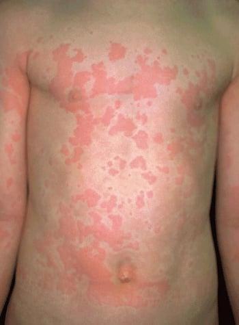
Urticaria (wheals or hives) are migratory, elevated, pruritic, erythematous lesions caused by local dermal edema.
Scale is heaped-up accumulations of cornified epithelium (stratum corneum) that occur in papulosquamous disorders (characterized by papules or plaques with overlying scale) such as psoriasis, seborrheic dermatitis, and fungal infections. Pityriasis rosea and chronic dermatitis of any type may be scaly.

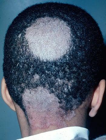
Scales are heaped-up accumulations of horny epithelium. Scaling is a characteristic feature of many dermatophytoses, including tinea capitis, resulting in the large bald patches. In this image, scale is especially noticeable at the nape of the neck.
Crusts (scabs) consist of dried serum, blood, and/or pus. Crusting can occur in inflammatory or infectious skin diseases (eg, impetigo).
Erosions are open areas of skin that result from loss of part or all of the epidermis. Erosions can be traumatic or can occur with various inflammatory or infectious skin diseases. An excoriation is a linear erosion caused by scratching, rubbing, or picking.
Ulcers result from loss of the epidermis and at least part of the dermis. Causes include venous stasis dermatitis, physical trauma with or without vascular compromise (eg, caused by pressure injuries or peripheral arterial disease), infections, and vasculitis.


In a few cases, cutaneous diphtheria may undergo central ulceration; occasionally, a grayish membrane forms.
Petechiae are nonblanchable punctate foci of hemorrhage. Causes include platelet abnormalities (eg, thrombocytopenia, platelet dysfunction), vasculitis, and infections (eg, meningococcemia, Rocky Mountain spotted fever, other rickettsioses).


This photo shows petechiae in a patient with Sjögren syndrome and biopsy-proven cutaneous small-vessel vasculitis.
Purpura is a larger area of hemorrhage that may be palpable. Purpura does not blanch. Palpable purpura is considered the hallmark of leukocytoclastic vasculitis. Purpura may indicate a coagulopathy. Small areas of purpura are called petechiae. Large areas of purpura may be called ecchymoses.


This photo shows an erythematous papular rash on the feet of a patient with immunoglobulin A–associated vasculitis.
Atrophy is thinning of the skin, which may appear dry and wrinkled. Atrophy may be caused by chronic sun exposure, aging, and some inflammatory and neoplastic skin diseases, including cutaneous T-cell lymphoma and lupus erythematosus. Atrophy also may result from long-term use of potent topical glucocorticoids.


This photo shows atrophied and depigmented areas on the abdomen of a patient.
Scars are areas of fibrosis that replace normal skin after injury. Some scars become hypertrophic or thickened and raised. Keloids are hypertrophic scars that extend beyond the original wound margin.


Keloids are distinct from hypertrophic scars because they extend beyond the borders of the original wound, invading normal skin. This photo shows multiple large keloid scars on the anterior abdominal wall that developed after intra-abdominal surgery.
Telangiectases are foci of small, permanently dilated blood vessels that may occur in areas of sun damage, rosacea, systemic diseases (especially systemic sclerosis), or inherited diseases (eg, ataxia-telangiectasia, hereditary hemorrhagic telangiectasia) or after long-term therapy with topical fluorinated glucocorticoids.

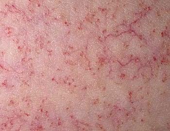
Telangiectasias are small, dilated blood vessels that are most often idiopathic.


This photo shows a close-up of the face of a patient with multiple telangiectasias secondary to hereditary hemorrhagic telangiectasia.
Lesion Configuration (Secondary Morphology)
Configuration is the shape of single lesions and the arrangement of clusters of lesions.
Linear lesions take on the shape of a straight line and are suggestive of some forms of contact dermatitis, linear epidermal nevi, and lichen striatus. Excoriations resulting from scratching are typically linear.
Annular lesions are rings with central clearing. Examples include granuloma annulare, some drug eruptions, some dermatophyte infections (eg, tinea [ringworm]), and secondary syphilis.
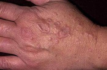
This photo shows the typical skin lesions of granuloma annulare. The lesions manifest as annular lesions with central clearing.
Nummular lesions are circular or coin-shaped; an example is nummular dermatitis (nummular eczema).

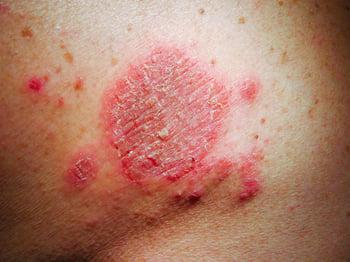
This photo shows discoid, erythematous, and scaly lesions consistent with nummular dermatitis in light skin.
Target (bull’s-eye or iris) lesions appear as rings with central duskiness and are classic for erythema multiforme. In patients with a dark skin tone, erythema may be less apparent, and lesions may appear violaceous or hyperpigmented with subtle peripheral contrast.

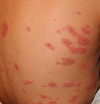
Target lesions (sometimes called iris lesions) manifest as annular lesions with a violaceous center and erythematous halo separated by a pale ring. Such lesions, which are typical of erythema multiforme, are distributed symmetrically.


This photo shows a target lesion with a violaceous to hyperpigmented center. In patients with more deeply pigmented skin, erythema may be less apparent, and peripheral contrast may be subtle.
Serpiginous lesions have linear, branched, and curving elements. Examples include some fungal and parasitic infections (eg, cutaneous larva migrans).


This photo shows a superficial, raised, serpiginous tract on the dorsal aspect of the foot.
Reticulated lesions have a lacy or net-like pattern. Examples include livedo reticularis (a reddish blue skin discoloration in a net-like pattern that typically resolves with rewarming) and livedo racemosa (a reddish blue skin discoloration in an irregular, broken, net-like pattern that does not resolve with rewarming).

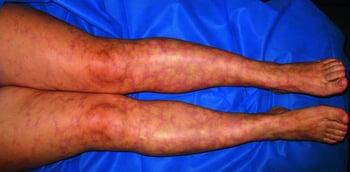
This photo shows the net-like pattern typical of livedo reticularis.
Herpetiform lesions are grouped papules or vesicles arranged like those of a herpes simplex infection.

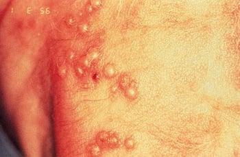
Herpetiform lesions manifest as grouped papules or vesicles in a formation resembling those of a herpes simplex infection. This photo shows clusters of vesicles on erythematous bases that are highly characteristic; however, inflammatory color may vary by skin tone, and configuration is the key identifying feature.
Zosteriform describes lesions clustered in a dermatomal distribution similar to those of herpes zoster.


This photo shows zosteriform lesions and erythematous vesicles arranged in a dermatomal distribution.
Texture of Skin Lesions
Some skin lesions have a visible or palpable texture that suggests a diagnosis.
Verrucous lesions have an irregular, pebbly, or rough surface. Examples include warts and seborrheic keratoses.

In a few cases, cutaneous diphtheria may undergo central ulceration; occasionally, a grayish membrane forms.
CDC
Lichenification is thickening of the skin with accentuation of normal skin markings; it results from repeated scratching or rubbing and is commonly seen in chronic inflammatory dermatoses (eg, atopic dermatitis).
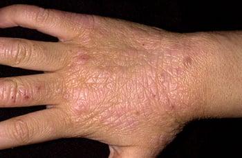
Lichenification is thickening of the skin with accentuation of normal skin markings; it is a result of chronic scratching or rubbing, which in this patient occurred during the chronic phase of atopic dermatitis.
Induration, or deep thickening of the skin, can result from edema, inflammation, or infiltration, including by cancer. Indurated skin has a hard, resistant feeling. Induration is characteristic of panniculitis, some skin infections, and cutaneous metastatic cancers. Peau d'orange is a clinical sign used to describe skin that has a dimpled, pitted appearance that resembles the surface of an orange. It is caused by edema surrounding tethered hair follicles and is sometimes observed in certain types of breast cancer (eg, inflammatory breast cancer).


Sometimes in inflammatory breast cancer and very advanced cancers, the breast appears inflamed, and the skin appears thickened, resembling orange peel (peau d’orange).
Umbilicated lesions have a central indentation and are usually viral. Examples include molluscum contagiosum and herpes simplex.


This photo shows the firm, dome-shaped, pearly papules with characteristic central umbilication of molluscum contagiosum.
Xanthomas, which are yellowish, waxy lesions, may be idiopathic or may occur in patients who have lipid disorders (eg, xanthelasma).


Patients with severe elevations of triglycerides can have eruptive xanthomas over the trunk, back, elbows, buttocks, knees, hands, and feet.
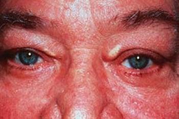
This photo shows yellow-white plaques on the upper medial eyelids in a patient with hypercholesterolemia. Xanthelasmas are not specific for familial hypercholesterolemia.
Location and Distribution of Skin Lesions
When examining the skin, it is important for clinicians to note whether:
Lesions are single or multiple.
Particular body parts are affected (eg, palms or soles, scalp, mucosal membranes).
Distribution is random or patterned, symmetric or asymmetric.
Lesions are on sun-exposed or protected skin.
Although few patterns are pathognomonic, some are consistent with certain diseases.
Psoriasis frequently affects the scalp, extensor surfaces of the elbows and knees, umbilicus, and the gluteal cleft.
Lichen planus frequently arises on the wrists, forearms, genitals, lower legs, and oral mucosa.
Vitiligo may be patchy and isolated or may group around the distal extremities and face, particularly around the eyes and mouth.
Discoid lupus erythematosus has characteristic lesions on sun-exposed skin of the face, especially the forehead, nose, and the conchal bowl of the ear.
Hidradenitis suppurativa involves skin containing a high density of apocrine glands, including intertriginous areas such as the axillae, groin, and under the breasts.
Color of Skin Lesions
When examining the color of skin, health care professionals should note that a patient's skin tone can impact the coloring of lesions and other skin signs.
Red skin (erythema) can result from inflammatory or infectious diseases, cutaneous tumors, or superficial vascular lesions such as . The degree and appearance of redness vary across skin tones.
Salmon-colored skin lesions may be observed in inflammatory conditions such as adult-onset systemic juvenile idiopathic arthritis (Still disease) or pityriasis rubra pilaris.
Orange skin is most often due to hypercarotenemia, a usually benign condition of carotene deposition after excess dietary ingestion of beta-carotene, and is typically most evident on the palms, soles, and nasolabial folds across skin tones.
Yellow skin is typical of jaundice, xanthelasmas and xanthomas, and pseudoxanthoma elasticum. In patients with a dark skin tone, scleral icterus and mucosal surfaces may provide clearer diagnostic contrast.
Violet (violaceous) skin lesions may result from cutaneous hemorrhage or vasculitis. Vascular lesions or tumors, such as Kaposi sarcoma and hemangiomas, can appear purple. A lilac color of the eyelids or heliotrope eruption is characteristic of dermatomyositis.
Shades of blue, silver, and gray can result from deposition of medications or metals in the skin, including minocycline, amiodarone, and silver (argyria). Ischemic skin appears purple to gray in color. Deep dermal nevi appear blue.
Black skin lesions may be melanocytic, including nevi and melanoma. Black eschars are collections of dead skin that can arise from infarction, which may be caused by infection (eg, anthrax, angioinvasive fungi including Rhizopus, meningococcemia), calciphylaxis, arterial insufficiency, or vasculitis.
(For information about discolored nail lesions, see Nail Dystrophies.)
Other Clinical Signs of Skin Lesions
Dermatographism (also known as dermographism) is a type of physical urticaria. It is the appearance of an urticarial wheal after the application of focal pressure (eg, stroking or scratching the skin) in the distribution of the pressure. Approximately 5% of patients may exhibit this sign in their lifetime (1).

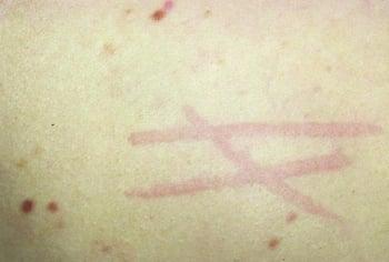
Dermatographism, or skin-writing, may occur when the skin is lightly scratched and results in raised red lines.


This photo shows band-like linear wheals on the torso induced by friction, which are characteristic of symptomatic dermatographism, with edema and texture change serving as key identifying features.
Darier sign refers to rapid swelling of a lesion when stroked. It occurs in patients with urticaria pigmentosa or mastocytosis.


This photo shows the characteristic finding of transient urticarial swelling around the lesion site, known as Darier sign.
Nikolsky sign is epidermal shearing that occurs with gentle lateral pressure on seemingly uninvolved skin in patients with toxic epidermal necrolysis and some autoimmune bullous diseases.
Auspitz sign is the appearance of pinpoint bleeding after scale is removed from plaques in psoriasis.
Koebner phenomenon describes the development of lesions within areas of trauma (eg, caused by scratching, rubbing, or injury). Psoriasis frequently exhibits this phenomenon, as may lichen planus, often resulting in linear lesions.
Reference
1. Kulthanan K, Bernstein JA, Rudenko M, et al. The Prevalence of Symptomatic Dermographism: Results of the International UCARE PREVALENCE-D Study. Allergy. 2026;81(2):468-479. doi:10.1111/all.70047
Drug Information for the Topic