Chronic obstructive pulmonary disease (COPD) is airflow limitation caused by an inflammatory response to inhaled toxins, often cigarette smoke. Alpha-1 antitrypsin deficiency and various occupational exposures are less common causes in patients who do not smoke. Symptoms are productive cough and dyspnea that develop over years; common signs include decreased breath sounds, prolonged expiratory phase of respiration, and wheezing. Severe cases may be complicated by weight loss, pneumothorax, frequent acute decompensation episodes, right heart failure, and/or acute or chronic respiratory failure. Diagnosis is based on history, physical examination, chest radiograph, and pulmonary function tests. Treatment is with bronchodilators, glucocorticoids, and, when necessary, oxygen and antibiotics. Biologics may be used for selected patients with eosinophilic inflammation and frequent exacerbations. Lung volume reduction procedures or transplantation are used in advanced disease. Survival in COPD is related to the severity of airflow limitation, the frequency of exacerbations, and the presence of comorbidities.
Chronic obstructive pulmonary disease (COPD) is defined as a heterogeneous lung condition characterized by chronic respiratory symptoms (dyspnea, cough, sputum production, and/or exacerbations) due to abnormalities of the airways (bronchitis, bronchiolitis) and/or alveoli (emphysema) that cause persistent, often progressive, airflow obstruction (1).
COPD comprises:
Chronic obstructive bronchitis (clinically defined)
Emphysema (pathologically or radiologically defined)
Many patients have features of both.
Chronic obstructive bronchitis is chronic bronchitis with airflow obstruction. Chronic bronchitis is defined as productive cough on most days of the week for a total of at least 3 months per year for 2 consecutive years. Chronic bronchitis becomes chronic obstructive bronchitis if spirometric evidence of airflow obstruction develops. Chronic asthmatic bronchitis is a similar, overlapping condition characterized by chronic productive cough, wheezing, and partially reversible airflow obstruction; it occurs predominantly in patients who smoke and have a history of asthma. When asthma and COPD co-exist in the same patient, pharmacologic treatment should follow guidelines for asthma (1).
Emphysema is destruction of lung parenchyma leading to a loss of elastic recoil, loss of alveolar septa, and radial airway traction, which increases the tendency for airway collapse. Lung hyperinflation, airflow limitation, and air trapping follow. Airspaces enlarge and may eventually develop blebs or bullae. Obliteration of small airways is thought to be the earliest lesion that precedes the development of emphysema.
Asthma and COPD can coexist in the same patient, a clinical presentation sometimes described as asthma-COPD overlap.
General reference
1. Global Initiative for Chronic Obstructive Lung Disease (GOLD): Diagnosis and assessment. Global Strategy for the Prevention, Diagnosis, and Management of COPD: 2026 report.
Epidemiology of COPD
The prevalence, incidence, and mortality rates of COPD increase with age. COPD may exhibit familial clustering independent of alpha-1 antitrypsin deficiency (alpha-1 antiprotease inhibitor deficiency).
Estimates of the global prevalence of COPD vary widely depending on the method of ascertainment. For example, the Global Burden of Disease study reported a prevalence of COPD of 2.5% in 2021 (1). In the United States, the National Health and Nutritional Examination Survey (NHANES) found a prevalence of airflow limitation in 13.4% of adults, although 71% of these individuals had not been diagnosed with COPD (2). In the National Health Interview Survey, the prevalence of self-reported COPD was higher in women (4.1%) than men (3.4%) (3).
COPD remains a leading cause of death worldwide (1, 4, 5, 6). Approximately 141,000 deaths from COPD occur annually in the United States (7).
Epidemiology references
1. Wang Z, Lin J, Liang L, et al. Global, regional, and national burden of chronic obstructive pulmonary disease and its attributable risk factors from 1990 to 2021: an analysis for the Global Burden of Disease Study 2021. Respir Res.2025;26(1):2. doi:10.1186/s12931-024-03051-2
2. Martinez CH, Mannino DM, Jaimes FA, et al. Undiagnosed Obstructive Lung Disease in the United States. Associated Factors and Long-term Mortality. Ann Am Thorac Soc. 2015;12(12):1788-1795. doi:10.1513/AnnalsATS.201506-388OC
3. Weeks JD, Elgaddal N. Chronic Obstructive Pulmonary Disease in Adults Age 18 and Older: United States, 2023. NCHS Data Brief. 2025;(529):1. doi:10.15620/cdc/174596
4. GBD 2023 Causes of Death Collaborators. Global burden of 292 causes of death in 204 countries and territories and 660 subnational locations, 1990-2023: a systematic analysis for the Global Burden of Disease Study 2023. Lancet. 2025;406(10513):1811-1872. doi:10.1016/S0140-6736(25)01917-8
5. World Health Organization (WHO). Fact Sheet. Chronic obstructive pulmonary disease (COPD). November 6, 2024. Accessed April 30, 2026.
6. Centers for Disease Control and Prevention. National Center for Health Statistics: Leading Causes of Death. Updated February 5, 2026. Accessed April 29, 2026.
7. Centers for Disease Control and Prevention. Chronic Obstructive Pulmonary Disease (COPD). Updated May 15, 2024. Accessed April 29, 2026.
Etiology of COPD
There are 2 main causes of COPD:
Smoking (and less often other inhalational exposures)
Genetic factors
Smoking and other inhalational exposures
Among all inhalational exposures, cigarette smoking is the primary risk factor in most countries, although only approximately 15% of people who smoke develop clinically apparent COPD (1). The risk for COPD increases with both duration (years of smoking) and cumulative dose (pack-years) (2). People who smoke and also have preexisting airway reactivity (defined by increased sensitivity to inhaled methacholine), even in the absence of clinically apparent asthma, are at greater risk of developing COPD than are those without airway reactivity.
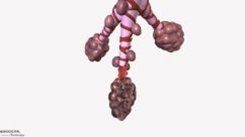
Smoke from indoor cooking and heating is an important causative factor in countries where indoor fires are commonly used for cooking or heating (3). The magnitude of risk of development of COPD from biomass cooking is highest in resource-limited countries and may be confounded by the other effects of poverty (4).
Exposure to passive cigarette smoke, air pollution, and occupational dust (eg, mineral dust, cotton dust) or inhaled chemicals (eg, cadmium) also contribute to the risk of COPD but are of less relevance compared to cigarette smoking. Low body weight and childhood respiratory disorders (eg, childhood wheezing, asthma) also contribute to the risk of COPD.
Genetic factors
The best-defined causative genetic disorder is alpha-1 antitrypsin deficiency, which is an important cause of emphysema in people who do not smoke and markedly increases susceptibility to development of COPD in people who do.
Over 80 genetic alleles have been found to be associated with COPD or decline in lung function in selected populations (5). However, the most consequential allele is alpha-1 antitrypsin.
Etiology references
1. Wheaton AG, Liu Y, Croft JB, et al. Chronic Obstructive Pulmonary Disease and Smoking Status - United States, 2017. MMWR Morb Mortal Wkly Rep. 2019;68(24):533-538. doi:10.15585/mmwr.mm6824a1
2. Bhatt SP, Kim YI, Harrington KF, et al. Smoking duration alone provides stronger risk estimates of chronic obstructive pulmonary disease than pack-years. Thorax. 2018;73(5):414-421. doi:10.1136/thoraxjnl-2017-210722
3. Ortiz-Quintero B, Martínez-Espinosa I, Pérez-Padilla R. Mechanisms of Lung Damage and Development of COPD Due to Household Biomass-Smoke Exposure: Inflammation, Oxidative Stress, MicroRNAs, and Gene Polymorphisms. Cells. 2022;12(1):67. doi:10.3390/cells12010067
4. Mortimer K, Montes de Oca M, Salvi S, et al. Household air pollution and COPD: cause and effect or confounding by other aspects of poverty? Int J Tuberc Lung Dis. 2022;26(3):206-216. doi:10.5588/ijtld.21.0570
5. Cho MH, Hobbs BD, Silverman EK. Genetics of chronic obstructive pulmonary disease: understanding the pathobiology and heterogeneity of a complex disorder. Lancet Respir Med. 2022;10(5):485-496. doi:10.1016/S2213-2600(21)00510-5
Pathophysiology of COPD
Various factors contribute to the pathophysiology of COPD, including a complex interplay of chronic inflammation, oxidative damage, and infection, leading to structural remodeling and progressive, incompletely reversible airflow limitation.
Inflammation
Inhalational exposures can trigger an inflammatory response in airways and alveoli that leads to disease in genetically susceptible people. The process is thought to be mediated by an increase in protease activity and a decrease in antiprotease activity. Lung proteases, such as neutrophil elastase, matrix metalloproteinases, and cathepsins, break down elastin and connective tissue during normal tissue repair. Their activity is normally balanced by antiproteases, such as alpha-1 antitrypsin, airway epithelium–derived secretory leukoproteinase inhibitor, elafin, and matrix metalloproteinase tissue inhibitor. In patients with COPD, activated neutrophils and other inflammatory cells (eg, macrophages) release proteases as part of the inflammatory process; protease activity exceeds antiprotease activity, and tissue destruction and mucus hypersecretion result.
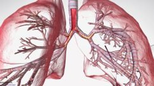
Activation of neutrophils and macrophages also leads to oxidative damage via the accumulation of free radicals, superoxide anions, and hydrogen peroxide, which inhibit antiproteases and cause bronchoconstriction, mucosal edema, and mucous hypersecretion. Neutrophil-induced oxidative damage, release of profibrotic neuropeptides (eg, bombesin), and reduced levels of vascular endothelial growth factor (VEGF) may contribute to apoptotic destruction of lung parenchyma.
The inflammation in COPD increases as disease severity increases, and, in severe (advanced) disease, inflammation does not resolve completely despite smoking cessation. This chronic inflammation may not respond to glucocorticoids, particularly in patients who continue to smoke cigarettes (1).
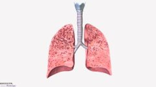
Mechanical stress on the alveoli from over-distension may make them susceptible to proteases, leading to alveolar septal destruction and progressive emphysema. This is particularly notable in the upper lobes of the lung (2).


This illustration of the lungs and bronchial branches shows bronchitis and emphysema. In emphysema (inset top right), the alveoli are destroyed, affecting lung elasticity. Chronic bronchitis (inset top left) is characterized by the persistent inflammation and mucus in the bronchi.
ILUSMEDICAL/SCIENCE PHOTO LIBRARY
Infection
Respiratory infections (to which patients with COPD are prone) may amplify progression of lung destruction.
Bacteria, especially Haemophilus influenzae, colonize the lower airways of approximately 30% of patients with COPD (3). In more severely affected patients (eg, those with previous hospitalizations), colonization with Pseudomonas aeruginosa or other gram-negative bacteria is common. Smoking and airflow obstruction may lead to impaired mucus clearance in lower airways, which predisposes to infection. Repeated bouts of infection increase the inflammatory burden that hastens disease progression. There is no direct evidence, however, that long-term use of antibiotics slows disease progression in COPD, despite the short-term benefits of some antibiotics (eg, azithromycin) in reducing exacerbation frequency and improving quality of life (4).
Viral infections, particularly rhinovirus, can account for up to 50% of COPD exacerbations and lead to progression of disease via intense airway epithelial inflammation (5).
Airflow limitation
The cardinal pathophysiologic feature of COPD is airflow limitation caused by airway narrowing and/or obstruction, loss of elastic recoil, or both.
There are 2 basic pathways by which COPD can develop and manifest with symptoms in later life:
In the first pathway, patients may have normal lung function in early adulthood, which is followed by a more rapid decline in the volume of air forcefully expired during the first second after taking a full breath (FEV1; ≥ 60 mL/year).
In the second pathway, patients have impaired lung function in early adulthood, often associated with asthma or other childhood respiratory disease. In these patients, COPD may present with a normal age-related decline in FEV1 (approximately 30 mL/year).
Although this second pathway model is conceptually helpful, a wide range of individual trajectories is possible (6). When the FEV1 falls below approximately 1 L, patients develop dyspnea during activities of daily living (although dyspnea is more closely related to the degree of dynamic hyperinflation [progressive hyperinflation due to incomplete exhalation] than to the degree of airflow limitation). When the FEV1 falls below approximately 0.8 L, patients are at risk of hypoxemia, hypercapnia, and cor pulmonale.
Airway narrowing and obstruction are caused by inflammatory mucus hypersecretion, mucus plugging, mucosal edema, bronchospasm, peribronchial fibrosis, and remodelling of small airways or a combination of these mechanisms. Alveolar septa are destroyed, reducing parenchymal attachments to the airways and thereby facilitating airway closure during expiration.
Enlarged alveolar spaces sometimes consolidate into bullae, defined as airspaces ≥ 1 cm in diameter. Bullae may be entirely empty or have strands of lung tissue traversing them in areas of locally severe emphysema; they occasionally occupy an entire hemithorax. These changes lead to loss of elastic recoil and lung hyperinflation.
Increased airway resistance increases the work of breathing. Lung hyperinflation, although it decreases airway resistance, also increases the work of breathing. Increased work of breathing may lead to respiratory muscle fatigue and alveolar hypoventilation with hypercapnia. Hypoxia and hypercapnia can also be caused by ventilation/perfusion (V/Q) mismatch.
Complications
In addition to airflow limitation and sometimes respiratory insufficiency, complications include:
Pulmonary hypertension
Respiratory infection
Weight loss and other comorbidities
Chronic alveolar hypoxia increases pulmonary vascular tone, which, if diffuse, causes pulmonary hypertension and cor pulmonale. The increase in pulmonary vascular pressure may be augmented by the destruction of the pulmonary capillary bed due to destruction of alveolar septa.
Viral or bacterial respiratory infections are common among patients with COPD and cause a large percentage of acute exacerbations. It is currently thought that acute bacterial infections are due to acquisition of new strains of bacteria rather than overgrowth of chronic colonizing bacteria.
Weight loss may occur, and has been consistently associated with the insufficient caloric intake and increased levels of circulating tumor necrosis factor (TNF)-alpha that is seen in patients with COPD (7, 8). A mismatch between caloric expenditure and nutritional intake typically occurs because caloric expenditure can be high in the presence of heightened inflammatory cytokines and hypoxemia.
Other coexisting or complicating disorders that adversely affect quality of life and/or survival include frailty, osteoporosis, depression, anxiety, coronary artery disease, arrhythmias, lung cancer and other cancers, muscle atrophy, and gastroesophageal reflux. Whether these disorders arise from COPD itself, smoking, or shared pathogenic mechanisms causing systemic inflammation remains uncertain.
Pathophysiology references
1. Adcock IM, Bhatt SP, Balkissoon R, Wise RA. The Use of Inhaled Corticosteroids for Patients with COPD Who Continue to Smoke Cigarettes: An Evaluation of Current Practice. Am J Med. 2022;135(3):302-312. doi:10.1016/j.amjmed.2021.09.006
2. Suki B, Sato S, Parameswaran H, Szabari MV, Takahashi A, Bartolák-Suki E. Emphysema and mechanical stress-induced lung remodeling. Physiology (Bethesda). 2013;28(6):404-413. doi:10.1152/physiol.00041.2013
3. Short B, Carson S, Devlin AC, et al: Non-typeable Haemophilus influenzae chronic colonization in chronic obstructive pulmonary disease (COPD). Crit Rev Microbiol. 2021;47(2):192-205. https://doi.org/10.1080/1040841X.2020.1863330
4. Janjua S, Mathioudakis AG, Fortescue R, et al. Prophylactic antibiotics for adults with chronic obstructive pulmonary disease: a network meta-analysis. Cochrane Database Syst Rev. 2021;1(1):CD013198. doi:10.1002/14651858.CD013198.pub2
5. Owuor N, Nalamala N, Gimenes JA Jr, Sajjan US. Rhinovirus and COPD airway epithelium. Pulm Crit Care Med. 2017;2(3):10.15761/PCCM.1000139. doi:10.15761/PCCM.1000139
6. Lange P, Celli B, Agusti A, et al: Lung-function trajectories leading to chronic obstructive pulmonary disease. N Engl J Med 373(2):111–122, 2015.
7. De Brandt J, Beijers RJHCG, Chiles J, et al. Update on the Etiology, Assessment, and Management of COPD Cachexia: Considerations for the Clinician. Int J Chron Obstruct Pulmon Dis. 2022;17:2957-2976. Published 2022 Nov 18. doi:10.2147/COPD.S334228
8. Takabatake N, Nakamura H, Abe S, et al. The relationship between chronic hypoxemia and activation of the tumor necrosis factor-alpha system in patients with chronic obstructive pulmonary disease. Am J Respir Crit Care Med. 2000;161(4 Pt 1):1179-1184. doi:10.1164/ajrccm.161.4.9903022
Symptoms and Signs of COPD
COPD takes years to develop and progress.
Productive cough is the initial symptom, typically developing among people who smoke and are in their 40s and 50s.
Dyspnea that is progressive, persistent, exertional, or worse during respiratory infection appears when patients are in their late 50s or 60s.
Other symptoms include chest tightness, fatigue, and activity limitation, all of which are more pronounced during an exacerbation. Symptoms usually progress quickly in patients who continue to smoke and in those who have a higher lifetime tobacco exposure. Morning headache develops in more advanced disease and signals nocturnal hypercapnia or hypoxemia.
Signs of COPD include wheezing, a prolonged expiratory phase of breathing, lung hyperinflation manifested as decreased heart and lung sounds, and increased anteroposterior diameter of the thorax (barrel chest).
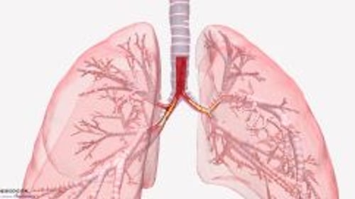
Signs of advanced disease include pursed-lip breathing, accessory respiratory muscle use, paradoxical inward movement of the lower rib cage during inspiration (Hoover sign), and cyanosis. Signs of cor pulmonale include neck vein (eg, jugular vein) distention, splitting of the second heart sound with an accentuated pulmonic component, tricuspid insufficiency murmur, and peripheral edema. Right ventricular heaves may be imperceptible in COPD because the lungs are hyperinflated.
Patients with advanced emphysema lose weight and experience muscle wasting.
Spontaneous pneumothorax may occur (possibly related to rupture of bullae) and should be suspected in any patient with COPD whose pulmonary status abruptly worsens.
The symptoms can be graded according to which activities cause dyspnea (see table ).
Breathlessness Measurement Using the Modified British Medical Research Council (mMRC) Questionnaire
Grade | Shortness of Breath |
|---|---|
0 | None except during strenuous exercise |
1 | Occurring when hurrying on level ground or walking up a slight incline |
2 | Resulting in walking more slowly than people of the same age on level ground or Resulting in stopping for breath when walking at own pace on level ground |
3 | Resulting in stopping for breath after walking about 100 meters or after a few minutes on level ground |
4 | Preventing the person from leaving the house or Occurring when dressing or undressing |
Adapted from Mahler DA, Wells CK. Evaluation of clinical methods for rating dyspnea. Chest. 1988;93(3):580-586. doi: 10.1378/chest.93.3.580 | |
Acute exacerbations
Acute exacerbations occur sporadically during the course of COPD and are heralded by increased symptom severity. The specific cause of any exacerbation is almost always impossible to determine, but exacerbations are often attributed to viral respiratory infections, acute bacterial bronchitis, or exposure to respiratory irritants. As COPD progresses, acute exacerbations tend to become more frequent, averaging approximately 1 to 3 episodes/year.
Diagnosis of COPD
Chest imaging
Pulmonary function testing
The diagnosis of COPD is suggested by a combination of history, physical examination, and chest imaging findings and is confirmed by pulmonary function tests (1).
Symptoms similar to those of COPD can be caused by asthma, heart failure, and bronchiectasis (see table ). COPD and asthma are sometimes easily confused and may coexist in an individual (also called asthma-COPD overlap).
Differential Diagnosis of COPD
Diagnosis | Onset | Imaging Results | Other Features |
|---|---|---|---|
COPD | Middle age | Sometimes lung hyperinflation, bullae, increased retrosternal air space, and/or bronchial wall thickening (seen on chest imaging); however, imaging is usually not helpful diagnostically and is done mainly to exclude other disorders | Slowly progressive symptoms History of smoking or exposure to tobacco or other types of smoke |
Early in life (often during childhood) | Usually normal or possibly hyperinflation (eg, during an asthma exacerbation) | Symptoms vary widely from day to day Variable but reversible airflow obstruction Symptoms often worse at night or early morning History of allergies, rhinosinusitis, or eczema Often family history of asthma | |
All ages, but most often in older or middle aged adults | Bronchial dilation and bronchial wall thickening (seen on chest radiograph or chest CT) | Often large amounts of purulent sputum Often history of chronic recurrent bacterial infection | |
Diffuse panbronchiolitis | Usually between ages 10 and 60 years (mean age of 40 years) | Diffuse small centrilobular nodular opacities and hyperinflation seen on chest radiograph and high-resolution CT | Mostly males who do not smoke Almost all patients have chronic sinusitis Predominately in individuals of Asian descent |
All ages, but most often in older or middle aged adults | Enlarged heart, pleural effusion, fluid in major lung fissure, sometimes pulmonary edema (seen on chest radiograph) | Volume restriction without airflow limitation (detected by pulmonary function tests) | |
Obliterative bronchiolitis | Onset at younger age | Peripheral hypodense areas (seen on chest CT during expiration) | History of lung or bone marrow transplantation Inspiratory squeaks (ie, high-pitched, end-inspiratory wheezes) on auscultation |
All ages | Lung infiltrates and cavitation (typically on the upper lobes) seen on chest radiograph | Confirmed by microbiologic testing (eg, NAAT, AFB staining, mycobacterial culture) Usually in areas with high prevalence of TB | |
Data adapted from Global Initiative for Chronic Obstructive Lung Disease (GOLD): Diagnosis and assessment. Global Strategy for the Prevention, Diagnosis, and Management of COPD: 2026 report. | |||
AFB = acid-fast bacillus; NAAT = nucleic acid amplification test. | |||
Systemic disorders that may have a component of airflow limitation may suggest COPD; they include HIV infection, abuse of illicit drugs (particularly cocaine and amphetamines), sarcoidosis, Sjögren syndrome, bronchiolitis obliterans, lymphangioleiomyomatosis, and eosinophilic granuloma. COPD can be differentiated from interstitial lung diseases by chest imaging, which shows increased interstitial markings in interstitial lung disease, and pulmonary function testing, which shows a restrictive rather than an obstructive ventilatory defect. In some patients, COPD and interstitial lung disease coexist (combined pulmonary fibrosis and emphysema [CPFE]) in which lung volumes are relatively preserved, but gas exchange is severely impaired.
Pulmonary function tests
Patients suspected of having COPD should undergo pulmonary function testing to confirm airflow limitation, to quantify its severity and reversibility, and to distinguish COPD from other disorders. Pulmonary function testing is also useful for following disease progression and monitoring response to treatment. The primary diagnostic parameters are:
FEV1: The volume of air forcefully expired during the first second after taking a full breath
Forced vital capacity (FVC): The total volume of air expired with maximal force
Flow-volume loops: Simultaneous spirometric recordings of airflow and volume during forced maximal expiration and inspiration
In patients with COPD, there are reductions of FEV1, FVC, and the ratio of FEV1/FVC are the hallmark of airflow limitation. Flow-volume loops show a concave pattern in the expiratory tracing (see figure ).
FEV1 and FVC are easily measured with office spirometry. Normal reference values are determined by patient age, sex, and height. It is recommended that race not be used for calculating predicted reference values (2). (See also Airflow, Lung Volumes, and Flow-Volume Loop.)


Airflow limitation severity in patients with COPD and FEV1/FVC < 0.70 can be classified based on post-bronchodilator FEV1 (1):
Mild: ≥ 80% of predicted
Moderate: 50% to 79% of predicted
Severe: 30% to 49% of predicted
Very severe: < 30% of predicted
Additional pulmonary function testing is necessary only in specific circumstances, such as before lung volume reduction procedures. Other test abnormalities may include:
Increased total lung capacity
Increased functional residual capacity
Increased residual volume
Decreased vital capacity
Decreased single-breath diffusing capacity for carbon monoxide (DLCO)
Findings of increased total lung capacity, functional residual capacity, and residual volume can help distinguish COPD from restrictive pulmonary disease, in which these measures are diminished.
Decreased DLCO is nonspecific and is reduced in other disorders that affect the pulmonary vascular bed, such as interstitial lung disease, but can help distinguish emphysema from asthma, in which DLCO is normal or elevated.
A determination of whether a patient's symptoms are a result of COPD or asthma is best made by comparison of pulmonary function test findings (see table ).
Pulmonary Function Test (PFT) Findings in Chronic Obstructive Pulmonary Disease (COPD) and Asthma
PFT Parameter | Typical COPD Finding | Typical Asthma Finding |
|---|---|---|
FEV1/FVC Ratio | < 0.70 | Typically normal < 0.80 during asthma exacerbation |
FEV1 | Little to no improvement with bronchodilator (< 12% and < 200 mL) | Significant improvement with bronchodilator (> 12% and > 200 mL) |
RV | Increased (> 120–140%) | Normal Increased only during asthma exacerbation |
TLC | Increased (> 120%) | Increased only during asthma exacerbation |
DLCO | Decreased | Normal or increased |
DLCO = diffusing capacity for carbon monoxide; FEV1 = volume of air forcefully expired during the first second after taking a full breath; FVC = forced vital capacity; RV = residual volume ; TLC = total lung capacity. | ||
Data adapted from Global Initiative for Chronic Obstructive Lung Disease (GOLD): Diagnosis and assessment. Global Strategy for the Prevention, Diagnosis, and Management of COPD: 2026 report and Global Initiative for Asthma (GINA). Global Strategy for Asthma Management and Prevention: 2026 report. | ||
Imaging tests
Chest radiograph may reveal characteristic findings. In patients with emphysema, changes can include lung hyperinflation manifested as a flat diaphragm (ie, increase in the angle formed by the sternum and anterior diaphragm on a lateral film from the normal value of 45° to > 90°), rapid tapering of hilar vessels, and bullae (ie, radiolucencies > 1 cm surrounded by arcuate, hairline shadows). Other typical findings include enlargement of the retrosternal airspace and a narrow cardiac shadow. Emphysematous changes occurring predominantly in the lung bases suggest alpha-1 antitrypsin deficiency. The lungs may look normal or have increased lucency secondary to loss of parenchyma. Among patients with chronic obstructive bronchitis, chest radiographs may be normal or may show a bibasilar increase in bronchovascular markings as a result of bronchial wall thickening.

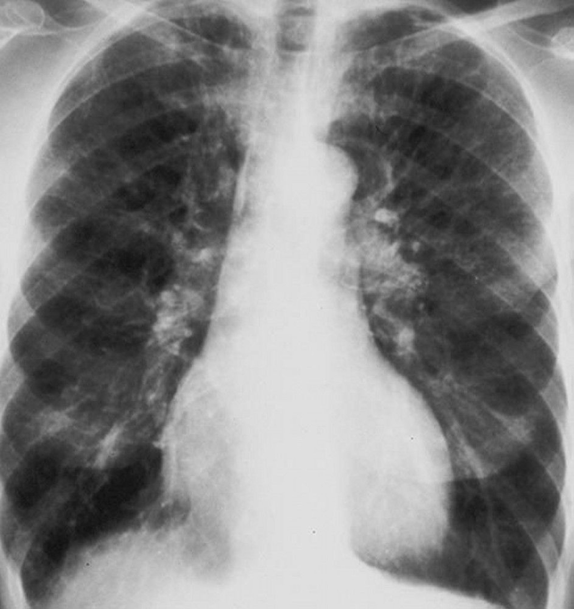
Chest radiograph of a patient with COPD. The lungs are hyperinflated, the diaphragm is flattened, vascular markings are increased, and the heart size is marginally increased.
By permission of the publisher. From Barnes P. In Bone's Atlas of Pulmonary and Critical Care Medicine. Edited by J Crapo. Philadelphia, Current Medicine, 2005.

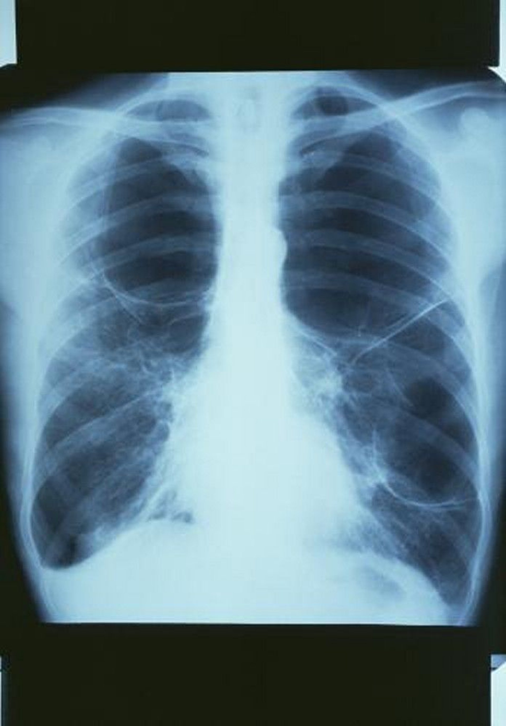
This chest radiograph shows a large bulla in the upper right lung and 2 large bullae in the left lung.
GJLP/CNRI/SCIENCE PHOTO LIBRARY
Prominent hila suggest large central pulmonary arteries that may signify pulmonary hypertension. Right ventricular enlargement that occurs in cor pulmonale may be masked by lung hyperinflation or may manifest as encroachment of the heart shadow on the retrosternal space or by widening of the transverse cardiac shadow in comparison with previous chest radiographs.
Chest CT may reveal abnormalities that are not apparent on the chest radiograph and may also suggest coexisting or complicating disorders, such as pneumonia, pneumoconiosis, or lung cancer. CT helps assess the extent and distribution of emphysema, estimated either by visual scoring or with analysis of the distribution of lung density. Indications for obtaining CT in patients with COPD include evaluation for lung volume reduction procedures, suspicion of coexisting or complicating disorders that are not clearly evident or excluded by chest radiograph, suspicion of lung cancer, and screening for lung cancer. Enlargement of the pulmonary artery diameter greater than the ascending aorta diameter suggests pulmonary hypertension (3).
Adjunctive tests
Alpha-1 antitrypsin levels should be measured in patients < 50 years with symptomatic COPD and in patients of any age with COPD who do not smoke to detect alpha-1 antitrypsin deficiency. Other indications of possible alpha-1 antitrypsin deficiency include a family history of premature COPD or unexplained liver disease, lower-lobe distribution of emphysema, and COPD associated with antineutrophil cytoplasmic antibody (ANCA)-positive vasculitis. If levels of alpha-1 antitrypsin are low, the diagnosis should be confirmed by genetic testing to establish the alpha-1 antitrypsin phenotype.
ECG, often done to exclude cardiac causes of dyspnea, typically shows diffusely low QRS voltage with a vertical heart axis caused by lung hyperinflation and increased P-wave voltage or rightward shifts of the P-wave vector caused by right atrial enlargement in patients with advanced emphysema. Findings of right ventricular hypertrophy include an R or R′ wave as tall as or taller than the S wave in lead V1; an R wave smaller than the S wave in lead V6; right-axis deviation > 110° without right bundle branch block; or some combination of these findings. Multifocal atrial tachycardia, an arrhythmia that can accompany COPD, manifests as a tachyarrhythmia with polymorphic P waves and variable PR intervals.
Echocardiography is occasionally useful for assessing right ventricular function and pulmonary hypertension, although air trapping makes it technically difficult in patients with COPD. Echocardiography is most often indicated when coexistent left ventricular or valvular heart disease is suspected.
Hemoglobin and hematocrit are of little diagnostic value in the evaluation of COPD but may show reactive erythrocytosis (hematocrit > 48%) if the patient has chronic hypoxemia. Patients with anemia (for reasons other than COPD) have disproportionately severe dyspnea. The differential white blood cell count (eg, neutrophilia, eosinophilia) may be helpful. Neutrophilia has been associated with a higher incidence of exacerbations and eosinophilia is well-established to predict response to inhaled glucocorticoids and anti-interleukin (IL)-5 biologic therapies.
Serum electrolytes are of little value but may show an elevated bicarbonate level (ie, compensatory metabolic alkalosis) if patients have chronic hypercapnia. Venous blood gases are useful for diagnosis of acute or chronic hypercapnia.
Evaluation of exacerbations
Patients with acute exacerbations usually have combination of increased cough, sputum, dyspnea, and work of breathing, as well as low oxygen saturation determined with pulse oximetry, diaphoresis, tachycardia, anxiety, and cyanosis. Patients with exacerbations accompanied by retention of carbon dioxide may be lethargic or somnolent.
All patients requiring hospitalization for an acute exacerbation should undergo testing to quantify hypoxemia and hypercapnia. Hypercapnia may exist without hypoxemia.
Findings of partial pressure of arterial oxygen (PaO2) < 50 mm Hg, or partial pressure of carbon dioxide in arterial blood (PaCO2) > 50 mm Hg, or partial pressure of carbon dioxide in venous blood (PvCO2) > 55 mm Hg in patients with respiratory acidemia (pH < 7.35) define acute respiratory failure. Some patients chronically manifest such levels of PaO2 and PaCO2 in the absence of acute respiratory failure.
A chest radiograph is often done to check for pneumonia or pneumothorax. Point-of-care ultrasound (POCUS) has been increasingly useful as an adjunctive procedure for rapid bedside diagnosis of pneumonia or pneumothorax. In patients with acute onset of symptoms, chest CT angiogram is done to check for pulmonary embolism. Very rarely, among patients receiving chronic systemic glucocorticoids, infiltrates may represent Aspergillus pneumonia.
Yellow or green sputum may be associated with neutrophils (mainly, because of the presence of the enzyme myeloperoxidase) in the sputum and raises suspicion for the presence of bacterial colonization or infection (4).
Sputum culture is usually done in hospitalized patients but is not usually necessary in outpatients. In samples from outpatients, Gram stain usually shows neutrophils with a mixture of organisms, often gram-positive diplococci (Streptococcus pneumoniae), gram-negative bacilli (H. influenzae), or both. However, culture and microscopic examination of sputum is usually not necessary for outpatients. Other oropharyngeal commensal organisms, such as Moraxella catarrhalis (formerly known as Branhamella catarrhalis), occasionally cause exacerbations. In hospitalized patients, resistant gram-negative organisms (eg, Pseudomonas) or, rarely, Staphylococcus may be identified in culture specimens.
During influenza season, a rapid influenza test may help guide treatment with anti-influenza agents (eg, oseltamivir), and a respiratory viral panel for respiratory syncytial virus (RSV), rhinovirus, and metapneumovirus may allow tailoring of antimicrobial therapy. Testing for COVID-19 and consideration of COVID-19–specific therapies is also indicated.
Serum C-reactive protein (CRP) is helpful in guiding the use of antibiotics during exacerbations. In clinical trials, the use of antibiotics can be decreased in patients with low CRP without evidence of harm (5, 6). Evidence regarding use of the inflammatory biomarker procalcitonin is conflicting; while it has been found to be beneficial in guiding antibiotic therapy in some outpatient settings, meta-analyses have largely been inconclusive and ICU studies have shown harm (7). Therefore, it is not recommended to use procalcitonin to guide antibiotic treatment during COPD exacerbations.
Diagnosis references
1. Global Initiative for Chronic Obstructive Lung Disease (GOLD): Diagnosis and assessment. Global Strategy for the Prevention, Diagnosis, and Management of COPD: 2026 report.
2. Bhakta NR, Bime C, Kaminsky DA, et al. Race and ethnicity in pulmonary function test interpretation: An Official American Thoracic Society Statement. Am J Respir Crit Care Med. 2023;207(8):978-995. doi:10.1164/rccm.202302-0310ST
3. Iyer AS, Wells JM, Vishin S, et al. CT scan-measured pulmonary artery to aorta ratio and echocardiography for detecting pulmonary hypertension in severe COPD. Chest. 2014;145(4):824-832. doi:10.1378/chest.13-1422
4. Spies R, Potter M, Hollamby R, et al. Sputum Color as a Marker for Bacteria in Acute Exacerbations of Chronic Obstructive Pulmonary Disease: A Systematic Review and Meta-analysis. Ann Am Thorac Soc. 2023;20(5):738-748. doi:10.1513/AnnalsATS.202204-319OC
5. Butler CC, Gillespie D, White P, et al. C-Reactive protein testing to guide antibiotic prescribing for COPD exacerbations. N Engl J Med. 2019;381(2):111-120. doi: 10.1056/NEJMoa1803185
6. Prins HJ, Duijkers R, van der Valk P, et al. CRP-guided antibiotic treatment in acute exacerbations of COPD in hospital admissions. Eur Respir J. 2019;53(5):1802014. doi: 10.1183/13993003.02014-2018
7. Chen K, Pleasants KA, Pleasants RA, et al. Procalcitonin for Antibiotic Prescription in Chronic Obstructive Pulmonary Disease Exacerbations: Systematic Review, Meta-Analysis, and Clinical Perspective. Pulm Ther. 2020;6(2):201-214. doi:10.1007/s41030-020-00123-8
Treatment of COPD
Smoking cessation
Inhaled bronchodilators, corticosteroids (glucocorticoids), or both
Supportive care (eg, oxygen therapy, pulmonary rehabilitation)
COPD management involves minimization of symptoms and prevention and treatment of exacerbations (1). Timely diagnosis and initiation of appropriate treatment, particularly smoking cessation, long-term oxygen therapy in patients with hypoxemia, and medications including triple-inhaled therapy (ie, anticholinergics, long-acting beta-agonists, and inhaled corticosteroids (glucocorticoids) in symptomatic patients with frequent exacerbations can reduce long-term mortality. Treatment of cor pulmonale, a common complication of long-standing, severe COPD, is discussed elsewhere.
Smoking cessation is critical in treatment of COPD.
Treatment of chronic stable COPD aims to prevent exacerbations and improve lung and physical function. Treatment relieves symptoms rapidly with primarily short-acting beta-adrenergic medications and decreases exacerbations with inhaled corticosteroids (glucocorticoids), long-acting bronchodilators or a combination (see table ).
Pulmonary rehabilitation includes structured and supervised exercise training, nutrition counseling, and self-management education.
Oxygen therapy is indicated for selected patients.
Treatment of exacerbations ensures adequate oxygenation and near-normal blood pH, reverses airway obstruction, and treats underlying disorders.
Immunization against influenza, pneumococcus, COVID-19, and respiratory syncytial virus (RSV, in patients age 50 years and older) should also be recommended as a preventive measure. Tetanus-diphtheria-pertussis (Tdap) immunization is recommended for patients who were not immunized in adolescence to prevent pertussis.
End-of-life care
In patients with very severe disease, exercise is inadvisable and activities of daily living are arranged to minimize energy expenditure. For example, patients may arrange to live on one floor of the house, have several small meals rather than fewer large meals, and avoid wearing shoes that must be tied. End-of-life care should be discussed, including whether to pursue mechanical ventilation, the use of palliative sedation, and appointment of a surrogate medical decision-maker in the event of the patient’s incapacitation.
Treatment reference
1. Global Initiative for Chronic Obstructive Lung Disease (GOLD): Diagnosis and assessment. Global Strategy for the Prevention, Diagnosis, and Management of COPD: 2026 report.
Prognosis for COPD
Severity of airway obstruction predicts survival in patients with COPD.
Measurement of body mass index (B), the degree of airflow obstruction (O, which is the FEV1), dyspnea (D, which is measured using the ), and exercise capacity (E, which is measured with a 6-minute walk test) is called the BODE index (1). The BODE index predicts mortality more accurately than FEV₁ alone. Higher scores on the BODE index predict a greater risk of death.
Older age and the presence of heart disease, anemia, resting tachycardia, hypercapnia, or hypoxemia also predict decreased survival, whereas a significant response to bronchodilators predicts improved survival. Risk factors for death in patients with acute exacerbation requiring hospitalization include older age, higher PaCO2, and use of maintenance oral glucocorticoids.
Patients at high risk of imminent death are those with progressive unexplained weight loss or severe functional decline (eg, those who experience dyspnea with even minimal exertion, such as dressing, bathing, or eating).
Mortality in COPD may result from comorbidities (eg, cardiovascular disease, cancer) or intercurrent illnesses rather than from progression of the underlying disorder in patients who have stopped smoking. Death in patients with COPD is generally due to acute respiratory failure, pneumonia, lung cancer, heart disease, or pulmonary embolism.
Prognosis reference
1. Celli BR, Cote CG, Marin JM, et al. The body-mass index, airflow obstruction, dyspnea, and exercise capacity index in chronic obstructive pulmonary disease. N Engl J Med. 2004;350(10):1005-1012. doi:10.1056/NEJMoa021322
Key Points
Cigarette smoking in susceptible people is the major cause of chronic obstructive pulmonary disease (COPD) in the resource-rich settings.
COPD should be diagnosed after differentiating from disorders that have similar characteristics (eg, asthma, heart failure) primarily by routine clinical information, such as symptoms (particularly time course), age at onset, risk factors, and results of routine tests (eg, chest radiograph, pulmonary function tests).
Reductions of FEV1, FVC, and the ratio of FEV1/FVC post-bronchodilator (also called fixed obstruction) are characteristic findings.
Patients should be categorized based on symptoms and exacerbation risk into one of 3 groups and use that category to guide treatment.
Treatment of acute symptoms is primarily with short-acting beta-adrenergic medications.
Treatment of exacerbations is with inhaled corticosteroids (glucocorticoids), long-acting beta-adrenergic medications, long-acting anticholinergic medications, or a combination.
The BODE (body mass index, obstruction [FEV1], dyspnea, exercise capacity) index is helpful in predicting survival in patients with COPD,
Smoking cessation using multiple interventions should be encouraged in all patients who smoke.
Immunization against influenza, pneumococcus, COVID-19, Tdap, and RSV (in patients age ≥ 50 years) should also be done as a preventive measure.
Drug Information for the Topic