

Abdominal pain is common and often inconsequential. Acute and severe abdominal pain, however, is almost always a symptom of intra-abdominal disease. It may be the sole indicator of the need for surgery and must be attended to swiftly: Gangrene and perforation of the gut can occur < 6 hours from onset of symptoms in certain conditions (eg, interruption of the intestinal blood supply due to a strangulating obstruction or an arterial embolus). Abdominal pain is of particular concern in patients who are very young or very old and those who have HIV infection or are taking immunosuppressants (including corticosteroids).
Textbook descriptions of abdominal pain have limitations because people react to pain differently. Some, particularly older people, are stoic, whereas others exaggerate their symptoms. Infants, young children, and some older people may have difficulty localizing the pain (1).
The term acute abdomen refers to abdominal symptoms and signs of such severity or concern that disorders requiring surgery should be considered.
General reference
1. Lyon C, Clark DC. Diagnosis of acute abdominal pain in older patients. Am Fam Physician. 2006;74(9):1537-1544.
Pathophysiology of Acute Abdominal Pain
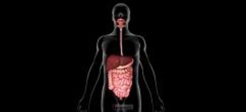
Visceral pain comes from the abdominal viscera, which are innervated by autonomic nerve fibers and respond mainly to the sensations of distention and muscular contraction—not to cutting, tearing, or local irritation. Visceral pain is typically vague, dull, and nauseating. It is poorly localized and tends to be referred to areas corresponding to the embryonic origin of the affected structure. Foregut structures (stomach, duodenum, liver, and pancreas) cause upper abdominal pain. Midgut structures (small bowel, proximal colon, and appendix) cause periumbilical pain. Hindgut structures (distal colon and genitourinary tract) cause lower abdominal pain.
Somatic pain comes from the parietal peritoneum, which is innervated by somatic nerves, which respond to irritation from infectious, chemical, or other inflammatory processes. Somatic pain is sharp and well localized.
Referred pain is pain perceived distant from its source and results from convergence of nerve fibers at the spinal cord. Common examples of referred pain are scapular pain due to biliary colic, groin pain due to renal colic, and shoulder pain due to blood or infection irritating the diaphragm.
Peritonitis
Peritonitis is inflammation of the peritoneal cavity.
The most serious cause is perforation of the gastrointestinal tract, which causes immediate chemical inflammation followed shortly by infection from intestinal organisms. Peritonitis can also result from any abdominal condition that causes marked inflammation (eg, appendicitis, diverticulitis, strangulating intestinal obstruction, pancreatitis, pelvic inflammatory disease, mesenteric ischemia).
Intraperitoneal blood from any source (eg, ruptured aneurysm, trauma, surgery, ectopic pregnancy) is irritating and results in peritonitis.
Barium causes severe peritonitis and should never be given to a patient with suspected gastrointestinal tract perforation. However, water-soluble contrast agents can be safely used.
Peritoneosystemic shunts, drains, and dialysis catheters in the peritoneal cavity predispose a patient to infectious peritonitis, as does ascitic fluid.
Rarely, spontaneous bacterial peritonitis occurs, in which the peritoneal cavity is infected by bloodborne bacteria. Spontaneous bacterial peritonitis occurs primarily in patients with cirrhosis and ascites.
Peritonitis causes fluid to shift into the peritoneal cavity and bowel, leading to severe dehydration and electrolyte disturbances. Acute respiratory distress syndrome can develop rapidly. Kidney failure, liver failure, and disseminated intravascular coagulation follow. Without treatment, death occurs within days.
Etiology of Acute Abdominal Pain
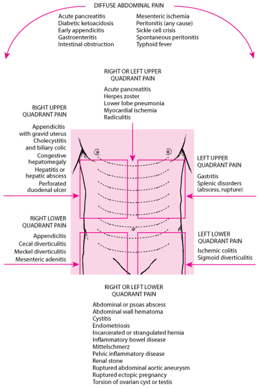
Many intra-abdominal disorders cause abdominal pain (see figure ); some are trivial, but some are immediately life threatening, requiring rapid diagnosis and surgery. These include ruptured abdominal aortic aneurysm (AAA), perforated viscus, mesenteric ischemia, and ruptured ectopic pregnancy. Others (eg, intestinal obstruction, appendicitis, severe acute pancreatitis) are also serious and nearly as urgent. Several extra-abdominal disorders also cause abdominal pain (see table ).
Location of Abdominal Pain and Possible Causes

Extra-Abdominal Causes of Abdominal Pain
Abdominal wall |
Rectus muscle hematoma |
Genitourinary |
Infectious |
Metabolic |
Thoracic |
Costochondritis Radiculitis |
Toxic |
Heavy metal poisoning Methanol poisoning |
Abdominal pain in neonates, infants, and young children has numerous causes not encountered in adults. These causes include
Meconium peritonitis
Volvulus of a gut with intestinal malrotation
Intestinal obstruction caused by atresia
Evaluation of Acute Abdominal Pain
Evaluation of mild and severe pain follows the same process, although with severe abdominal pain, therapy sometimes proceeds simultaneously and involves early consultation with a surgeon. History and physical examination usually exclude all but a few possible causes, with final diagnosis confirmed by judicious use of laboratory and imaging tests. Life-threatening causes should always be ruled out before focusing on less serious diagnoses.
In seriously ill patients with severe abdominal pain, the most important diagnostic measure may be expeditious surgical exploration.
In mildly ill patients, watchful waiting and a diagnostic evaluation may be best.
History
A thorough history usually suggests the diagnosis (see table ). Of particular importance are pain location (see figure ) and characteristics, history of similar symptoms, and associated symptoms. Concomitant symptoms such as heartburn, nausea, vomiting, diarrhea, constipation, jaundice, melena, hematuria, hematemesis, weight loss, and mucus or blood in the stool help direct subsequent evaluation.
A drug history should include details concerning prescription medication and illicit drug use as well as alcohol. Many medications cause gastrointestinal upset. Prednisone or immunosuppressants may inhibit the inflammatory response to perforation or peritonitis and result in less pain, tenderness, or leukocytosis than might otherwise be expected. Anticoagulants can increase the chances of bleeding and hematoma formation. Alcohol predisposes to pancreatitis.
History in Patients with Acute Abdominal Pain
Question | Potential Responses and Indications |
|---|---|
Where is the pain? | See figure Location of Abdominal Pain and Possible Causes. |
What is the pain like? | Acute waves of sharp constricting pain that “take the breath away” (renal or biliary colic) Waves of dull pain with vomiting (intestinal obstruction) Colicky pain that becomes steady (appendicitis, strangulating intestinal obstruction, mesenteric ischemia) Sharp, constant pain, worsened by movement (peritonitis) Tearing pain (dissecting aneurysm) Dull ache (appendicitis, diverticulitis, pyelonephritis) |
Have you had it before? | Yes suggests recurrent problems such as ulcer disease, gallstone colic, diverticulitis, or mittelschmerz |
Was the onset sudden? | Sudden: “Like a light switching on” (perforated ulcer, renal stone, ruptured ectopic pregnancy, ovarian torsion, testicular torsion, some ruptured aneurysms) Less sudden: Most other causes |
How severe is the pain? | Severe pain (perforated viscus, kidney stone, peritonitis, pancreatitis) Pain out of proportion to physical findings (mesenteric ischemia) |
Does the pain travel to any other part of the body? | Right scapula (gallbladder pain) Left shoulder region (ruptured spleen, pancreatitis) Pubis or vagina (renal pain) Back (ruptured aortic aneurysm, pancreatitis, sometimes perforated ulcer) |
What relieves the pain? | Antacids (peptic ulcer disease) Lying as quietly as possible (peritonitis) |
What other symptoms occur with the pain? | Vomiting precedes pain and is followed by diarrhea (gastroenteritis) Delayed vomiting, absent bowel movement and flatus (acute intestinal obstruction; the delay increases with a lower site of obstruction) Severe vomiting precedes intense epigastric, left chest, or shoulder pain (emetic perforation of the intra-abdominal esophagus) |
Known medical conditions and previous abdominal surgeries are important to ascertain.
Female patients of child-bearing age should be asked whether they are or might be pregnant.
Physical examination
The general appearance is important. A happy, comfortable-appearing patient rarely has a serious problem, unlike one who is anxious, pale, diaphoretic, or in obvious pain. Blood pressure, pulse, state of consciousness, and other signs of peripheral perfusion must be evaluated. However, the focus of the examination is the abdomen, beginning with inspection and auscultation, followed by percussion and palpation. Rectal examination and pelvic examination (for women and possibly adolescent girls) to locate tenderness, masses, and blood are essential.
Palpation begins gently, away from the area of greatest pain, detecting areas of particular tenderness, as well as the presence of guarding, rigidity, and rebound (all suggesting peritoneal irritation) and any masses. Guarding is an involuntary contraction of the abdominal muscles that is slightly slower and more sustained than the rapid, voluntary flinch exhibited by sensitive or anxious patients. Rebound is a distinct flinch upon brisk withdrawal of the examiner’s hand. The inguinal area and all surgical scars should be palpated for hernias.
Red flags
Certain findings raise suspicion of a more serious etiology:
Severe pain
Signs of shock (eg, tachycardia, hypotension, diaphoresis, confusion)
Signs of peritonitis
Abdominal distention
Interpretation of findings
Distention, especially when surgical scars, tympany to percussion, and high-pitched peristalsis or borborygmi in rushes are present, strongly suggests bowel obstruction.
Severe pain in a patient with a silent abdomen who is lying as still as possible suggests peritonitis; location of tenderness suggests etiology (eg, right upper quadrant suggests cholecystitis, right lower quadrant suggests appendicitis) but may not be diagnostic.
Back pain with shock suggests ruptured abdominal aortic aneurysm, particularly if there is a tender, pulsatile mass.
Shock and vaginal bleeding in a pregnant woman suggest ruptured ectopic pregnancy.
Ecchymoses of the costovertebral angles (Grey Turner sign) or around the umbilicus (Cullen sign) suggest hemorrhagic pancreatitis but are not very sensitive for this disorder.
History is often suggestive (see table ). Mild to moderate pain in the presence of active peristalsis of normal pitch suggests a nonsurgical disease (eg, gastroenteritis) but may also be the early manifestations of a more serious disorder. A patient who is writhing around trying to get comfortable is more likely to have an obstructive mechanism (eg, renal or biliary colic).
Previous abdominal surgery makes obstruction caused by adhesions more likely.
Generalized atherosclerosis increases the possibility of myocardial infarction, abdominal aortic aneurysm, and mesenteric ischemia.
HIV infection makes infectious causes more likely.
Testing
Tests are selected based on clinical suspicion:
Urine pregnancy test for all female patients of childbearing age
Selected imaging tests based on suspected diagnosis
Standard tests (eg, complete blood count, chemistries, urinalysis) are often done but are of little value due to poor specificity; patients with significant disease may have normal results. Abnormal results do not provide a specific diagnosis (the urinalysis in particular may show pyuria or hematuria in a wide variety of conditions), and they can also occur in the absence of significant disease. An exception is serum lipase, which strongly suggests a diagnosis of acute pancreatitis.
A bedside urine pregnancy test should be done for all women of childbearing age because a negative result effectively excludes ruptured ectopic pregnancy.
An abdominal series, consisting of flat and upright abdominal radiographs and upright chest radiographs (left lateral recumbent abdomen and anteroposterior chest radiograph for patients unable to stand), should be done when perforation or obstruction is suspected. However, these plain radiographs are seldom diagnostic for other conditions and do not need to be otherwise automatically done.
Ultrasound should be done for suspected biliary tract disease or ectopic pregnancy (transvaginal probe) and for suspected appendicitis in children. Ultrasound can also detect abdominal aortic aneurysm but cannot reliably identify rupture.
Noncontrast helical CT is the modality of choice for suspected renal stones (1). CT with oral and IV contrast is diagnostic in approximately ≥ 95% of patients with significant abdominal pain and has markedly lowered the negative laparotomy rate. However, advanced imaging must not be allowed to delay surgery in patients with definitive symptoms and signs.
Evaluation reference
1. Expert Panel on Urological Imaging, Gupta RT, Kalisz K, et al. ACR Appropriateness Criteria® Acute Onset Flank Pain-Suspicion of Stone Disease (Urolithiasis). J Am Coll Radiol. 2023;20(11S):S315-S328. doi:10.1016/j.jacr.2023.08.020
Treatment of Acute Abdominal Pain
Some clinicians feel that providing pain relief before a diagnosis is made interferes with their ability to evaluate. However, moderate doses of IV analgesics (eg, fentanyl 50 to 100 mcg, morphine 4 to 6 mg) do not mask peritoneal signs and, by diminishing anxiety and discomfort, often make examination easier (Some clinicians feel that providing pain relief before a diagnosis is made interferes with their ability to evaluate. However, moderate doses of IV analgesics (eg, fentanyl 50 to 100 mcg, morphine 4 to 6 mg) do not mask peritoneal signs and, by diminishing anxiety and discomfort, often make examination easier (1).
Treatment reference
1. Manterola C, Vial M, Moraga J, Astudillo P. Analgesia in patients with acute abdominal pain. Cochrane Database Syst Rev. 2011;(1):CD005660. Published 2011 Jan 19. doi:10.1002/14651858.CD005660.pub3
Key Points
Look for life-threatening causes first.
Rule out pregnancy in women of childbearing age.
Seek signs of peritonitis, shock, and obstruction.
Blood tests are of minimal value.


