Acute pancreatitis is acute inflammation of the pancreas (and, sometimes, adjacent tissues). The most common triggers are gallstones and alcohol intake. The severity of acute pancreatitis is classified as mild, moderately severe, or severe based on the presence of local complications and transient or persistent organ failure. Diagnosis is based on clinical presentation, serum amylase and lipase levels, and imaging studies. Treatment is supportive with IV fluids, analgesics, and nutritional support. Although overall mortality of acute pancreatitis is low, morbidity and mortality are significant in severe cases.
(See also Overview of Pancreatitis.)
Acute pancreatitis is a common disorder, causing abdominal pain that frequently requires hospital admission.
Pancreatitis is acute inflammation caused by inappropriate release of pancreatic enzymes resulting in autodigestive injury to the gland itself. Damaged tissue activates the complement system and the inflammatory cascade, producing cytokines causing inflammation and edema. Although overall morbidity and mortality is low, in severe cases and recurrent cases necrosis can occur, resulting in abscess formation, peritonitis, and shock.
Etiology of Acute Pancreatitis
Most cases of acute pancreatitis are due to gallstones (40 to 70%) and alcohol consumption (25 to 35%) (1).
The remaining cases result from myriad causes (see table ).
Some Causes of Acute Pancreatitis
Cause | Examples |
|---|---|
Medications | ACE inhibitors, asparaginase, azathioprine, 2´,3´-dideoxyinosine, furosemide, GLP-1 receptor agonists, 6-mercaptopurine, pentamidine, sulfa medications, valproate |
Infectious | Coxsackievirus B, cytomegalovirus, mumps, hepatitis A, hepatitis E |
Inherited | Multiple known gene mutations, including a small percentage of patients with cystic fibrosis |
Mechanical/structural | Gallstones, endoscopic retrograde cholangiopancreatography (ERCP), trauma, pancreatic cancer or periampullary cancer, choledochal cyst, sphincter of Oddi stenosis, pancreas divisum |
Metabolic | Hypertriglyceridemia, hypercalcemia (including hyperparathyroidism), estrogen use associated with high lipid levels |
Toxins | Alcohol, methanol |
Other | Cigarette smoking, pregnancy, postrenal transplant, ischemia caused by hypotension or atheroembolism, tropical pancreatitis |
Gallstones
Gallstones are the most common etiology of acute pancreatitis.
The precise mechanism of gallstone pancreatitis is unclear but likely involves increased pressure in the pancreatic duct caused by obstruction at the ampulla secondary to a stone or edema caused by the passage of a stone (2). Ductal hypertension results in aberrant activation of digestive enzymes from acinar cells. It has been also proposed that reflux of bile acid itself on acinar cells might cause pancreatitis (3).
Gallstone pancreatitis is rare in pregnancy and can occur during any trimester, but occurs most commonly in the third trimester (4).
Alcohol
Alcohol intake is the second most common etiology of acute pancreatitis. The risk of developing pancreatitis generally increases with increasing doses of alcohol although there is significant variation in the amount. Acute pancreatitis attacks may occur in susceptible patients after short periods of high alcohol intake. Low or moderate levels of alcohol consumption are associated with progression from acute to chronic pancreatitis. Approximately 10 to 30% of patients will develop recurrent acute pancreatitis and 35% of those patients will progress to develop chronic pancreatitis (5). However, < 5% of patients with chronic alcohol consumption develop acute pancreatitis, suggesting additional triggers or cofactors are needed to precipitate pancreatitis (6).
Pancreatic acinar cells metabolize alcohol into toxic metabolites via both oxidative and nonoxidative pathways and exhibit effects that predispose the cells to autodigestive injury and predispose the pancreas to necrosis, inflammation, and cell death (7). These effects include increased enzyme content, destabilization of lysosomal and zymogen granules, sustained increase in calcium overload, and activation of pancreatic stellate cells. Another theory proposes that alcohol increases the propensity of formation of protein plugs within pancreatic ducts by altering the level of lithogenic proteins and increasing the viscosity of pancreatic secretions, causing obstruction, and, eventually, acinar atrophy.
Other causes
Hypertriglyceridemia is a rare cause of pancreatitis and accounts for approximately 1 to 4% of all cases of pancreatitis; the risk is highest if triglyceride levels are ≥ 1000 mg/dL (8).
A number of genetic mutations predisposing to pancreatitis have been identified. An autosomal dominant mutation of the cationic trypsinogen gene causes pancreatitis in 80% of carriers; an obvious familial pattern is present (9). Other mutations have lesser penetrance and are not readily apparent clinically except through genetic testing. The gene that causes cystic fibrosis increases the risk of recurrent acute pancreatitis as well as chronic pancreatitis (10).
Acute pancreatitis is a complication that develops after endoscopic retrograde cholangiopancreatography (ERCP) in approximately 5 to 8% of patients and may be as high as 16.5% based on an inpatient database (13). Most events are of mild-to-moderate severity (11, 12).
Etiology references
1. Tenner S, Vege SS, Sheth SG, et al. American College of Gastroenterology Guidelines: Management of Acute Pancreatitis. Am J Gastroenterol. 2024;119(3):419-437. doi:10.14309/ajg.0000000000002645
2. McDermott J, Kao LS, Keeley JA, et al. Management of Gallstone Pancreatitis: A Review. JAMA Surg. 2024;159(7):818-825. doi:10.1001/jamasurg.2023.8111
3. Siqin D, Wang C, Zhou Z, Li Y. The key event of acute pancreatitis: pancreatic duct obstruction and bile reflux, not a single one can be omitted. Med Hypotheses. 2009;72(5):589-591. doi:10.1016/j.mehy.2008.12.009
4. Eddy JJ, Gideonsen MD, Song JY, et al. Pancreatitis in pregnancy. Obstet Gynecol. 2008;112(5):1075-1081. doi:10.1097/AOG.0b013e318185a032
5. Strand DS, Law RJ, Yang D, et al. AGA Clinical Practice Update on the Endoscopic Approach to Recurrent Acute and Chronic Pancreatitis: Expert Review. Gastroenterology. 2022;163(4):1107-1114. doi:10.1053/j.gastro.2022.07.079
6. Aghdassi AA, Weiss FU, Mayerle J, et al. Genetic susceptibility factors for alcohol-induced chronic pancreatitis. Pancreatology. 2015;15(4 Suppl):S23-S31. doi:10.1016/j.pan.2015.05.476
7. Apte MV, Pirola RC, Wilson JS. Mechanisms of alcoholic pancreatitis. J Gastroenterol Hepatol. 2010;25(12):1816-1826. doi:10.1111/j.1440-1746.2010.06445.x
8. Tsuang W, Navaneethan U, Ruiz L, et al. Hypertriglyceridemic pancreatitis: presentation and management. Am J Gastroenterol. 2009;104(4):984-991. doi:10.1038/ajg.2009.27
9. Rebours V, Levy P, Ruszniewski P. An overview of hereditary pancreatitis. Dig Liver Dis. 2012;44(1):8-15. doi:10.1016/j.dld.2011.08.003
10. De Boeck K, Weren M, Proesmans M, Kerem E. Pancreatitis among patients with cystic fibrosis: correlation with pancreatic status and genotype. Pediatrics. 2005;115(4):e463-e469. doi:10.1542/peds.2004-1764
11. Kröner PT, Bilal M, Samuel R, et al. Use of ERCP in the United States over the past decade. Endosc Int Open. 2020;8(6):E761-E769. doi: 10.1055/a-1134-4873
12. Andriulli A, Loperfido S, Napolitano G, et al. Incidence rates of post-ERCP complications: a systematic survey of prospective studies. Am J Gastroenterol. 2007;102(8):1781-1788. doi: 10.1111/j.1572-0241.2007.01279.x
13. Kochar B, Akshintala VS, Afghani E, et al. Incidence, severity, and mortality of post-ERCP pancreatitis: a systematic review by using randomized, controlled trials. Gastrointest Endosc. 2015;81(1):143-149.e9. doi: 10.1016/j.gie.2014.06.045
Pathogenesis of Acute Pancreatitis
Regardless of the etiology, an early event in the pathogenesis of acute pancreatitis is intra-acinar activation of pancreatic enzymes (including trypsin, phospholipase A2, and elastase), leading to the autodigestive injury of the gland itself (1). The enzymes can damage tissue and activate the complement system and the inflammatory cascade, producing cytokines and causing inflammation and edema. This process causes necrosis in a few cases. Acute pancreatitis increases the risk of infection by compromising the gut barrier, leading to bacterial translocation from the gut lumen to the circulation.
Activated enzymes and cytokines that enter the peritoneal cavity cause a chemical burn and third spacing of fluid; those that enter the systemic circulation cause a systemic inflammatory response that can result in acute respiratory distress syndrome and acute kidney injury. The systemic effects are mainly the result of increased capillary permeability and decreased vascular tone, which result from the released cytokines and chemokines. Phospholipase A2 is thought to injure alveolar membranes of the lungs.
Pathogenesis reference
1. Mederos MA, Reber HA, Girgis MD. Acute Pancreatitis: A Review. JAMA. 2021;325(4):382-390. doi:10.1001/jama.2020.20317
Classification of Acute Pancreatitis
Acute pancreatitis is categorized by type and severity (1).
Types of pancreatitis
Types of acute pancreatitis include:
Interstitial edematous pancreatitis
Necrotizing pancreatitis
Interstitial edematous pancreatitis is defined by the presence of an enlarged pancreas on imaging. Peripancreatic stranding may be seen and is a sign of inflammation. Most patients develop this type of pancreatitis. The majority of cases are self-limited.
Necrotizing pancreatitis is defined by the presence of pancreatic and/or peripancreatic necrosis. It is best seen on contrast-enhanced cross-sectional imaging. Necrotizing pancreatitis occurs in approximately 5 to 10% of patients with acute pancreatitis and is associated with a prolonged and more severe disease course.
Severity of pancreatitis
Severity of acute pancreatitis can be classified as:
Mild
Moderately severe
Severe
In mild acute pancreatitis, inflammation is confined to the pancreas and its close vicinity. Patients do not have organ failure or systemic or local complications. Mortality is rare.
In moderately severe acute pancreatitis, patients have local or systemic complications but no organ failure, or only transient organ failure (resolves within 48 hours).
In severe acute pancreatitis, there is persistent single or multiorgan failure (> 48 hours).
Classification reference
1. Banks PA, Bollen TL, Dervenis C, et al. Classification of acute pancreatitis 2012: Revision of the Atlanta classification and definitions by international consensus. Gut. 2013;62:102–111. doi: 10.1136/gutjnl-2012-302779
Complications of Acute Pancreatitis
Complications of acute pancreatitis can include (1):
Local: Pancreatic and peripancreatic fluid collections, splenic vein thrombosis, pseudoaneurysm formation, and gastric outlet dysfunction
Systemic: Shock, organ failure
Local complications include pancreatic and peripancreatic collections of enzyme-rich pancreatic fluid, which can occur early in the disease course. The collections contain only fluid or only fluid and necrotic material. Most of these collections resolve spontaneously, but if they do not resolve after approximately 4 weeks, they develop a fibrous capsule that can be seen on cross-sectional imaging. Such encapsulated collections are termed pancreatic pseudocysts or walled-off necrosis according to their contents; a pseudocyst contains only fluid, and walled-off necrosis contains both fluid and solid necrotic material. Approximately one-third of pseudocysts resolve spontaneously. In approximately one-third of patients with pancreatic necrosis the fluid becomes infected by gut bacteria, which leads to very high morbidity and mortality.
Systemic complications include acute single or multiple organ failure (eg, cardiovascular and/or respiratory failure, acute kidney injury) and shock. Risk is increased in patients with underlying comorbidities and/or a persistent systemic inflammatory response syndrome (SIRS).
SIRS, in the appropriate clinical setting, is defined as the presence of two or more of the following (2):
Temperature > 38.3° C or < 36.0° C
Heart rate > 90/minute
Respiratory rate > 20/minute or PACO2 < 32 mmHg
White blood cell count > 12,000/mcL (12 × 109/L), < 4,000/mcL (4 × 10⁹/L) or with > 10% bands
Organ failure is defined using the modified Marshall scoring system (see table ) (1), which is based on laboratory and vital sign indicators of respiratory, renal, and cardiovascular impairment.
Modified Marshall Organ Failure Score
Organ System | Score 0* | Score 1 | Score 2 | Score 3 | Score 4 |
Cardiovascular (systolic blood pressure)† | > 90 mm Hg | < 90 mm Hg (fluid responsive) | < 90 mm Hg (not fluid responsive) | < 90 mm Hg (pH < 7.3) | < 90 mm Hg (pH < 7.2) |
Renal (serum creatinine)‡ | < 1.4 mg/dL (≤ 134 micromol/L) | 1.4–1.8 mg/dL (134–169 micromol/L) | 1.9–3.6 mg/dL (170–310 micromol/L) | 3.6–4.9 mg/dL (311–439 micromol/L) | > 4.9 mg/dL (> 439 micromol/L) |
Respiratory (PaO2/FiO2 ratio)§ | > 400 mm Hg | 301–400 mm Hg | 201–300 mm Hg | 101–200 mm Hg | ≤ 101 mm Hg |
* A score of 2 or more in any organ system indicates organ failure. | |||||
† Off inotropic support. | |||||
‡ A score for patients with preexisting chronic kidney disease depends on the extent of further deterioration of baseline renal function. No formal correction exists for a baseline serum creatinine ≥ 1.5 mg/dL (≥ 134 micromol/L). | |||||
§ PaO2 in mm Hg; FiO2 is fractional inspired O2 in decimal fraction (eg, 0.5, rather than 50%). For nonventilated patients, the FiO2 can be estimated from the following: Supplemental oxygen: FiO2 (decimal fraction)
| |||||
Data from Banks PA, Bollen TL, Dervenis C, et al. Classification of acute pancreatitis 2012: Revision of the Atlanta classification and definitions by international consensus. Gut. 2013;62:102–111. doi: 10.1136/gutjnl-2012-302779 | |||||
Complications references
1. Banks PA, Bollen TL, Dervenis C, et al. Classification of acute pancreatitis 2012: Revision of the Atlanta classification and definitions by international consensus. Gut. 2013;62:102–111. doi: 10.1136/gutjnl-2012-302779
2. Bone RC, Balk RA, Cerra FB, et al. Definitions for sepsis and organ failure and guidelines for the use of innovative therapies in sepsis. The ACCP/SCCM Consensus Conference Committee. American College of Chest Physicians/Society of Critical Care Medicine. Chest. 1992;101(6):1644-1655. doi: 10.1378/chest.101.6.1644
Symptoms and Signs of Acute Pancreatitis
An acute pancreatitis attack causes steady, boring upper abdominal pain, typically severe enough to require parenteral opioids. The pain is typically in the epigastric region or left upper quadrant and can radiates through to the back. Pain usually develops suddenly in gallstone pancreatitis; in alcoholic pancreatitis, pain develops over a few days. The pain usually persists for several days. Sitting up and leaning forward may reduce pain, but coughing, vigorous movement, and deep breathing may accentuate it. Nausea and vomiting are common.
The patient tends to appear acutely ill and sweaty. Tachycardia, tachypnea, and hypotension can occur in moderate and severe cases. The lungs may have limited diaphragmatic excursion and evidence of atelectasis. Over 60% of patients develop a fever (1). Sensorium may be blunted to the point of obtundation. Scleral icterus is occasionally present because of obstruction of the bile duct by a gallstone or inflammation and swelling of the pancreatic head.
Patients may have an ileus, resulting in decreased bowel sounds and abdominal distention. Marked abdominal tenderness occurs, most often in the upper abdomen. Rarely, severe peritoneal irritation results in a rigid and boardlike abdomen. Pancreatic duct disruption may cause ascites (pancreatic ascites). The Grey Turner sign (ecchymoses of the flanks) and the Cullen sign (ecchymoses of the umbilical region) indicate extravasation of hemorrhagic exudate, occur in < 3% of cases, and portend a poor prognosis (2).
Infection in the pancreas or in an adjacent fluid collection should be suspected if the patient has a generally toxic appearance with fever and an elevated white blood cell count or if deterioration follows an initial period of stabilization. Patients with severe disease can develop multiorgan failure (cardiovascular, renal, and respiratory).
Symptoms and signs references
1. Bohidar NP, Garg PK, Khanna S, Tandon RK. Incidence, etiology, and impact of Fever in patients with acute pancreatitis. Pancreatology. 2003;3(1):9-13. doi:10.1159/000069146
2. Bem J, Bradley EL 3rd. Subcutaneous manifestations of severe acute pancreatitis. Pancreas. 1998;16(4):551-555. doi:10.1097/00006676-199805000-00015
Diagnosis of Acute Pancreatitis
Serum markers (amylase, lipase)
Imaging studies
Pancreatitis is suspected whenever severe unexplained abdominal pain occurs, especially in a patient with significant alcohol consumption or known gallstones.
The diagnosis of acute pancreatitis is established by the presence of at least 2 of the following (1):
Abdominal pain consistent with the disease
Serum amylase and/or lipase > 3 times the upper limit of normal (normal range of amylase and lipase levels can differ depending on the assay used)
Characteristic findings on contrast-enhanced cross-sectional imaging studies
The differential diagnosis of the symptoms of acute pancreatitis includes:
Perforated gastric or duodenal ulcer
Mesenteric infarction
Biliary colic
Inferior wall myocardial infarction
Hematoma of the abdominal muscles or spleen
To exclude other causes of abdominal pain and to diagnose metabolic complications of acute pancreatitis, a broad range of tests is usually performed at initial evaluation. These include laboratory and imaging tests.
Laboratory tests
Serum amylase and lipase concentrations increase on the first day of acute pancreatitis and typically return to normal in 3 to 5 days (2). Lipase is more specific for pancreatitis, but both enzymes may be increased in renal failure and various abdominal conditions (eg, perforated ulcer, mesenteric vascular occlusion, intestinal obstruction). Other causes of increased serum amylase include salivary gland dysfunction, macroamylasemia, and tumors that secrete amylase. Fractionation of total serum amylase into pancreatic type (p-type) isoamylase and salivary-type (s-type) isoamylase increases the accuracy of serum amylase.
Both amylase and lipase levels may remain normal if destruction of acinar tissue during previous episodes results in pancreatic fibrosis and reducing production and release of enzymes. The normal range of amylase and lipase levels can differ depending on the assay used. The serum of patients with hypertriglyceridemia may contain a circulating inhibitor that must be diluted before an elevation in serum amylase can be detected.
Serum amylase levels may be chronically elevated in macroamylasemia where amylase is bound to a serum immunoglobulin to form a complex that is filtered slowly from the blood by the kidneys (3).
A urine dipstick test for trypsinogen-2 has sensitivity and specificity of > 90% for acute pancreatitis.


The white blood cell count usually increases to 12,000 to 20,000/mcL (12 to 20 × 109/L). Third-space fluid losses may increase the hematocrit to as high as 50 to 55% and elevate the blood urea nitrogen (BUN), indicating severe inflammation. Persistent elevation in BUN despite resuscitation is an indicator of increased morbidity and mortality.
Hyperglycemia and hypocalcemia may occur. Patients may have abnormal liver function test results, including elevated serum bilirubin, due to a retained stone in the bile duct or to compression of the bile duct by pancreatic edema. Patients with shock may have an elevated anion gap metabolic acidosis or other electrolyte abnormalities. Hypomagnesemia needs to be excluded in patients with hypocalcemia.
Imaging studies
CT with IV contrast is the imaging study of choice to establish the diagnosis of acute pancreatitis and to assess for local complications. It is performed early in the disease course if the diagnosis of acute pancreatitis is uncertain or to exclude other causes for the patient's symptoms. Also, once pancreatitis has been diagnosed, CT is performed later in the disease course to identify the complications of acute pancreatitis such as necrosis, fluid collection, or pseudocysts, particularly if abdominal symptoms persist. Necrotic pancreatic tissue does not enhance after IV contrast is given. Generally, MRI is preferred over CT for detecting choledocholithiasis and pancreatic duct injury and necrosis (2).

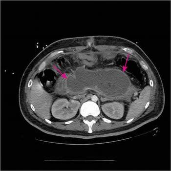
This CT scan with IV and oral contrast shows a large acute necrotizing collection (arrows) replacing most of the pancreatic tissue < 4 weeks from initial presentation of necrotizing pancreatitis.
Abdominal ultrasound should be performed if gallstone pancreatitis is suspected (and another cause is not obvious) to detect gallstones or dilation of the common bile duct, which indicates biliary tract obstruction. Edema of the pancreas may be visible, but overlying gas frequently obscures the pancreas.
If performed, plain radiographs of the abdomen may disclose calcifications within pancreatic ducts (evidence of prior inflammation and hence chronic pancreatitis), calcified gallstones, localized ileus of a segment of small intestine in the left upper quadrant or the center of the abdomen (a “sentinel loop”), or the colon cutoff sign (absence of air in left colonic flexure or descending colon) in more severe disease. However, the value of routine abdominal radiographs is unclear.
Chest radiographs should be performed and may reveal atelectasis or a pleural effusion (usually left-sided or bilateral but rarely confined to the right pleural space), which are signs of severe disease.
The role of endoscopic ultrasound is limited in acute pancreatitis. Endoscopic ultrasound has a comparable specificity to magnetic resonance cholangiopancreatography (MRCP) for detecting common bile duct stones, but MRCP has the advantage of being noninvasive (4). MRCP is performed if liver tests, particularly direct bilirubin and alanine aminotransferase (ALT), are elevated from their baseline and abdominal ultrasound does not exclude gallstones in the common bile duct as the etiology for gallstone acute pancreatitis.
Endoscopic retrograde cholangiopancreatography (ERCP) to relieve bile duct obstruction should be done expeditiously in patients with gallstone pancreatitis who have increasing serum bilirubin and signs of cholangitis.
Diagnosis references
1. Banks PA, Bollen TL, Dervenis C, et al. Classification of acute pancreatitis 2012: Revision of the Atlanta classification and definitions by international consensus. Gut. 2013;62:102–111. doi: 10.1136/gutjnl-2012-302779
2. Tenner S, Vege SS, Sheth SG, et al. American College of Gastroenterology Guidelines: Management of Acute Pancreatitis. Am J Gastroenterol. 2024;119(3):419-437. doi:10.14309/ajg.0000000000002645
3. Berk JE, Kizu H, Wilding P, Searcy RL. Macroamylasemia: a newly recognized cause for elevated serum amylase activity. N Engl J Med. 1967;277(18):941-946. doi:10.1056/NEJM196711022771801
4. Afzalpurkar S, Giri S, Kasturi S, et al. Magnetic resonance cholangiopancreatography versus endoscopic ultrasound for diagnosis of choledocholithiasis: an updated systematic review and meta-analysis. Surg Endosc. 2023;37(4):2566-2573. doi:10.1007/s00464-022-09744-3
Treatment of Acute Pancreatitis
Supportive measures
For severe acute pancreatitis and complications, antibiotics and therapeutic interventions as needed
Treatment of acute pancreatitis is typically supportive. Patients who develop complications may require specific additional treatment (1, 2).
The basic treatment of acute pancreatitis includes:
Resuscitation with moderate fluid volume of lactated Ringers solution
Analgesia
Nutritional support
Fluid resuscitation should be initiated with lactated Ringers solution at 1.5 mL/kg/h (1). In addition, fluid bolus of 10 mL/kg can be given if patients show signs of hypovolemia, unless contraindicated by cardiovascular, renal, or other related comorbid factors. Adequacy of fluid replacement can be assessed by reduction in hematocrit and blood urea nitrogen levels over the first 24 hours, particularly if they were high at the onset. Other parameters include improvement in vital signs and maintenance of adequate urine output. Guidelines also recommend that fluid requirements be reassessed at frequent intervals in the first 6 hours of admission and for the next 24 to 48 hours (1). Patients undergoing volume resuscitation should have pulse oximetry monitored continuously, receive supplemental oxygen as needed, and have their fluid intake and output strictly monitored. (1, 2, 3).
Pain relief requires use of parenteral opioids such (eg, hydromorphone, morphine, fentanyl), which should be given in adequate doses (4). Because morphine can theoretically increase pressure in the sphincter of Oddi, hydromorphone is usually preferred to morphine. Antiemetic medications should be given to relieve nausea and vomiting.
Early enteral nutrition is recommended because it is associated with lower morbidity compared to delayed or no nutrition (1). Patients with mild pancreatitis can begin an oral low-residue, low-fat, soft diet as soon as it can be tolerated. If oral nutrition cannot be initiated early in the disease course, enteral feeding is preferred over total parenteral nutrition because parenteral nutrition is associated with increased risk of infectious complications and organ failure.
Severe acute pancreatitis and complications
Treatment of severe acute pancreatitis and complications includes:
Intensive care unit (ICU) care
Enteral nutrition preferred over parenteral nutrition
Antibiotics for extrapancreatic infections and infected necrosis
Necrosectomy (removal of necrotic tissue) for infected necrosis
Endoscopic retrograde cholangiopancreatography (ERCP) for acute pancreatitis and concurrent acute cholangitis
Drainage of pseudocysts
The management of patients with severe acute pancreatitis and its complications should be individualized using a multidisciplinary approach including therapeutic endoscopists, interventional radiologists, and a surgeon. Patients with severe acute pancreatitis should be monitored closely in the first 24 to 48 hours in an ICU. Patients with worsening condition or widespread local complications requiring intervention should be transferred to centers of excellence focusing on pancreatic disease (if available).
Patients with severe acute pancreatitis may need artificial nutritional support, although the optimal starting time and duration of nutritional support are still unclear. Guidelines recommend using enteral nutrition and giving parenteral nutrition only if the enteral route is not available, not tolerated, or not meeting caloric requirements (1). The enteral route is preferred because it:
Helps maintain the intestinal mucosal barrier
Prevents the intestinal atrophy that can occur with prolonged bowel rest (and helps prevent translocation of bacteria that can seed pancreatic necrosis)
Avoids the risk of infection of a central IV catheter
Is less expensive
If enteral nutrition needs to be administered by a feeding tube, it was previously thought that the nasojejunal route was preferred to avoid stimulating the gastric phase of the digestive process; however, Guidelines recommend that nasogastric feeding tubes should be utilized because of comparable safety and efficacy with the advantage of easier placement (not requiring interventional radiology or ultrasound). If patients are at risk for aspiration, they should be placed in an upright position to decrease the risk.
According to guidelines prophylactic antibiotics are not recommended in patients with acute pancreatitis, regardless of the type or disease severity (1, 5). However, antibiotics should be started if patients develop an extrapancreatic infection (eg, cholangitis, pneumonia, bloodstream infection, urinary tract infection) or infected pancreatic necrosis.
Infection (pancreatic or extrapancreatic) should be suspected in patients who have signs of deterioration (eg, fever, hypotension, tachycardia, altered mental status, increasing white blood cell count) or who fail to improve after 7 to 10 days of hospitalization. Most infections in pancreatic necrosis are caused by single bacterial species from the gut. The most common organisms are gram-negative bacteria; gram-positive bacteria and fungi are rare. In patients with infected necrosis, antibiotics known to penetrate pancreatic necrosis, such as carbapenems, fluoroquinolones, and metronidazole, are recommended.
For necrosectomy (removal of infected tissue), a minimally invasive approach is preferred over an open surgical approach and should be attempted initially. Guidelines recommend that drainage of infected necrosis (radiologic, endoscopic, or surgical approach) should be delayed, preferably for > 4 weeks in patients who are stable, to allow liquefication of the contents and the development of a fibrous capsule around the necrosis (walled-off necrosis) (1).
Most patients with gallstone pancreatitis pass the stone spontaneously and do not require endoscopic retrograde cholangiopancreatography (ERCP). Patients with acute pancreatitis and concurrent acute cholangitis should undergo early ERCP. Patients with mild gallstone pancreatitis who spontaneously improve should undergo cholecystectomy before discharge to prevent recurrent attacks.
A pseudocyst that is expanding rapidly, infected, bleeding, or likely to rupture requires drainage. Whether drainage is percutaneous, surgical, or via endoscopic ultrasound–guided cystogastrostomy depends on location of the pseudocyst and institutional expertise.
Treatment references
1. Tenner S, Vege SS, Sheth SG, et al. American College of Gastroenterology Guidelines: Management of Acute Pancreatitis. Am J Gastroenterol. 2024;119(3):419-437. doi:10.14309/ajg.0000000000002645
2. Trikudanathan G, Yazici C, Evans Phillips A, et al. Diagnosis and Management of Acute Pancreatitis. Gastroenterology. 2024;167(4):673-688. doi:10.1053/j.gastro.2024.02.052
3. de-Madaria E, Buxbaum JL, Maisonneuve P, et al. Aggressive or Moderate Fluid Resuscitation in Acute Pancreatitis. N Engl J Med. 2022;387(11):989-1000. doi: 10.1056/NEJMoa2202884
4. Basurto Ona X, Rigau Comas D, Urrútia G. Opioids for acute pancreatitis pain. Cochrane Database Syst Rev. 2013;2013(7):CD009179. Published 2013 Jul 26. doi:10.1002/14651858.CD009179.pub2
5. Crockett SD, Wani S, Gardner TB, et al.; American Gastroenterological Association Institute Clinical Guidelines Committee. American Gastroenterological Association Institute Guideline on Initial Management of Acute Pancreatitis. Gastroenterology. 2018;154(4):1096-1101. doi:10.1053/j.gastro.2018.01.032
Prognosis for Acute Pancreatitis
The severity of acute pancreatitis is determined by the presence of organ failure, local and systemic complications, or a combination. Using patient-related risk factors to assess severity early in the course of disease can help identify patients at increased risk of developing organ dysfunction and other complications. These patients can then be given maximal supportive therapy at presentation to improve outcomes and decrease morbidity and mortality.
For initial risk assessment, patient-related risk factors that predict a severe course include the following:
Age ≥ 60 years
Comorbid health problems
Obesity with body mass index > 30
Long-term, heavy alcohol consumption
Presence of systemic inflammatory response syndrome (SIRS)
Laboratory markers of hypovolemia (eg, elevated BUN, elevated hematocrit)
Presence of pleural effusions and/or infiltrates on admission chest radiography
Altered mental status
Severity scoring systems require multiple measurements and may delay appropriate management. Some of these can be performed at admission to assist in triage of patients, whereas others are not accurate until 48 to 72 hours after presentation.
Ranson criteria (1): This scoring system is cumbersome and requires data from a 48-hour period to compute but has good negative predictive value.
The APACHE II score: This system is complex and cumbersome to use but has good negative predictive value.
Systemic inflammatory response syndrome score: This is inexpensive, readily available, and can be applied at the bedside.
Bedside index of severity in acute pancreatitis (BISAP) score: This score is simple and calculated during the first 24 hours.
Harmless acute pancreatitis score: This simple score is calculated within 30 minutes of admission.
Organ failure–based scores: These scores do not directly measure the severity of acute pancreatitis.
CT severity index (Balthazar score): This score is based on the degree of necrosis, inflammation, and the presence of fluid collections on CT.
Mortality in the early phase (within 1 week) of acute pancreatitis usually results from multiorgan failure, whereas mortality in the late phase (> 1 week) usually results from a combination of factors, including multiorgan failure, infection of pancreatic necrosis, and complications of surgical and endoscopic interventions.
The long-term risks after acute pancreatitis include the risks of recurrent attacks and development of chronic pancreatitis. Risk factors include the severity and amount of pancreatic necrosis in the initial episode of acute pancreatitis as well as the etiology; long-term, heavy alcohol consumption and cigarette smoking increase the risk of developing chronic pancreatitis.
Prognosis reference
1. Ranson JH, Rifkind KM, Roses DF, et al. Prognostic signs and the role of operative management in acute pancreatitis. Surg Gynecol Obstet 1974;139(1):69-81.
Key Points
There are many causes of acute pancreatitis, but the most common are gallstones and alcohol intake.
Inflammation is confined to the pancreas in mild cases, but, with increasing severity, a severe systemic inflammatory response may develop, resulting in shock and/or organ failure.
Once pancreatitis is diagnosed, assess risk using clinical criteria and scoring systems to triage appropriate patients to more intensive care and aggressive therapy and to help estimate prognosis.
Treatment includes IV fluid resuscitation, pain control, and nutritional support.
Complications, including pseudocyst and infected pancreatic necrosis, need to be identified and treated appropriately (eg, drainage of pseudocyst, necrosectomy).
Drug Information for the Topic