

In the United States, the collection, storage, and transport of blood and its components are standardized and regulated by the Food and Drug Administration (FDA), by the AABB (Association for the Advancement of Blood and Biotherapies), and sometimes by state or local health authorities. Donor screening includes a health questionnaire and interview, vital signs assessment, minimum weight of 50 kg, and hemoglobin between 12.5 and 20 g/dL (125 to 200 g/L). Some potential donors are deferred either temporarily or permanently (see table ). Criteria for deferral protect prospective donors from possible ill effects of donation and protect recipients from disease. Although transfusion is probably safer than ever, risk (and the public’s perception of risk) mandates informed consent whenever practical.
Whole blood donation is allowed every 56 days; apheresis red blood cell (RBC) donation every 112 days; and apheresis platelet donation every 72 hours (maximum 24/year) (1). Blood donors are generally unpaid.
In a standard blood donation, approximately 450 mL of whole blood is collected in a plastic bag containing an anticoagulant preservative. Whole blood or packed RBCs preserved with citrate-phosphate-dextrose-adenine may be stored for 35 days. Over 95% of packed RBCs are stored for 42 days when an adenine-dextrose-saline solution is added.
Autologous donation of blood for subsequent transfusion into the same donor is generally a less preferred method of transfusion. For elective surgery, 3 to 4 units may be collected 2 to 3 weeks preoperatively with iron supplementation. Autologous donation is reserved for patients with rare blood types or multiple alloantibodies when a compatible donor blood is difficult to obtain. It is most commonly used prior to elective surgery when there is a potential for significant blood loss. Intraoperative and post-traumatic blood salvage can also be used for autotransfusion.
Some Reasons for Blood Donation Deferral or Denial*
Reason | Donation Outcome | Comment |
|---|---|---|
Infections and risks of infection | ||
Denial | Includes any individual who has ever had a positive test for HIV infection Includes any individual who has ever taken any medication to treat HIV infection | |
Activities that increase risk of HIV infection† | Deferral | Wait for 2 years from last use of any medication given by injection to prevent HIV infection (ie, long-acting antiviral PrEP or PEP) Wait for 3 months from the last use of any medication by mouth (oral) to prevent HIV infection (ie, antiviral PrEP or PEP) Wait for 3 months from last time high-risk activity has taken place. Activities include:
|
Denial | Denial based on possible persistent infection, high risk of transmission, and lack of screening test | |
Hepatitis exposure | Deferral | Wait 12 months after possible exposure |
Denial | Ineligible to donate if ever diagnosed with viral hepatitis B or C, or if ever tested positive for viral hepatitis B or C | |
Malaria or exposure to malaria | Deferral | Wait 3 years after treatment for malaria or living in an area in which malaria is endemic; wait 3 months after visit to an area in which malaria is endemic |
Transfusion | Deferral | Wait 3 months after a transfusion done in the United States |
Vaccinations (selected) | Deferral | Waiting period depends on vaccination:
|
Deferral | For recent Zika virus infection, the US FDA recommends a 120-day deferral from resolution of symptoms or the last positive test, whichever is longer | |
Cancer | ||
Cancers involving blood cells (eg, leukemia, lymphoma, myeloma) | Denial | Such people cannot donate even if they are cancer-free |
Other cancers (ie, not involving blood cells) | Deferral | People may donate if they are cancer-free and treatment was completed more than 12 months previously People with mild, treatable forms (eg, small skin cancers) that have been completely removed may be able to donate before 12 months |
Other | ||
Deferral | Donation permitted after anemia resolves | |
Denial | — | |
Heart disease | Deferral | Any heart disease must be medically evaluated and treated, and the person should have no heart-related symptoms within the last 6 months |
Deferral | Defer donation until blood pressure is controlled | |
Major surgery, if recent | Deferral | Deferral based mainly on likely perioperative transfusion or the underlying reason for surgery |
Medications (selected) | Deferral | Waiting period depends on medication, for example:
|
Pregnancy | Deferral | Wait 6 weeks after giving birth |
* Includes data from American Red Cross Blood Donor Eligibility FAQs. | ||
† Reflects FDA May 2023 Guidance document: Recommendations for Evaluating Donor Eligibility Using Individual Risk-Based Questions to Reduce the Risk of Human Immunodeficiency Virus Transmission by Blood and Blood Products. | ||
‡ These vaccines include anthrax, cholera, diphtheria, hepatitis A, hepatitis B, influenza, Lyme disease, paratyphoid, pertussis, plague, pneumococcal polysaccharide, polio (Salk), Rocky Mountain spotted fever, tetanus, and typhoid injection. | ||
§ Recipients of other live-attenuated viral or bacterial vaccines may be deferred 2 or 4 weeks, depending on the vaccine. | ||
FDA = US Food and Drug Administration; PEP = post-exposure prophylaxis; PrEP = pre-exposure prophylaxis. | ||
General reference
1. American Red Cross. Blood Services. Eligibility Requirements. Requirements by Donation Type. Accessed January 30, 2026.
Pretransfusion Testing
Donor blood testing includes:
ABO and Rh(D) antigen typing
Antibody screening
Testing for infectious disease markers (see table )
Compatibility testing:
Tests the recipient’s RBCs for antigens A, B, and Rh(D)
Screens the recipient’s plasma for antibodies against RBC antigens
Includes a cross-match to ensure that the recipient’s plasma is compatible with antigens on donor RBCs
Compatibility testing is done before a transfusion; however, in an emergency, testing is done after releasing blood from the blood bank. Testing can also help in diagnosing transfusion reactions.
The addition of a cross-match to ABO/Rh typing and antibody screening increases detection of incompatibility by only 0.01%. Therefore, most hospitals do computerized electronic cross-matches rather than physical cross-matches in a test tube in patients who have negative antibody screening. If the recipient has a clinically significant anti-RBC antibody, donor blood is restricted to RBC units negative for the corresponding antigen; further testing for compatibility is done by combining recipient plasma, donor RBCs, and antihuman globulin. In recipients without clinically significant anti-RBC antibodies, an immediate spin cross-match, which omits the antiglobulin phase, confirms ABO compatibility when electronic cross-match is not feasible.
Emergency transfusion is done when not enough time (generally < 60 minutes) is available for thorough compatibility testing because the patient is in hemorrhagic shock. When time permits (approximately 10 minutes is needed), ABO/Rh type-specific blood may be given. In urgent situations when the patient’s blood type is unknown, type O RBCs are transfused. If Rh status is also unknown, Rh-negative blood is used for females of child-bearing age to prevent alloimmunization; in other patients, either Rh-negative or Rh-positive type O blood may be given.
“Type and screen” may be requested in circumstances that are not likely to require transfusion, as in surgery in which the risk of bleeding is low. The patient’s blood is typed for ABO/Rh antigens and screened for antibodies. If antibodies are absent and the patient needs blood, ABO/Rh type–specific or compatible RBCs may be released without the antiglobulin phase of the cross-match. If an unexpected antibody is present, full compatibility testing is required. "Type and cross" is requested for procedures in which there is a high bleeding risk (eg, cardiac bypass surgery), and the number of units for cross matching is specified.
ABO and Rh typing
ABO typing of donor and recipient blood is performed to prevent transfusion of incompatible RBCs (see figure ). As a rule, blood for transfusion should be of the same ABO type as that of the recipient. In urgent situations or when the correct ABO type is in doubt or unknown, type O Rh-negative packed RBCs (not whole blood—see Acute Hemolytic Transfusion Reaction), which contains neither A nor B antigens, may be used for patients of any ABO type.
Compatible RBC Types

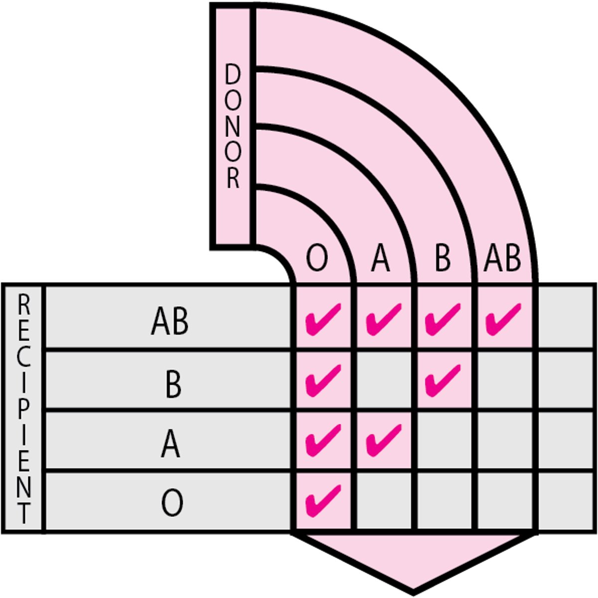
Rh typing determines whether the Rh factor D antigen is present on (Rh-positive) or absent from (Rh-negative) the RBCs. Rh-negative patients should always receive Rh-negative blood except in life-threatening emergencies when Rh-negative blood is unavailable. Rh-positive patients may receive Rh-positive or Rh-negative blood. Occasionally, RBCs from some Rh-positive people react weakly on standard Rh typing due to less expression of D antigen (weak D), but these people are still considered Rh-positive.
Antibody screening
Antibody screening for unexpected anti-RBC antibodies is routinely done on blood from prospective recipients and prenatally on maternal specimens. Unexpected anti-RBC antibodies are specific for RBC blood group antigens other than A and B [eg, Rh(D), Kell (K), Duffy (Fy)]. Early detection is important because such antibodies can cause serious hemolytic transfusion reactions or hemolytic disease of the fetus and neonate, and they may greatly complicate compatibility testing and delay procurement of compatible blood.
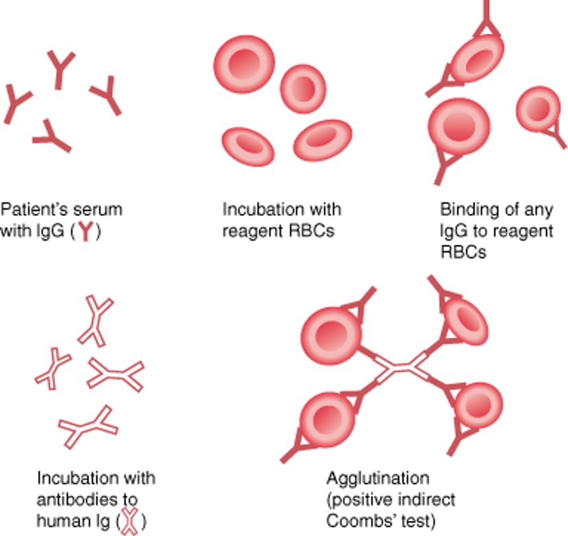
Indirect antiglobulin testing (the indirect Coombs test) is used to screen for unexpected anti-RBC antibodies (see figure ). This test may be positive in the presence of an unexpected blood group antibody or when free (non-RBC–attached) antibody is present in autoimmune hemolytic anemias. Reagent RBCs are mixed with the patient’s plasma or serum, incubated at 37oC, washed, tested with antihuman globulin, and observed for agglutination. Once an antibody is detected, its specificity is determined. Knowing the specificity of the antibody is helpful for assessing its clinical significance, selecting compatible blood, and managing hemolytic disease of the fetus and neonate.
Indirect Antiglobulin (Indirect Coombs) Test
The indirect antiglobulin (indirect Coombs) test is used to detect IgG antibodies against RBCs in a patient's plasma. The patient's serum or plasma is incubated with reagent RBCs; then Coombs serum (antibodies to human IgG, or human anti-IgG) is added. If agglutination occurs, IgG antibodies (autoantibodies or alloantibodies) against RBCs are present. This test is also used to determine the specificity of an alloantibody. |

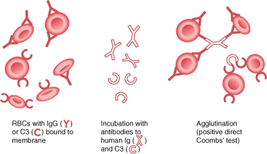
Direct antiglobulin testing (the direct Coombs test) detects antibodies that have coated the patient’s RBCs in vivo (see figure ). It is used when immune-mediated hemolysis is suspected. Patients’ RBCs are directly tested with antihuman globulin and observed for agglutination. A positive result, if correlated with clinical findings and laboratory indicators of hemolysis, suggests autoimmune hemolytic anemia, drug-induced hemolysis, a transfusion reaction, or hemolytic disease of the newborn.
Direct Antiglobulin (Direct Coombs) Test (DAT)
The direct antiglobulin (direct Coombs) test is used to determine whether IgG antibody or complement (C3d) is present on red blood cell (RBC) membranes. The patient's RBCs are incubated with antibodies to human IgG and C3d. If IgG or C3d is bound to RBC membranes, agglutination occurs—a positive result. A positive result suggests the presence of autoantibodies to RBCs. A positive result due only to C3d suggests the presence of a cold autoantibody (usually IgM, which is not detected in the DAT since the Coombs serum is against IgG only). A positive test result does not always equate with hemolysis. Thus, results should always be correlated with clinical signs and symptoms. |

Antibody titration is done when a clinically significant, unexpected anti-RBC antibody is identified in the plasma of a patient who is pregnant or in a patient with cold agglutinin disease. The maternal antibody titer correlates fairly well with the severity of hemolytic disease in the fetus with the corresponding antigen and is often used to guide treatment in hemolytic disease of the fetus and neonate along with ultrasound and amniotic fluid study.
Infectious disease testing
Donated blood products are tested for the presence of a number of infectious agents (see table ).
Infectious Disease Transmission Testing*
Infectious Agent | Type of Testing |
|---|---|
Babesia microti (babesiosis) | Nucleic acid and antibody testing (in Babesia endemic regions) |
Nucleic acid and antibody testing (particularly for high-risk, immunocompromised or neonatal recipients) | |
HBsAg, anti HBc, and nucleic acid testing | |
Nucleic acid and antibody testing | |
HIV-1 and HIV-2 | Nucleic acid and antibody testing |
Human T-cell lymphotropic viruses 1 and 2 | Antibody testing |
Treponema pallidum (syphilis) | Antigen testing |
Trypanosoma cruzi (Chagas disease) | Antibody testing |
Nucleic acid testing | |
Nucleic acid testing | |
* Blood is also cultured for a wide range of bacteria | |
Anti-HBc = antibody to hepatitis B core; HBsAg = hepatitis B surface antigen; HIV = human immunodeficiency virus. | |
More Information
The following English-language resources may be useful. Please note that The Manual is not responsible for the content of these resources.
Centers for Disease Control and Prevention. Blood Safety. Clinical Testing Guidance for Blood Safety, September 24, 2025.:
FDA May 2023 Guidance document. Recommendations for Evaluating Donor Eligibility Using Individual Risk-Based Questions to Reduce the Risk of Human Immunodeficiency Virus Transmission by Blood and Blood Products.


