Allergic (including atopic) and other hypersensitivity disorders are pathological and/or exaggerated immune reactions to foreign or self antigens. Pathological immune reactions include those that are misdirected against intrinsic body components (self), leading to autoimmune disorders. This topic focuses on type I hypersensitivity reactions.
Classification of Hypersensitivity Reactions
Hypersensitivity reactions are divided into 4 types (I through IV) by the Gell and Coombs classification (1). Hypersensitivity disorders can sometimes involve more than 1 type.
Type I hypersensitivity reactions
Type I hypersensitivity reactions (immediate hypersensitivity) are IgE-mediated. Antigen binds to IgE that is bound to tissue mast cells and blood basophils primarily via the Fc epsilon receptors (Fc-epsilon-R1, Fc-epsilon-R2), triggering the degranulation and release of preformed mediators (eg, histamine, proteases, chemotactic factors) and synthesis of other mediators (eg, prostaglandins, leukotrienes, platelet-activating factor, cytokines). These mediators cause vasodilation, increased capillary permeability, mucus hypersecretion, smooth muscle spasm, and tissue infiltration with eosinophils, type 2 helper T (Th2) cells, and other inflammatory cells.

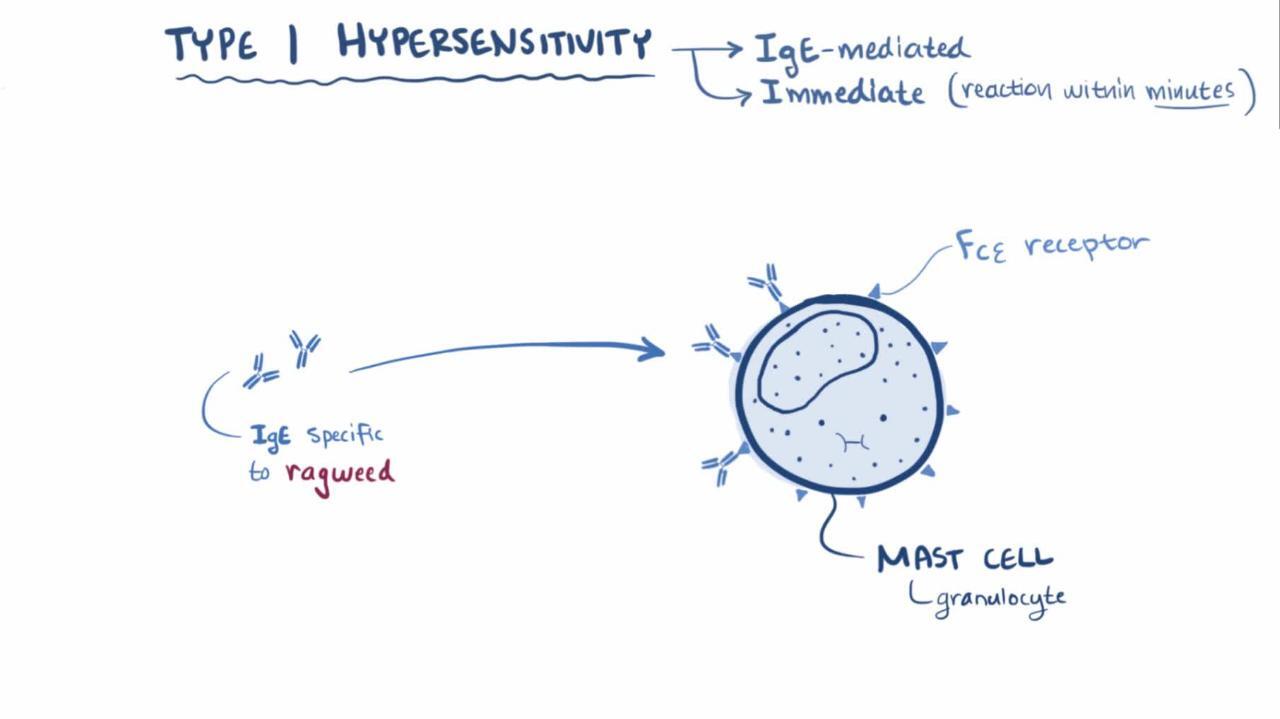
Type I hypersensitivity reactions develop < 1 hour after exposure to antigen.
Type I reactions underlie all atopic disorders (eg, atopic dermatitis, allergic asthma, rhinitis, conjunctivitis) and many allergic disorders (eg, anaphylaxis, some cases of angioedema, urticaria, latex and some food allergies). The terms atopy and allergy are often used interchangeably but are different:
Atopy is an exaggerated IgE-mediated immune response; all atopic disorders are type I hypersensitivity disorders.
Allergy is any exaggerated immune response to a foreign antigen regardless of mechanism.
Thus, all atopic disorders are considered allergic, but many allergic disorders (eg, hypersensitivity pneumonitis) are not atopic.
Atopic disorders most commonly affect the nose, eyes, skin, and lungs. These disorders include conjunctivitis, extrinsic atopic dermatitis (the most common type of eczema), immune-mediated urticaria, some forms of angioedema, acute , some allergic lung disorders (eg, allergic asthma, IgE-mediated components of allergic bronchopulmonary aspergillosis), allergic rhinitis, and allergic reactions to venomous stings.
Type II hypersensitivity reactions
Type II hypersensitivity reactions (antibody-dependent cytotoxic hypersensitivity) result when antibody binds to cell surface antigens or to a molecule coupled to a cell surface. The surface-bound antigen-antibody structure (as opposed to the circulating antigen-antibody complex in type III hypersensitivity) activates cells (eg, natural killer cells, eosinophils, macrophages) that participate in antibody-dependent cell-mediated cytotoxicity (ADCC), complement, or both. The result is cell and tissue damage.
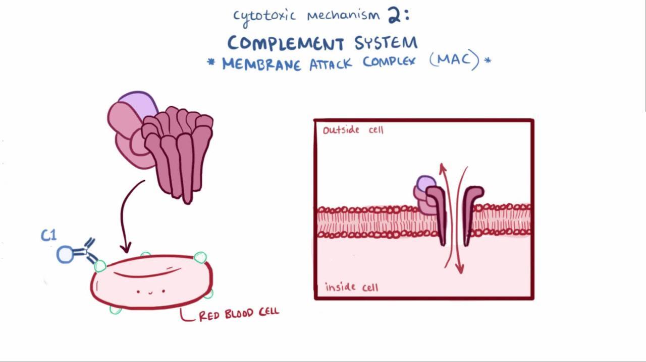
Disorders involving type II reactions include hyperacute graft rejection of an organ transplant, Coombs-positive hemolytic anemias, Hashimoto thyroiditis, and anti-glomerular basement membrane (Anti-GBM) disease.
Type III hypersensitivity reactions
Type III hypersensitivity reactions (immune complex disease) cause inflammation in response to circulating antigen-antibody immune complexes that have been deposited in vessels or tissue. These complexes can activate the complement system or bind to and activate certain immune cells, resulting in release of inflammatory mediators.
Consequences of immune complex formation depend in part on the relative proportions of antigen and antibody in the immune complex and the anatomic site of complex localization. Early in the immune response, there is excess antigen with small antigen-antibody complexes, which do not efficiently activate complement. Later, when antigen and antibody are more balanced (ie, when the immune system has had time to produce more antibodies), immune complexes are larger and are usually deposited in various tissues (eg, glomeruli, blood vessels), causing systemic reactions. The isotype of induced antibodies (eg, IgM, IgG) changes during an immune response, and the isotype, glycosylation, size, and charge of the complex’s components contribute to the clinical response.
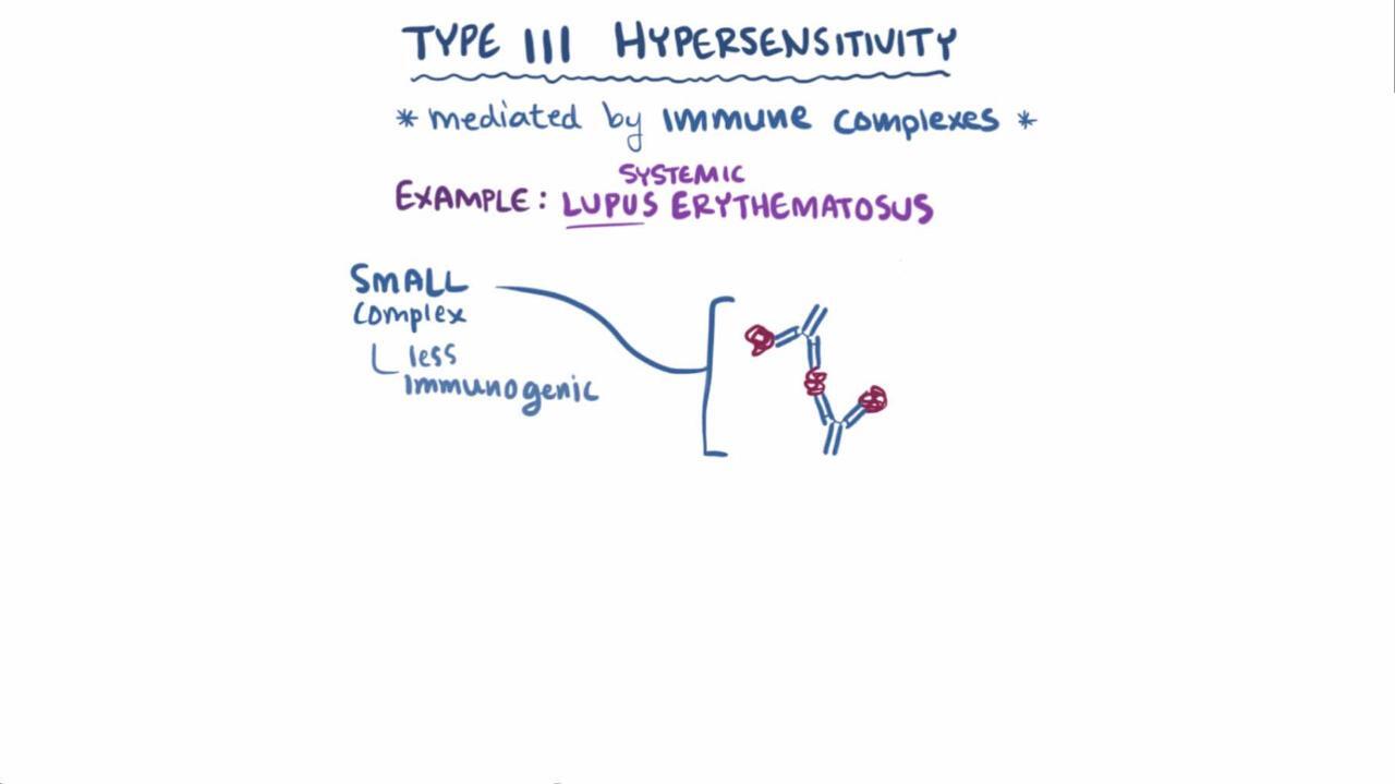
Type III disorders include serum sickness, systemic lupus erythematosus (SLE), rheumatoid arthritis (RA), leukocytoclastic vasculitis, cryoglobulinemia, acute hypersensitivity pneumonitis, and several types of glomerulonephritis.
Type III reactions may develop hours, days, or weeks after exposure to antigen and, if the presence of the antigen continues, can become chronic (2).
Type IV hypersensitivity reactions
Type IV hypersensitivity reactions (delayed hypersensitivity) do not involve antibodies (ie, humoral immunity) but are mediated by T cells (ie, cell-mediated immunity).
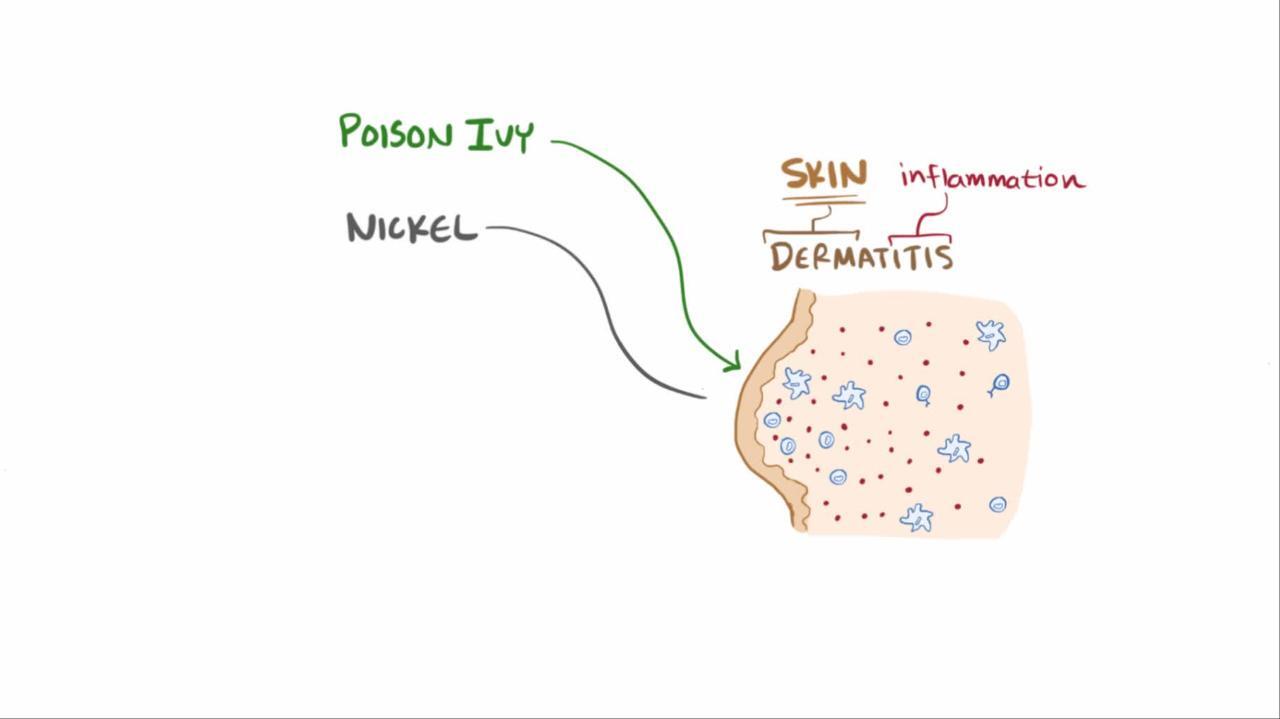
T cells are initially sensitized after contact with a specific antigen. They are then activated by continued exposure or reexposure to the antigen (see figure ).
They can damage tissue by direct toxic effects (eg, CD8 cytotoxic lymphocytes) or through release of cytokines, which can activate eosinophils (eg, interleukin [IL]-5), monocytes and macrophages (eg, interferon [IFN]-gamma), neutrophils (eg, IL-8), or natural killer cells (IL-2).
Disorders involving type IV reactions include contact dermatitis (eg, poison ivy), Stevens-Johnson syndrome, toxic epidermal necrolysis (SJS/TEN), drug rash with eosinophilia and systemic symptoms (DRESS), subacute and chronic hypersensitivity pneumonitis, acute and chronic allograft rejection, the immune response to tuberculosis, and many forms of drug hypersensitivity.
For more information, see T cells and the figure .
Classification references
1. Dispenza MC. Classification of hypersensitivity reactions. Allergy Asthma Proc. 2019;40(6):470-473. doi:10.2500/aap.2019.40.4274
2. Schneider G, Kachroo S, Jones N, et al. A systematic review of validated methods for identifying hypersensitivity reactions other than anaphylaxis (fever, rash, and lymphadenopathy), using administrative and claims data. Pharmacoepidemiol Drug Saf. 2012;21 Suppl 1:248-255. doi:10.1002/pds.2333
Etiology of Allergic and Atopic Disorders
Complex interplays of genetic, environmental, and anatomic site-specific factors contribute to development of IgE-mediated allergies.
Genetic factors are involved, as suggested by familial inheritance of disease, association between atopy and specific human leukocyte antigen (HLA) loci, and polymorphisms of several genes, including those for the high-affinity IgE receptor (Fc-epsilon-R1) beta-chain, IL-4 receptor alpha-chain, interleukin (IL)-4, IL-13, CD14, dipeptidyl-peptidase 10 (DPP10), and a disintegrin and metalloprotease domain 33 (ADAM33).
Environmental factors interact with genetic factors to maintain type 2 helper T (Th2) cell–directed immune responses. Th2 cells activate eosinophils, promote IgE production, and are proallergic. Early childhood exposure to bacterial and viral infections and bacterially derived endotoxins (eg, lipopolysaccharide) may shift inherent Th2-cell responses to type 1 helper T (Th1)–cell responses, which cause counter-regulation and suppression of Th2 cells; therefore discouraging allergic responses. Regulatory T (Treg) cells (eg, CD4+, CD25+, Foxp3+), which are capable of suppressing Th2-cell responses, and IL-12–secreting dendritic cells, which drive Th1-cell responses, are usually also involved (1). General trends in resource-rich countries that favor smaller families with fewer children, cleaner indoor environments, and more widespread use of antibiotics may limit children's exposure to the infectious agents that drive a predominantly Th1-cell response; such trends may explain the increased prevalence of some allergic disorders. This phenomenon has been called the hygiene hypothesis.
Other environmental factors thought to contribute to allergy development include chronic allergen exposure and sensitization, diet, and environmental pollutants.
Anatomic site-specific factors include adhesion molecules in bronchial epithelium and skin and molecules in the gastrointestinal (GI) tract that direct Th2 cells to target tissues. The composition of the GI tract, respiratory tract, and skin microbiota appears to strongly influence the development of allergy. These microbiota may provide new targets for allergy therapy.
Allergens
By definition, an allergen induces type I IgE-mediated or type IV T-cell–mediated immune responses. Allergic triggers are typically proteins; many of them can become attached to airborne particles. Carbohydrate moieties (eg, polysaccharides), or carbohydrate-protein complexes (eg, glycoproteins) and low-molecular-weight chemicals (also called haptens) may also be allergenic.
Sources of allergens that most commonly cause acute and chronic allergic reactions (type I and type IV) include:
House dust mite feces
Animal dander
Pollens (tree, grass, weed)
Molds
Insect saliva and venom (transmitted by bites and stings)
Household chemicals (eg, hydroxyisohexyl 3-cyclohexene carboxaldehyde, cinnamal, eugenol)
Environmental chemical exposures (eg, nickel, fragrances, preservatives) are the most common trigger of type IV hypersensitivity reactions, of which allergic contact dermatitis is the most common manifestation (2, 3). For most other type IV hypersensitivity reactions, medications are the most common trigger.
Etiology references
1. Venuprasad K, Kong YC, Farrar MA. Control of Th2-mediated inflammation by regulatory T cells. Am J Pathol. 2010;177(2):525-531. doi:10.2353/ajpath.2010.090936
2. Alinaghi F, Bennike NH, Egeberg A, Thyssen JP, Johansen JD. Prevalence of contact allergy in the general population: A systematic review and meta-analysis. Contact Dermatitis. 2019;80(2):77-85. doi:10.1111/cod.13119
3. Fonacier L, Bernstein DI, Pacheco K, et al. Contact dermatitis: a practice parameter-update 2015. J Allergy Clin Immunol Pract. 2015;3(3 Suppl):S1-S39. doi:10.1016/j.jaip.2015.02.009
Pathophysiology of Allergic and Atopic Disorders
Mast cells are widely distributed throughout vascularized tissues but are most concentrated in tissues directly exposed to the external environment (eg, skin, lungs, GI mucosa) rather than in blood, where mature mast cells do not normally circulate. When an allergen binds to IgE-sensitized mast cells or basophils, histamine is released from their intracellular granules via degranulation. Histamine is the primary mediator of clinical atopy and the associated inflammatory response. Physical disruption of tissue and various substances (eg, tissue irritants, opiates, surface-active agents, the anaphylatoxin complement components C3a and C5a) can also trigger histamine release directly, independent of IgE.
Histamine causes the following:
Local vasodilation (causing erythema)
Increased capillary permeability and edema (producing a wheal)
Vasodilation of surrounding arterioles mediated by neuronal reflex mechanisms (causing flare—the redness around a wheal)
Stimulation of sensory nerves (causing pruritus)
Smooth muscle contraction in the airways (bronchoconstriction) and in the GI tract (increasing GI motility)
Increased nasal, salivary, and bronchial gland secretions
When released systemically, histamine is a potent arteriolar dilator and can cause extensive peripheral pooling of blood and hypotension; cerebral vasodilation may be a factor in vascular headache. Histamine increases capillary permeability; the resulting loss of plasma and plasma proteins from the vascular space can worsen circulatory collapse. This loss triggers a compensatory activation of the sympathetic nervous system and a catecholamine surge via adrenal chromaffin cells. In severe cases (eg, peanut ingestion in a patient with peanut allergy), anaphylactic shock can occur.
Symptoms and Signs of Allergic and Atopic Disorders
Common symptoms of type I hypersensitivity allergic disorders include:
Rhinorrhea, sneezing, and nasal congestion (upper respiratory tract)
Wheezing and dyspnea (lower respiratory tract)
Pruritus (eyes, nose, skin)
Urticarial or other rash (skin)
Swelling of the lips, tongue, face, or intra-abdominal organs (subcutaneous tissue, GI tract)
Signs may include nasal turbinate edema, sinus pain with facial palpation, wheezing, conjunctival hyperemia and edema, urticaria, angioedema, dermatitis, and (if chronic) skin lichenification.
Stridor, wheezing, and hypotension are life-threatening signs of anaphylaxis.
Diagnosis of Allergic and Atopic Disorders
History and physical examination
Sometimes complete blood count (to check for eosinophilia), serum tryptase (to assess mast cell activation), and occasionally serum IgE levels (nonspecific tests)
Often skin testing (allergen-specific test)
Serum allergen-specific IgE testing and component-resolved diagnostic testing (antigen-specific test)
Rarely provocative testing (eg, to food, medications, physical urticaria)
A thorough history is required. History should include:
Questions about the age and/or timing of onset, frequency and duration of attacks and changes over time
Triggering factors if identifiable
Relation to seasonal or situational settings (eg, predictably occurring during pollen seasons; after exposure to animals, hay, or dust; during exercise; or in particular places)
Family history of similar symptoms or of atopic disorders
Responses to attempted treatments
Travel history (to exclude parasitic infection)
Age at onset may be important in asthma because childhood asthma is likely to be atopic.
Nonspecific tests
Certain adjunctive, nonspecific tests can suggest an allergic origin of symptoms.
Complete blood count (CBC) may be done to detect eosinophilia if patients are not taking glucocorticoids, which reduce the eosinophil count. However, CBC is of limited value because although eosinophils may be increased in atopy or other conditions (eg, drug hypersensitivity, cancer, inflammatory bowel disease, parasitic infection, some systemic inflammatory diseases), a normal eosinophil count does not exclude allergy. Total white blood cell count is usually normal. Anemia and thrombocytosis are not typical of allergic responses and should prompt consideration of a systemic inflammatory disorder.
Conjunctival or nasal secretions or sputum can be examined for leukocytes; finding any eosinophils suggests that localized Th2-mediated inflammation is likely.
Serum tryptase is a marker of mast cell activation. It may be elevated during anaphylaxis and can support the diagnosis when measured within 2 hours of symptom onset. Persistently elevated baseline levels may suggest an underlying mast cell disorder. However, normal tryptase levels do not exclude an allergic reaction, and routine measurement is not useful in most patients with common atopic disorders.
Total serum IgE levels are elevated in atopic disorders but are of little help in making a specific allergic diagnosis because they may also be elevated in parasitic infections, infectious mononucleosis, some autoimmune disorders, drug reactions, immunodeficiency disorders (hyper-IgE syndrome and Wiskott-Aldrich syndrome), IgG4-related disease, and in some forms of multiple myeloma. IgE levels are helpful for monitoring response to therapy in allergic bronchopulmonary aspergillosis.
Specific tests
Skin testing uses standardized concentrations of antigen introduced directly into skin and is indicated when a detailed history and physical examination do not identify the cause and triggers for persistent or severe symptoms (1). Skin testing has higher positive predictive values for diagnosing allergic rhinitis and conjunctivitis than for diagnosing allergic asthma or food allergy; the negative predictive value for food allergy is high (2).
The most commonly used challenge antigens are pollens (tree, grass, weed), molds, house dust mite feces, animal danders and sera, insect venom, foods, and beta-lactam antibiotics. Choice of antigens to include is based on patient history and geographic prevalence.
Two skin test techniques can be used:
Percutaneous (prick)
Intradermal
The prick test can detect most common environmental allergies; it is usually done first. The intradermal test is more sensitive but less specific; it is typically used to evaluate sensitivity to allergens when prick test results are negative or equivocal, particularly in the evaluation of environmental, and medication allergies. Alternatively, intradermal testing may primarily be used for confirming stinging insect venom allergy in patients with a history of systemic reactions to stinging insects of the order Hymenoptera (eg, honeybees, yellow-jackets, hornets, paper-wasps, and some fire ants); it is considered more sensitive than routine skin prick testing or serum allergen-specific IgE for this purpose. Intradermal testing is generally not recommended for food allergy evaluation due to high false-positive rates.


WLADIMIR BULGAR/SCIENCE PHOTO LIBRARY
For the prick test, a drop of antigen extract is placed on the skin, which is then tented up and pricked or punctured through the extract with the tip of a 27-gauge needle held at a 20° angle or with a commercially available prick device.
If no allergen is identified in the prick test, an intradermal test is done.


This illustration shows an example of a skin prick method of allergy testing. Drops of allergen extracts, such as pollen and dog hair, are placed on the volar surface of the forearm. A lancet or similar device is then used to prick through each drop, gently lifting the epidermis to introduce the allergen to cutaneous mast cells. If the person is allergic to any of the allergens, a wheal-and-flare reaction appears at the site of application, as shown in the magnified view.
MAURIZIO DE ANGELIS/SCIENCE PHOTO LIBRARY
For the intradermal test, just enough extract to produce a 1- or 2-mm bleb (typically 0.02 mL) is injected intradermally with a 0.5- or 1-mL syringe and a 27-gauge short-bevel needle.
Both prick and intradermal skin testing should include the diluent alone (usually saline) as a negative control and histamine (10 mg/mL for prick tests, 0.1 mg/mL solution for intradermal tests) as a positive control. For patients who have had a recent (< 1 year) generalized reaction to the test antigen, testing begins with the standard reagent diluted 100-fold, then 10-fold, and then the standard concentration.


This photo shows an intradermal allergy test. For this test, just enough extract to produce a 1- or 2-mm bleb (typically 0.02 mL) is injected intradermally with a 0.5- or 1-mL syringe and a 27-gauge short-bevel needle.
EDWIGE/SCIENCE PHOTO LIBRARY
A test is considered positive if a wheal and flare reaction occurs and wheal diameter is 3 mm greater than that of the negative control after 15 to 20 minutes.
False-positive results occur in patients with dermatographism (a wheal and flare reaction provoked by stroking or scraping the skin). False-negative test results occur when allergen extracts have been stored improperly or have expired.
The use of certain medications can also interfere with results, and these medications should be stopped a few days to a week before testing. These medications include over-the-counter (OTC) and prescription antihistamines, tricyclic antidepressants, omalizumab, and monoamine oxidase inhibitors. Some clinicians suggest that testing should be avoided in patients taking beta-blockers because these patients are more likely to have risk factors for severe reactions. These risk factors tend to predict limited cardiopulmonary reserve and include coronary artery disease, arrhythmias, and older age. Also, beta-blockers can interfere with the treatment of severe reactions by blocking responsiveness to beta-adrenergic agonists such as epinephrine.
Allergen-specific serum IgE tests use an enzyme-labeled anti-IgE antibody to detect binding of serum IgE to a known allergen. These tests are done when skin testing might be ineffective or risky—for example, when medications that interfere with test results cannot be temporarily stopped before testing or when a skin disorder such as eczema, dermatographism, or psoriasis would make skin testing difficult. For allergen-specific serum IgE tests, the allergen is immobilized on a synthetic surface. After incubation with patient serum and enzyme-labeled anti-IgE antibody, a substrate for the enzyme is added; the substrate provides colorimetric, fluorescent, or chemiluminescent detection of binding.
Component-resolved diagnostic testing is a molecular diagnostic approach that identifies specific antigenic protein components within an allergen that are responsible for an IgE-mediated immune response. It is an important adjunct to allergen-specific IgE testing for some allergens. Component-resolved diagnostic testing is used primarily to distinguish true allergies from cross-reactivity to a related allergen (eg, determine whether a nut or fruit allergy is a primary allergy or results from cross-reactivity with a botanically related tree pollen allergen). It can also help in predicting risk stratification of allergic reactions and optimizing selection of immunotherapy (3). Component-resolved diagnostic testing is available for foods (eg, peanuts, tree nuts, cow milk, egg, seafood, grains and seeds), stinging insect venoms, natural rubber latex, and some environmental aero-allergens.
Provocative testing includes an oral challenge, which involves direct exposure of the mucosae to allergen; it is indicated for patients who must document their reaction (eg, for occupational or disability claims) and for excluding an IgE-mediated allergy in patients thought to be at low risk of allergy. This test is frequently done to exclude food and medication allergy. Other types of provocative testing include various direct or indirect tests to diagnose asthma (see Bronchoprovocation Challenge and Diagnosis of Drug Hypersensitivity).
Various provocative tests for physical urticaria (particularly chronic inducible urticaria) can be performed in the office; they include (4):
Cold urticaria: Cold provocation and threshold test (eg, ice cube or ice pack on forearm for 5 minutes)
Heat urticaria: Heat provocation test (eg, using a heated cylinder or heating-element based device)
Symptomatic dermographism: Elicit dermographism using a firm object (eg, tongue blade, ballpoint pen) or a standardized dermographometer
Delayed pressure urticaria: Pressure test (eg, 7-kg weight hung over shoulder for 10 to 15 minutes)
Solar urticaria: Exposure to ultraviolet (UV) and visible light of different wavelengths
Vibratory urticaria: Tested with vibration (eg, vortex mixer applied to forearm)
Cholinergic urticaria: Graded provocation test using pulse-controlled ergometry or partial hot water immersion (42° C)
Aquagenic urticaria: Water compress at 35° C applied to skin of upper body for 30 minutes
Nasal and bronchial challenge are primarily research tools, but bronchial challenge is sometimes used when the clinical significance of a positive skin test is unclear or when no antigen extracts are available (eg, for work-related asthma).
Diagnosis references
1. Bernstein IL, Li JT, Bernstein DI, et al. Allergy diagnostic testing: an updated practice parameter. Ann Allergy Asthma Immunol. 2008;100(3 Suppl 3):S1-S148. doi:10.1016/s1081-1206(10)60305-5
2. Patel G, Saltoun C. Skin testing in allergy. Allergy Asthma Proc. 2019;40(6):366-368. doi:10.2500/aap.2019.40.4248
3. Panaitescu C, Haidar L, Buzan MR, et al. Precision medicine in the allergy clinic: the application of component resolved diagnosis. Expert Rev Clin Immunol. 2022;18(2):145-162. doi:10.1080/1744666X.2022.2034501
4. Zuberbier T, Aberer W, Asero R, et al. The EAACI/GA²LEN/EDF/WAO guideline for the definition, classification, diagnosis and management of urticaria. Allergy. 2018;73(7):1393-1414. doi:10.1111/all.13397
Treatment of Allergic and Atopic Disorders
Emergency treatment
Removal or avoidance of allergic triggers
H1 blockers
Mast cell stabilizers
Anti-inflammatory glucocorticoids and leukotriene receptor antagonists
Immunotherapy (desensitization)
Biologic therapies
Emergency treatment
Severe allergic reactions (eg, anaphylaxis) require prompt emergency treatment with epinephrine.
If the airways are affected (eg, in angioedema), airway management is the highest priority. Treatment may include epinephrine and/or endotracheal intubation.
Patients who have severe allergic reactions should be advised to always carry a prefilled, self-injecting syringe of epinephrine (also called an autoinjector) and oral antihistamines and, if a severe reaction occurs, to use these treatments as quickly as possible and then go to the emergency department. There, patients can be closely monitored and treatment can be repeated or adjusted as needed.
Environmental control
Removal or avoidance of allergic triggers is the primary treatment and preventive strategy for allergy. However, when patients are sensitized to multiple allergens, complete avoidance can be very challenging.
Antihistamines
Antihistamines block histamine receptors; they do not affect histamine production or metabolism. There are 4 different histamine receptors: H1, H2, H3, and H4.
Antihistamines that block the H1 receptor (H1 blockers) are a mainstay of treatment for allergic disorders. H2 blockers are used primarily for gastric acid suppression and but are useful for certain allergic reactions; they may be indicated as adjunctive therapy for treatment of anaphylaxis, mastocytosis, and certain atopic disorders, especially chronic spontaneous urticaria. H3 receptors are found primarily on cells in the central nervous system, and H4 receptors are found on a variety of immune cells. The precise downstream molecular mechanisms of both H3 and H4 receptors and their therapeutic efficacy via receptor blockade has yet to be established.
Oral H1 blockers relieve symptoms in various atopic and allergic disorders (eg, seasonal hay fever, allergic rhinitis, conjunctivitis, urticaria, other dermatoses, minor reactions to blood transfusion incompatibilities); they are less effective for allergic bronchoconstriction and systemic vasodilation. Onset of action is usually 15 to 30 minutes, with peak effects in 1 hour; duration of action is usually 3 to 6 hours.
Products that contain an oral H1 blocker and a sympathomimetic (eg, pseudoephedrine) may be used with caution in adults and children ≥ 12 years because of the risk of adverse effects, including cardiovascular effects and urinary retention. These products are particularly useful when both an antihistamine and an intranasal decongestant are needed; however, they are sometimes contraindicated (eg, if patients are taking a monoamine oxidase inhibitor [MAOI]). Decongestants should not be used for more than 3 to 5 days.
Oral H1 blockers are classified as:
First-generation: Sedating
Second-generation: Nonsedating (better thought of as less sedating)
First-generation antihistamines are widely available without prescription. They easily cross the blood-brain barrier. All have significant sedative and anticholinergic properties; they pose particular problems for older patients and for patients with glaucoma, benign prostatic hyperplasia, constipation, orthostatic hypotension, delirium, or dementia. Antihistamines can cause a paradoxical excitation in some patients, resulting in nervousness, restlessness, and agitation.
Second-generation antihistamines are also widely available without prescription. They do not cross the blood-brain barrier as easily as first-generation antihistamines. Second-generation antihistamines are usually preferred.
Antihistamines may be given by various routes, including:
Oral (multiple—see table )
Intranasal (azelastine or olopatadine to treat rhinitis)
Ocular (eg, azelastine, ketotifen, olopatadine; emedastine, levocabastine, pemirolast [available outside the United States] to treat conjunctivitis)
Cutaneous (eg, diphenhydramine, doxepin)
Oral H1 Blockers
Medication | Available Preparations |
|---|---|
First-generation* | |
Brompheniramine† | Available only as combination products with a decongestant or an antitussive, which are not recommended for children < 6 years 4-, 8-, and 12-mg tablets 2 mg/5 mL elixir 8- and 12-mg tablets (sustained-release, single-entity) |
Chlorpheniramine† | Available only as combination products with a decongestant or an antitussive, which are not recommended for children < 6 years 2-mg chewable tablets 4-, 8-, and 12-mg tablets Extended-release: 12-mg tablets or capsules 2 mg/5 mL syrup |
Clemastine | 1.34- and 2.68-mg tablets 0.67 mg/5 mL syrup |
Cyproheptadine | 4-mg tablets 2 mg/5 mL syrup |
Dexchlorpheniramine | 2-mg tablets 2 mg/5 mL syrup 4- and 6-mg tablets (extended-release) |
Diphenhydramine | 25- and 50-mg capsules or tablets 12.5-mg chewable tablet 12.5 mg/5 mL elixir 12.5 mg/mL syrup |
Hydroxyzine | 25-, 50-, and 100-mg capsules 10-, 25-, 50-, and 100-mg tablets 10 mg/5 mL syrup 25 mg/5 mL oral suspension |
Promethazine | Safer alternatives preferred for allergic disorders in children 12.5-, 25-, and 50-mg tablets 6.25 mg/5 mL and 25 mg/5 mL syrup |
Second-generation | |
Bilastine‡ | 20-mg tablet 10-mg disintegrating oral tablets 2.5 mg/mL syrup |
Cetirizine | 5- and 10-mg tablets 1 mg/mL syrup |
Desloratadine | 5-mg tablets 0.5 mg/mL syrup |
Ebastine‡ | 10-mg tablets 20-mg tablets 20-mg disintegrating oral tablets 1 mg/mL oral solution |
Fexofenadine | 30-, 60-, and 180-mg tablets 30-mg orally disintegrating tablets 6 mg/mL oral suspension |
Levocetirizine | 5-mg tablets 0.5 mg/mL oral solution |
Loratadine | 10-mg tablets and capsules 5-mg chewable tablets 5- and 10-mg disintegrating oral tablets 1 mg/mL syrup 1 mg/mL oral solution |
Mizolastine‡ | 10-mg tablets |
Rupatadine‡ | 10-mg tablets 1 mg/mL oral solution |
Cetirizine/pseudoephedrine | 5-mg cetirizine plus 120-mg pseudoephedrine tablets |
Fexofenadine/pseudoephedrine | 60-mg fexofenadine plus 120-mg pseudoephedrine tablets 180-mg fexofenadine plus 240-mg pseudoephedrine tablets |
Loratadine/pseudoephedrine | 5-mg loratadine plus 120-mg pseudoephedrine tablets 10-mg loratadine plus 240-mg pseudoephedrine tablets |
Acrivastine/pseudoephedrine‡ | 8-mg acrivastine plus 60-mg pseudoephedrine capsules |
* Most first-generation antihistamines have strong anticholinergic properties. Generally, they should not be used in older adults or in patients with glaucoma, benign prostatic hyperplasia, constipation, delirium, dementia, or orthostatic hypotension. These medications commonly cause sedation, dry mouth, blurred vision, urinary retention, constipation, and orthostatic hypotension. Long-term use of some first-generation antihistamines has been linked to cognitive decline and dementia. In children, first-generation antihistamines should be used only as needed given their potential adverse effects, particularly when combined with other medications; second-generation antihistamines are preferred for allergic rhinitis. | |
† Usually, brompheniramine and chlorpheniramine are available only in combination with other medications (eg, decongestants, cough suppressants). Brompheniramine and chlorpheniramine combined with other medications (eg, dextromethorphan, phenylephrine) should not be used in children < 6 years. | |
‡ Not available in the United States. | |
Mast cell stabilizers
Mast cell stabilizers block the release of mediators from mast cells.
Mast cell stabilizers are used when other medications (eg, antihistamines, topical glucocorticoids) are ineffective or not well-tolerated. They are also frequently used in patients with mast cell disease when mast cell activation and release of mediators overwhelm conventional antihistamines.
These medications may be given:
Orally (cromolyn, ketotifen)
Intranasally (eg, azelastine, cromolyn)
Ocularly (eg, azelastine, cromolyn, lodoxamide, ketotifen, nedocromil, olopatadine, pemirolast)
Several ocular and intranasal medications (eg, azelastine, ketotifen, olopatadine, pemirolast) are dual-acting mast cell stabilizers/antihistamines.
Anti-inflammatory medications
Glucocorticoids can be given intranasally (see tables and ) or orally.
Topical glucocorticoids are used in the treatment of inflammatory cutaneous disorders such as atopic dermatitis and contact dermatitis. See table .
Oral glucocorticoids are indicated for the following:
Allergic disorders that are severe but self-limited and not easily treated with topical glucocorticoids (eg, acute asthma exacerbations, severe widespread contact dermatitis)
Disorders refractory to other measures
Ocular glucocorticoids are generally used only when an ophthalmologist is involved because infection, increased intraocular pressure (leading to angle-closure glaucoma), and posterior subcapsular cataracts are potential adverse effects.
Nonsteroidal anti-inflammatory drugs (NSAIDs) are typically not useful, with the exception of topical forms used to relieve conjunctival injection and pruritus due to allergic conjunctivitis.
Intranasal Glucocorticoids
Medication | Dose per Spray |
|---|---|
Beclomethasone | 42 mcg |
40 mcg | |
80 mcg | |
Budesonide | 32 mcg |
Flunisolide | 25 mcg |
Fluticasone furoate | 27.5 mcg |
Fluticasone propionate* | 50 mcg 93 mcg |
Mometasone* | 25 mcg 50 mcg |
Triamcinolone | 55 mcg |
* Combinations of azelastine/fluticasone (137 mcg/50 mcg) and olopatadine/mometasone (665 mcg/25 mcg) are available. | |
Intranasal Mast Cell Stabilizers
Medication | Dose per Spray |
|---|---|
Azelastine*,† | 137 mcg in 0.1% solution 205.5 mg in 0.15% solution |
Cromolyn | 5.2 mg |
Olopatadine*,† | 665 mcg |
* Azelastine and olopatadine are dual-acting mast cell stabilizers/antihistamines. | |
† Combinations of azelastine/fluticasone (137 mcg/50 mcg) and olopatadine/mometasone (665 mcg/25 mcg) are available. | |
Leukotriene modifiers
Leukotriene modifiers (eg, montelukast, zafirlukast, zileuton) are sometimes adjunctively indicated for treatment of the following (see also table ):
Mild persistent asthma: Montelukast, zafirlukast, or zileuton
Seasonal allergic rhinitis: Montelukast
Urticaria: Montelukast or zafirlukast
Zafirlukast may require baseline and sometimes periodic liver test monitoring, specifically to assess serum transaminase levels because of the risks of rare but severe hepatotoxicity. Montelukast carries a boxed warning because of the risk of severe neuropsychiatric reactions; however, evidence from longitudinal data has been mixed, and 2 large cohort studies did not find an increased risk of neuropsychiatric adverse effects (1, 2).
Biologic therapies in allergic disorders
Several biologic therapies are used in the management of allergic conditions driven by Th2 inflammation, targeting key mediators including IgE, IL-4, IL-5, IL-13, anti-thymic stromal lymphopoietin (TSLP), and some of their receptors. These medications are generally reserved for moderate-to-severe disease that has been inadequately controlled by other therapies.
Anti-IgE antibody (omalizumab) is used to treat the following:
Moderately persistent or severe asthma refractory to standard treatment
Chronic spontaneous urticaria refractory to antihistamine therapy
Chronic rhinosinusitis with nasal polyps
Type 1 allergic reactions (including anaphylaxis) that may occur with accidental exposure to 1 or more foods in patients ≥ 1 year old with IgE-mediated food allergy
Anti interleukin (IL)-4/-13 (IL-4R alpha) (eg, dupilumab) is used to treat the following allergic conditions:
Allergic fungal rhinosinusitis
Anti IL-5 antibodies (eg, mepolizumab) is used to treat the following allergic conditions (3):
Anti IL-5R antibodies (eg, benralizumab) is used to treat (4):
Anti-thymic stromal lymphopoietin (TSLP) antibody (eg, tezepelumab) is indicated for the following allergic condition (5):
Anti IL-13 antibodies (eg, tralokinumab, lebrikizumab) are used to treat (6):
Immunotherapy
Exposure to allergen in gradually increasing doses (also known as hyposensitization or desensitization) subcutaneously, sublingually, or orally (for food allergens) can induce tolerance. Allergen immunotherapy is indicated when allergen exposure cannot be avoided and pharmacotherapy proves inadequate.
Allergen immunotherapy generally involves induction of the following:
IgG antibodies, which compete with IgE for allergen or block IgE from binding with mast cell IgE receptors
Interferon-gamma, IL-12, and cytokines secreted by Th1 cells
Regulatory T cells
Immunotherapy is initially given subcutaneously once or twice a week, typically in a setting where patients can be monitored (eg, outpatient clinic) (7). Dose typically starts at 0.1 to 1.0 biologically active units (BAU), depending on the allergen and level of sensitization of the patient, and is increased weekly or every 2 weeks by ≤ 2 times with each injection until the maximum tolerated dose (the dose that begins to elicit moderate adverse effects) is established. This is called the build-up phase. Patients should be observed for about 30 minutes postinjection during dose escalation because anaphylaxis may occur after injection. Subsequently, injections of the maximum tolerated dose should be given every 2 to 4 weeks year-round; this is called the maintenance phase. Year-round subcutaneous immunotherapy is considered more effective than preseasonal or coseasonal treatment, even for seasonal allergies.
Allergens used are those that typically cannot be avoided:
Pollens
Animal dander, particularly from household pets
House dust mite feces
Pests (eg, rodents, cockroaches)
Molds
Venom of stinging insects
Insect venoms are standardized by weight. Oral immunotherapy to peanut has been successful in approximately 60 to 80% of the cases (8). Desensitization for penicillin and certain other medications and for foreign (xenogeneic) serum can be performed. Desensitization for medication allergies is successful in most cases (9).
Guidelines for venom immunotherapy (also called stinging insect immunotherapy) recommend desensitization for adults with severe anaphylactic reactions but generally not for adults with solely cutaneous reactions (10). Rush regimens may be used for initiating therapy because they been shown to have the same or lower risk of systemic adverse reactions compared to traditional slower regimens.
Adverse effects are most commonly related to overdose, occasionally via inadvertent IM or IV injection of a dose that is too high, and range from mild cough or sneezing to generalized urticaria, severe asthma, anaphylactic shock, and, rarely, death. Adverse effects can be prevented by the following:
Increasing the dose in small increments
Repeating or decreasing the dose if local reaction to the previous injection is large (≥ 2.5 cm in diameter)
Reducing the dose when a fresh extract is used
Reducing the dose of pollen extract during pollen season is recommended. Epinephrine, oxygen, and resuscitation equipment should be immediately available for prompt treatment of anaphylaxis.
Sublingual immunotherapy with several commercially available allergen extracts can be used for allergic rhinitis and allergic (atopic) asthma when it is induced by these allergens (11, 12). Substantial evidence supports the efficacy and tolerability of sublingual immunotherapy in improving symptoms and reducing medication use (13, 14). Sublingual immunotherapy for grass pollen, ragweed, and house dust mite are commercially available in the United States. In some jurisdictions (eg, Europe), sublingual immunotherapy to these as well as many other allergens is widely used as a preferred route of immunotherapy (15). The first dose is given in a health care setting; patients should be observed for 30 minutes after administration because anaphylaxis may occur. If the first dose is tolerated, patients take subsequent doses daily at home. In adults, the initial dose is not increased, but in children and adolescents aged 10 to 17 years, the dose is increased over the first 3 days. In patients with grass pollen or ragweed allergy, treatment is initiated at least 3 months before the onset of each grass pollen or ragweed season and maintained throughout the season. In patients with a dust mite allergy, treatment can be initiated at any time of year and should be given year-round.
The main adverse effects of sublingual immunotherapy are localized (eg, lip and tongue tingling). The anaphylaxis rate of commercially available sublingual immunotherapy extracts is very low, and has been variably reported to be 0.02 to 0.03% vs near 0.1% with subcutaneous immunotherapy (SCIT) (16).
Oral immunotherapy is used for desensitization to food allergies but is not available in all centers. The anti-IgE monoclonal antibody omalizumab is sometimes used alone or in conjunction with oral immunotherapy for several food allergens (eg, peanut, some tree nuts, wheat, milk, and eggs) (17). (Also see Treatment of Food Allergies.)
Allergy treatment during pregnancy and breastfeeding
For pregnant patients with environmental allergies and sinopulmonary symptoms, avoidance of the allergen is recommended. Allergen immunotherapy is generally not initiated during pregnancy. However, immunotherapy may be continued if begun before pregnancy occurs. If symptoms are severe, an antihistamine intranasal spray is recommended. An oral antihistamine should be used only if antihistamine intranasal sprays are inadequate.
During breastfeeding, nonsedating antihistamines are preferred (eg, loratadine). Antihistamine intranasal sprays are preferred to oral antihistamines. If oral antihistamines are essential for controlling symptoms, they should be taken immediately after breastfeeding.
Treatment references
1. Sansing-Foster V, Haug N, Mosholder A, et al. Risk of Psychiatric Adverse Events Among Montelukast Users. J Allergy Clin Immunol Pract. 2021;9(1):385-393.e12. doi:10.1016/j.jaip.2020.07.052
2. Wintzell V, Brenner P, Halldner L, Rhedin S, Gong T, Almqvist C. Montelukast Use and the Risk of Neuropsychiatric Adverse Events in Children. JAMA Pediatr. 2025;179(4):418-427. doi:10.1001/jamapediatrics.2024.5429
3. Pavord ID, Bel EH, Bourdin A, et al. From DREAM to REALITI-A and beyond: Mepolizumab for the treatment of eosinophil-driven diseases. Allergy. 2022;77(3):778-797. doi:10.1111/all.15056
4. Bleecker ER, FitzGerald JM, Chanez P, et al. Efficacy and safety of benralizumab for patients with severe asthma uncontrolled with high-dosage inhaled corticosteroids and long-acting β2-agonists (SIROCCO): a randomised, multicentre, placebo-controlled phase 3 trial. Lancet. 2016;388(10056):2115-2127. doi:10.1016/S0140-6736(16)31324-1
5. Brusselle GG, Koppelman GH. Biologic Therapies for Severe Asthma. N Engl J Med. 2022;386(2):157-171. doi:10.1056/NEJMra2032506
6. Silverberg JI, Guttman-Yassky E, Thaçi D, et al. Two Phase 3 Trials of Lebrikizumab for Moderate-to-Severe Atopic Dermatitis. N Engl J Med. 2023;388(12):1080-1091. doi:10.1056/NEJMoa2206714
7. Cox L, Nelson H, Lockey R, et al. Allergen immunotherapy: a practice parameter third update. J Allergy Clin Immunol. 2011;127(1 Suppl):S1-S55. doi:10.1016/j.jaci.2010.09.034
8. Alghamdi R, Alshaier R, Alotaibi A, et al. Immunotherapy Effectiveness in Treating Peanut Hypersensitivity: A Systemic Review. Cureus. 2022;14(2):e21832. doi:10.7759/cureus.21832
9. Brennan PJ, Rodriguez Bouza T, Hsu FI, Sloane DE, Castells MC. Hypersensitivity reactions to mAbs: 105 desensitizations in 23 patients, from evaluation to treatment. J Allergy Clin Immunol. 2009;124(6):1259-1266. doi:10.1016/j.jaci.2009.09.009
10. Golden DBK. Update in stinging insect hypersensitivity. Allergy Asthma Proc. 2025;46(5):382-387. doi:10.2500/aap.2025.46.250060
11. Dykewicz MS, Wallace DV, Amrol DJ, et al. Rhinitis 2020: A practice parameter update. J Allergy Clin Immunol. 2020;146(4):721-767. doi:10.1016/j.jaci.2020.07.007
12. Greenhawt M, Oppenheimer J, Nelson M, et al. Sublingual immunotherapy: A focused allergen immunotherapy practice parameter update. Ann Allergy Asthma Immunol. 2017;118(3):276-282.e2. doi:10.1016/j.anai.2016.12.009
13. Lin SY, Erekosima N, Kim JM, et al. Sublingual immunotherapy for the treatment of allergic rhinoconjunctivitis and asthma: a systematic review. JAMA. 2013;309(12):1278-1288. doi:10.1001/jama.2013.2049
14. Wang Z, Wang N, Liang X, et al. Efficacy and safety of Sublingual immunotherapy for allergic rhinitis: an overview of systematic reviews and meta analyses. Eur Arch Otorhinolaryngol. 2026;283(3):1301-1318. doi:10.1007/s00405-025-09664-7
15. Linkov G, Toskala E. Sublingual immunotherapy: what we can learn from the European experience. Curr Opin Otolaryngol Head Neck Surg. 2014;22(3):208-210. doi:10.1097/MOO.0000000000000042
16. Zeiger RS, Schatz M, Pomichowski ME, et al. Real-world assessment of anaphylaxis and eosinophilic esophagitis with 12 SQ house dust mite SLIT-tablet sublingual immunotherapy. J Allergy Clin Immunol Glob. 2024;3(3):100250. doi:10.1016/j.jacig.2024.100250
17. Wood RA, Togias A, Burk CM, et al. Treatment of Multifood Allergy With Omalizumab or Multiallergen Oral Immunotherapy: A Randomized Clinical Trial. JAMA Pediatr. Published online July 27, 2026. doi:10.1001/jamapediatrics.2026.2910
Prevention of Allergic and Atopic Disorders
Allergic triggers should be removed or avoided. Strategies include the following:
Removing items that collect dust, such as knickknacks, magazines, books, and soft toys
Using synthetic fiber pillows and impermeable mattress covers
Frequently washing bed sheets, pillowcases, and blankets in hot water
Frequently cleaning the house, including dusting, vacuuming, and wet-mopping
Removing upholstered furniture and carpets or frequently vacuuming upholstered furniture and carpets
Replacing draperies and shades with blinds
Exterminating pests (eg, cockroaches, mice) to eliminate exposure
Using dehumidifiers in basements and other poorly aerated, damp rooms
Using high-efficiency particulate air (HEPA) vacuums and filters
Avoiding food or medication triggers
Limiting pets to certain rooms or keeping them out of the house
For people with severe seasonal allergies, possibly moving to an area that does not have the allergen
Adjunctive nonallergenic triggers (eg, cigarette smoke, strong odors, irritating fumes, air pollution, cold temperatures, high humidity) should also be avoided or controlled to the extent possible.
Key Points
Atopic reactions (commonly caused by mite feces, animal dander, pollen, or mold) are IgE-mediated (type 1 hypersensitivity) allergic reactions that trigger histamine release.
Take a thorough history, including a detailed description of the timing of onset, frequency and duration of attacks, relationship of symptoms to seasons or situations, family history, possible triggers, and responses to attempted treatments.
When the history and examination do not identify the cause, skin tests or an allergen-specific serum IgE test may help identify the allergen.
Eliminating or avoiding the allergen is key to treatment and prevention; to relieve symptoms, use H1 blockers, topical glucocorticoids, and/or mast cell stabilizers.
If the allergen cannot be avoided and other treatments are ineffective, immunotherapy may be needed.
More Information
The following English-language resources may be useful. Please note that The Manual is not responsible for the content of these resources.
Drug Information for the Topic