


Most patients requiring an artificial airway can be managed with orotracheal intubation. Orotracheal intubation is performed via direct laryngoscopy or video laryngoscopy (see How To Do Orotracheal Intubation Using Video Laryngoscopy).
Nasotracheal intubation is a rarely used technique for patients who are awake and breathing spontaneously or for situations in intubation via the mouth is not possible (1).
(See also Overview of Respiratory Arrest and Airway Establishment and Control.)


General reference
1. Desilva M, Maan R, Helwany M, Shah SS. Refining the Clinical Pathway for Nasotracheal Intubation: An Updated Decision Making Algorithm. J Clin Med. 2025;14(21):7746. Published 2025 Oct 31. doi:10.3390/jcm14217746
Before Intubation
Evaluating the airway
When the clinical situation allows, the clinician should perform an airway risk assessment and elicit a focused medical history to identify potential factors that may indicate a difficult airway. These include history of difficult intubation, known distorted airway anatomy, snoring, obstructive sleep apnea, diabetes mellitus (1) and history of choking or aspiration (2).
Airway physical examination includes examining facial and jaw features (eg, trauma, arthritis) that may impede mouth opening. Additionally, narrow mouth opening, short hyoid-to-mental and short thyroid-to-mental distances sometimes predict difficult intubation (3).
However, anatomic assessments and scores to predict difficult laryngoscopy (eg, Mallampati scoring) are not able to be obtained in time-limited or critical situations so clinicians should always be prepared to use an alternate technique (eg, video laryngoscopy, supraglottic airways, bag-valve-mask ventilation, surgical airway) if an unanticipated difficult airway is encountered. Clinicians should use difficult airway algorithms appropriate for their clinical setting (eg, operating room, emergency department, critical care unit) that include alternative airways (used as primary and rescue airways) as well as emergency cricothyrotomy/front of neck access techniques (2, 4).
Preparing for intubation
During cardiac arrest, pauses for chest compressions should be minimized, including for intubation attempts (5). Alternatively, bag-valve-mask ventilation can be continued, or a supraglottic airway can be placed.
Maneuvers to open the airway by positioning or with airway adjuncts and to ventilate and oxygenate the patient are always indicated before attempting tracheal intubation. Patients who are difficult to ventilate with a bag-valve-mask are ten times more likely to undergo unsuccessful tracheal intubation (4). Once a decision to intubate has been made, preparatory measures include:
Correction of patient positioning (see figure )
Preoxygenation with 100% oxygen
Preparation of necessary equipment (including suction devices, laryngoscopes, backup devices)
Sometimes administration of medications
Preoxygenation with 100% oxygen denitrogenates healthy patients and significantly prolongs the safe apneic time (effect is less in patients with severe cardiopulmonary disorders).
Suction should be immediately available with a rigid tonsil-tip suction device to clear secretions and other material from the airway.
Anterior cricoid pressure (Sellick maneuver) has previously been recommended before and during intubation to prevent passive regurgitation. However, this maneuver may be less effective than once thought and may compromise laryngeal view during laryngoscopy.
Medications to aid intubation, including sedatives, muscle relaxants, and sometimes vagolytics, are typically given to conscious or semiconscious patients before laryngoscopy and intubation.
If the patient with an anticipated difficult airway is not in impending respiratory arrest, awake intubation should be considered, because it allows for the preservation of spontaneous respiration (2). Employing noninvasive ventilation for preoxygenation during emergency endotracheal intubation results in significantly fewer instances of hypoxemia associated with intubation compared to using an oxygen mask only (6, 7). Patients who are in shock prior to intubation are at risk of hemodynamic collapse during intubation, and clinicians should anticipate the need for fluids, vasoactive medications, and potentially CPR (cardiopulmonary resuscitation) or other rescue therapy.
Before intubation references
1. L'Hermite J, Nouvellon E, Cuvillon P, Fabbro-Peray P, Langeron O, Ripart J. The Simplified Predictive Intubation Difficulty Score: a new weighted score for difficult airway assessment. Eur J Anaesthesiol. 2009;26(12):1003-1009. doi:10.1097/EJA.0b013e32832efc71
2. Apfelbaum JL, Hagberg CA, Connis RT, et al. 2022 American Society of Anesthesiologists Practice Guidelines for Management of the Difficult Airway. Anesthesiology. 2022;136(1):31-81. doi:10.1097/ALN.0000000000004002
3. Brown III CA: Identification of the failed airway. In Brown III CA, Sakles JC, Mick NW, Mosier JM, Braude DB (Eds). Walls Manual of Emergency Airway Management, 6th ed, Wolters Kluwer, 2022.
4. Ahmad I, El-Boghdadly K, Iliff H, et al. Difficult Airway Society 2025 guidelines for management of unanticipated difficult tracheal intubation in adults. Br J Anaesth. 2026;136(1):283-307. doi:10.1016/j.bja.2025.10.006
5. Wigginton JG, Agarwal S, Bartos JA, et al. Part 9: Adult Advanced Life Support: 2025 American Heart Association Guidelines for Cardiopulmonary Resuscitation and Emergency Cardiovascular Care. Circulation. 2025;152(16_suppl_2):S538-S577. doi:10.1161/CIR.0000000000001376
6. Gibbs KW, Semler MW, Driver BE, et al. Noninvasive Ventilation for Preoxygenation during Emergency Intubation. N Engl J Med. 2024;390(23):2165-2177. doi:10.1056/NEJMoa2313680
7. Prekker ME, Driver BE, Trent SA, et al. Video versus Direct Laryngoscopy for Tracheal Intubation of Critically Ill Adults. N Engl J Med. 2023;389(5):418-429. doi:10.1056/NEJMoa2301601
Tube Selection and Preparation for Intubation
Most adults can accept a tube with an internal diameter of ≥ 8 mm; these tubes are preferable to smaller ones because they:
Have lower airflow resistance (reducing the work of breathing)
Facilitate suctioning of secretions
Allow passage of a bronchoscope
May aid in liberation from mechanical ventilation
For infants and children ≥ 1 year, uncuffed tube size is calculated by (patient’s age + 16)/4; thus, a 4-year-old should have a (4 + 16)/4 = 5 mm endotracheal tube. The tube size suggested by this formula should be reduced by 0.5 (1 tube size) if a cuffed tube is to be used (1). Reference charts (see table ) or devices such as the Broselow pediatric emergency tape or Pedi-Wheel can rapidly identify appropriate-sized laryngoscope blades and endotracheal tubes for infants and children.
For adults (and sometimes for children), a rigid stylet should be placed in the tube, taking care to stop the stylet 1 to 2 cm before the distal end of the endotracheal tube, so that the tube tip remains soft. The stylet should then be used to make the tube straight to the beginning of the distal cuff; from that point, the tube is bent upward about 35° to form a hockey stick shape. This straight-to-cuff shape improves tube delivery and avoids blocking the operator’s view of the vocal folds during tube passage. Routine testing of the endotracheal tube cuff prior to insertion is not required. If this technique is used, then all of the air must be removed before insertion.

Tube selection reference
1. Weiss M, Dullenkopf A, Fischer JE, Keller C, Gerber AC, European Paediatric Endotracheal Intubation Study Group. Prospective randomized controlled multi-centre trial of cuffed or uncuffed endotracheal tubes in small children. Br J Anaesth. 2009;103(6):867-873. doi:10.1093/bja/aep290
Insertion Technique for Intubation
Successful intubation on the first attempt is important. Repeated laryngoscopy (≥ 3 attempts) is associated with much higher rates of significant hypoxemia, aspiration, and cardiac arrest (1). In addition to correct positioning (see figure ), several other general principles are critical for success:
Visualizing the epiglottis
Visualizing the posterior laryngeal structures (ideally, the vocal folds)
Not passing the tube unless tracheal insertion is ensured
The conventional laryngoscope is designed to be held in the left hand, and the blade is inserted into the mouth and used as a retractor to displace the mandible and tongue up and away from the laryngoscopist, revealing the posterior pharynx. Avoiding contact with the incisors and not placing undue pressure on laryngeal structures are important.
The importance of identifying the epiglottis cannot be overstated. Identifying the epiglottis allows the operator to recognize critical airway landmarks and correctly position the laryngoscope blade. The epiglottis may rest against the posterior pharyngeal wall, where it blends in with the other pink mucus membranes or gets lost in the pool of secretions that invariably exists in the airway of a patient with cardiac arrest.
Once the epiglottis is found, the operator may use one of 2 techniques to lift it:
Typical straight blade approach: The operator picks up the epiglottis with the tip of the laryngoscope blade
Typical curved blade approach: The operator indirectly lifts the epiglottis and moves it out of the line of sight by advancing the blade into the vallecula and pressing against the hyoepiglottic ligament
Success with the curved blade depends on the proper positioning of the blade tip in the vallecula and the direction of the lifting force (see figure ). Lifting the epiglottis by either technique reveals the posterior laryngeal structures (arytenoid cartilages, interarytenoid notch), glottis, and vocal folds. If the tip of the blade is too deep, laryngeal landmarks may be entirely bypassed, and the dark, round hole of the esophagus may be mistaken for the glottis opening.
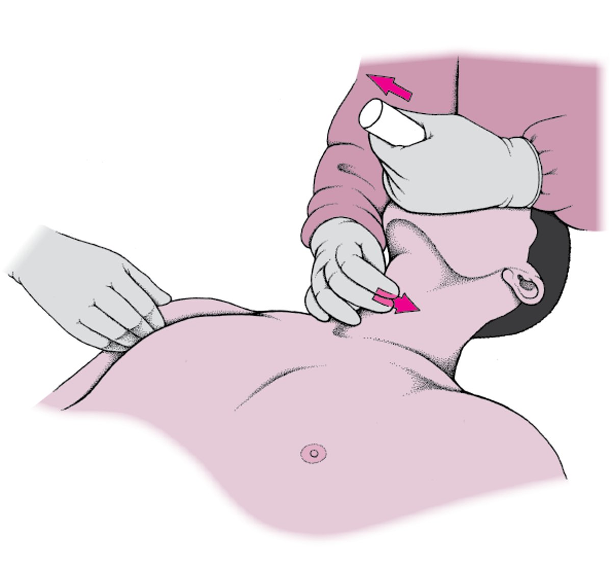
If identifying structures is difficult, manipulating the larynx with the right hand placed on the anterior neck (while continuing to hold the laryngoscope in the left hand ) may optimize the laryngeal view (see figure ). An assistant can help manipulate the larynx also. Another technique involves lifting the head higher (lifting at the occiput, not atlanto-occipital extension), which distracts the jaw and improves the line of sight. Head elevation is inadvisable in patients with potential cervical spine injury and is difficult in people who have severe obesity (who must be placed in a ramped or head-elevated position beforehand).
In an optimal view, the vocal folds are clearly seen. If the vocal folds are not seen, at a minimum, the posterior laryngeal landmarks must be viewed and the tip of the tube must be seen passing above the interarytenoid notch and posterior cartilages. Operators must clearly identify laryngeal landmarks to avoid potentially fatal esophageal intubation. If operators are not confident that the tube is going into the trachea, the tube should not be inserted.


In this illustration, a cuffed endotracheal tube has been placed using a laryngoscope with a curved blade to hold the tongue out of the way, lift the epiglottis, and allow for clear visualization of the vocal folds (round inset).
Sue Seif/SCIENCE PHOTO LIBRARY
Once an optimal view has been achieved, the right hand inserts the tube through the larynx into the trachea (if operators have been applying anterior laryngeal pressure with the right hand, an assistant should continue applying this pressure). If the tube does not pass easily, a 90° counter-clockwise twist of the tube may help it pass more smoothly over the anterior tracheal rings. After the cuff of the endotracheal tube has passed 1 to 2 cm past the vocal cords, the balloon should be inflated and the stylet removed. Before withdrawing the laryngoscope, operators should confirm that the tube is passing between the cords. Appropriate tube depth is usually 21 to 23 cm in adults and 3 times the endotracheal tube size in children (for a 4.0-mm endotracheal tube, 12 cm; for a 5.5-mm endotracheal tube, 16.5 cm). In adults, the tube, if inadvertently advanced, typically migrates into the right mainstem bronchus.
Intubation is unsuccessful in up to 30% of critically ill patients, so contingency plans should be established ( 2). When intubation fails, oxygenation should be prioritized and alternative interventions should be used while minimizing the number of airway interventions in order to minimize trauma and other procedural complications (3).
Bimanual Laryngoscopy
Pressure is applied on the neck opposite the direction of lift of the laryngoscope. Arrows show the direction for lift of the laryngoscope and for anterior neck pressure. |

Insertion technique references
1. Mort TC. Emergency tracheal intubation: complications associated with repeated laryngoscopic attempts. Anesth Analg. 2004;99(2):607-613. doi:10.1213/01.ANE.0000122825.04923.15
2. Higgs A, McGrath BA, Goddard C, et al. Guidelines for the management of tracheal intubation in critically ill adults. Br J Anaesth. 2018;120:323-352. doi: 10.1016/j.bja.2017.10.021
3. Ahmad I, El-Boghdadly K, Iliff H, et al. Difficult Airway Society 2025 guidelines for management of unanticipated difficult tracheal intubation in adults. Br J Anaesth. 2026;136(1):283-307. doi:10.1016/j.bja.2025.10.006
Alternative Intubation Devices
A number of devices and techniques can be used for intubation after failed laryngoscopy or as a primary means of intubation. Devices include:
Laryngeal mask airways (LMAs) with a passage that allows tracheal intubation
Fiberoptic scopes and optical stylets
Tube introducers
Each device has its own subtleties; clinicians who are skilled in standard laryngoscopic intubation techniques should not assume they can use one of these devices (especially after the patient has been given muscle relaxants) without becoming thoroughly familiar with it.
Video laryngoscopes enable practitioners to look around the curvature of the tongue and usually provide excellent laryngeal views. Video laryngoscopy may increase intubation success rate and shorten the time to successful intubation in adults with difficult airways (1) or in critially ill adults (2). However, some video laryngoscopes require that the endotracheal tube have an exaggerated bend angle to go around the tongue and thus may be more difficult to manipulate and insert. Guidelines suggest using a video laryngoscope for tracheal intubation in adults, particularly when a difficult airway is anticipated (3). In children, video laryngoscopy reduces the risk of failed first intubation attempts compared to direct laryngoscopy. However, more evidence is needed to determine whether all video laryngoscope devices provide the same benefit (4).
Some laryngeal mask airways have a passage to allow endotracheal intubation. To pass an endotracheal tube through a laryngeal mask airway, clinicians must understand how to optimally position the mask over the laryngeal inlet; there are sometimes mechanical difficulties passing the endotracheal tube. Other supraglottic airways can allow placement of an airway if an optimal laryngeal view cannot be obtained or an endotracheal tube cannot be passed through the vocal folds (eg, abnormal anatomy, subglottic stenosis). Intubation through a supraglottic airway is a high-risk maneuver and should not be performed blindly (5).
Flexible fiberoptic scopes and optical stylets are very maneuverable and can be used in patients with abnormal anatomy. However, practice is required to recognize laryngeal landmarks from a fiberoptic perspective. Compared with video laryngoscopes, fiberoptic scopes are more difficult to master and are more susceptible to problems with blood and secretions; also, they do not separate and divide tissue but instead must be moved through open channels.
Tube introducers (commonly called gum elastic bougies) are semirigid stylets that can be used when laryngeal visualization is suboptimal (eg, the epiglottis is visible, but the laryngeal opening is not). In such cases, the introducer is passed along the undersurface of the epiglottis; from this point, it is likely to enter the trachea. Tracheal entry is suggested by the tactile feedback, noted as the tip bounces over the tracheal rings. An endotracheal tube is then advanced over the introducer. During passage over a tube introducer, the tube tip sometimes catches the right aryepiglottic fold. Rotating the tube 90° counterclockwise often frees the endotracheal tube tip and allows it to pass smoothly.
Alternative intubation devices references
1. Su YC, Chen CC, Lee YK, Lee JY, Lin KJ. Comparison of video laryngoscopes with direct laryngoscopy for tracheal intubation: a meta-analysis of randomised trials. Eur J Anaesthesiol. 2011;28(11):788-795. doi:10.1097/EJA.0b013e32834a34f3
2. Prekker ME, Driver BE, Trent SA, et al. Video versus Direct Laryngoscopy for Tracheal Intubation of Critically Ill Adults. N Engl J Med. 2023;389(5):418-429. doi:10.1056/NEJMoa2301601
3. Ahmad I, El-Boghdadly K, Iliff H, et al. Difficult Airway Society 2025 guidelines for management of unanticipated difficult tracheal intubation in adults. Br J Anaesth. 2026;136(1):283-307. doi:10.1016/j.bja.2025.10.006
4. Perkins EJ, Begley JL, Brewster FM, Hanegbi ND, Ilancheran AA, Brewster DJ. The use of video laryngoscopy outside the operating room: A systematic review. PLoS One. 2022;17(10):e0276420. doi:10.1371/journal.pone.0276420
5. de Carvalho CC, Regueira SLPA, Souza ABS, et al. Videolaryngoscopes versus direct laryngoscopes in children: Ranking systematic review with network meta-analyses of randomized clinical trials. Paediatr Anaesth. 2022;32(9):1000-1014. doi:10.1111/pan.14521
Confirming Tube Placement
The stylet is removed and the balloon cuff is inflated with air using an appropriately sized syringe; a manometer, when available, can be used to verify that balloon pressure is < 30 cm water. The size of the endotracheal tube and cuff size affect the volume of air necessary for the correct pressure.
After balloon inflation, tube placement should be checked by one or more of the following methods:
Inspection and auscultation
Carbon dioxide detection
Esophageal detector devices
Sometimes chest radiograph
When a tube is correctly placed, manual ventilation should produce symmetric chest rise, good breath sounds over both lungs, and no gurgling over the upper abdomen that would indicate esophageal intubation.
Exhaled air should contain carbon dioxide and gastric air should not; detecting carbon dioxide with quantitative waveform capnography confirms tracheal placement. Continuous waveform capnography for the detection of end tidal carbon dioxide (ETCO2) is the standard of care and preferred method when available (1). Colorimetric devices are acceptable alternatives but are less comprehensive options for confirming and monitoring endotracheal tube placement. Nonetheless, both methods can be unreliable in cases of low cardiac output. Quantitative waveform capnography may not detect ETCO2 initially if cardiac arrest is prolonged prior to intubation. ETCO2, however, may be detected later if resuscitative efforts are successful. Detection of ETCO2 or rising ETCO2 levels reflect effective chest compressions and ventilation.
In prolonged cardiac arrest (ie, with little or no metabolic activity), ETCO2 may not be detectable even with correct tube placement. In such cases, an esophageal detector device may be used. These devices use an inflatable bulb or a large syringe to apply negative pressure to the endotracheal tube. The flexible esophagus collapses, and little or no air flows into the device; in contrast, the rigid trachea does not collapse, and the resultant airflow confirms tracheal placement.
Tube placement is typically also confirmed with a chest radiograph once the patient is not undergoing active resuscitation.
After correct placement is confirmed, the tube should be secured using a commercially available device or adhesive tape. Adapters connect the endotracheal tube to a resuscitator bag, T-piece supplying humidity and oxygen, or a mechanical ventilator.
Endotracheal tubes can be displaced, particularly in chaotic resuscitation situations, so tube position should be checked frequently. If breath sounds are absent on the left, right mainstem bronchus intubation is probably more likely than a left-sided tension pneumothorax, but both should be considered.
Confirming tube placement reference
1. Wigginton JG, Agarwal S, Bartos JA, et al. Part 9: Adult Advanced Life Support: 2025 American Heart Association Guidelines for Cardiopulmonary Resuscitation and Emergency Cardiovascular Care. Circulation. 2025;152(16_suppl_2):S538-S577. doi:10.1161/CIR.0000000000001376
Nasotracheal Intubation
Nasotracheal intubation is a technique, used infrequently, in patients who are awake and breathing spontaneously, or for situations in which the mouth must be avoided or cannot open widely enough (eg, edema, limited jaw motion) or the patient must be awake and/or upright until the airway is controlled (eg, large anterior mediastinal mass) (1). A serious complication of nasopharyngeal intubation is epistaxis. Blood in the airway can obscure the laryngoscopic view and complicate the intubation.
If patients are spontaneously breathing, nasotracheal intubation can be used in certain emergency situations, such as when patients have severe oral or cervical disorders (eg, injuries, edema, limitation of motion) that make laryngoscopy difficult. Nasotracheal intubation is absolutely contraindicated in patients with midface fractures or known or suspected basal skull fractures. Additional problems with nasal intubation include sinusitis (universal after 3 days), and the fact that tubes large enough to permit bronchoscopy (eg, ≥ 8 mm) can rarely be inserted nasotracheally.
When nasotracheal intubation is performed, a vasoconstrictor (eg, phenylephrine) and topical anesthetic (eg, benzocaine, lidocaine) must be applied to the nasal mucosa and the larynx to prevent bleeding and to blunt protective reflexes. Some patients may also require IV sedatives, opioids, or dissociative medications. A soft nasopharyngeal airway may be used to ensure adequate patency of the selected nasal passage, to serve as a conduit for topical anesthetic delivery to the pharynx and larynx, and to dilate the nasopharynx.
Complications of Tracheal Intubation
Complications of tracheal intubation include:
Direct trauma
Esophageal intubation
Tracheal erosion or stenosis
Laryngoscopy can damage lips, teeth, tongue, and supraglottic and subglottic areas.
Tube placement in the esophagus, if unrecognized, causes failure to ventilate and potentially death or hypoxic injury. Insufflating a tube in the esophagus causes regurgitation, which can result in aspiration, compromise subsequent bag-valve-mask ventilation, and obscure visualization in subsequent intubation attempts.
Any translaryngeal tube injures the vocal cords somewhat; sometimes ulceration, ischemia, and prolonged vocal fold paralysis occur. Subglottic stenosis can occur later (usually 3 to 4 weeks).
Erosion of the trachea is uncommon. It results more commonly from excessively high cuff pressure (> 30 cm water).
Rarely, hemorrhage from major vessels (eg, innominate artery), fistulas (especially tracheoesophageal), and tracheal stenosis occur. Using high-volume, low-pressure cuffs with tubes of appropriate size and measuring cuff pressure frequently (every 8 hours) to maintain it at < 30 cm water decrease the risk of ischemic pressure necrosis, but patients in shock, with low cardiac output, or with sepsis remain especially vulnerable.
In pediatric patients, laryngospasm may occasionally occur and can be prevented with topical or intravenous lidocaine and treated with propofol and/or succinylcholine (1).
Complications of intubation reference
1. von Ungern-Sternberg B, Sims C. Airway Management in Children. In: Sims C, Weber D, Johnson C (eds). A Guide to Pediatric Anesthesia. Springer, Cham. 2020. pp. 77–114. https://doi.org/10.1007/978-3-030-19246-4_4
Drug Information for the Topic


