

In the United States, injury is the most common cause of death for people aged 1 to 44 years (1). In 2023, there were 300,900 injury deaths, approximately 74% of which were accidental. Of intentional injury deaths, more than 65% were due to self-harm. In addition to deaths, injury results in over 40 million emergency department visits annually.
Patients whose injuries are serious but not immediately fatal benefit the most from treatment in designated trauma centers, hospitals that have special staffing and protocols to provide immediate care to critically injured patients. Criteria for such designation (and for the necessity of transport to them) vary but usually follow government or medical society guidelines, such as those from the American College of Surgeons Committee on Trauma.
Many traumatic injuries are discussed elsewhere in The Manual:
General reference
1. Centers for Disease Control and Prevention, National Center for Health Statistics: All injuries. FastStats. Available at https://www.cdc.gov/nchs/fastats/injury.htm. Accessed March 30, 2026.
Etiology of Trauma
Most physical trauma is due to blunt or penetrating injury. Blunt injury involves a forceful impact (eg, blow, kick, strike with an object, fall, motor vehicle crash, blast). Penetrating injury involves breach of the skin or other tissues by an object (eg, knife, broken glass) or projectile (eg, bullet, shrapnel from an explosion).
Other mechanisms of injury include thermal and chemical burns, toxic inhalations or ingestions, and radiation injury.
Pathophysiology of Trauma
All cases of trauma involve direct injury (immediate tissue damage from mechanical force at the time of impact), the nature and extent of which depend on the anatomic site, mechanism, and amount of force. Severe direct tissue damage to critical organs (eg, to the heart, brain, spinal cord) causes most immediate deaths due to trauma.
Trauma may also involve indirect injury (delayed tissue damage caused by the physiological consequences of the initial injury). This may include tissue ischemia–reperfusion injury, compartment syndrome, inflammation, infection, or other sequelae.
Vascular disruption may occur due to direct or indirect injury to blood vessels and hemorrhage may occur, which can be external or internal and either confined (eg, contusion, hematoma) or free (bleeding into a body cavity [eg, peritoneal cavity, thorax]). Small volumes of hemorrhage (ie, < 10% of blood volume) are tolerated well by most patients. Larger volumes cause hypovolemia with progressive declines in blood pressure and organ perfusion (ie, shock), leading to cellular dysfunction, organ failure, and eventually death.
Hemorrhagic shock and brain injury cause most short-term (ie, within hours) deaths, and multiple organ failure due to prolonged shock causes many of the near-term (ie, first 14 days) deaths (1). Additional near-term deaths result from infection because of disruption of normal anatomic barriers and immune system dysfunction.
Etiology reference
1. Cole E, Gillespie S, Vulliamy P, et al; Organ Dysfunction in Trauma (ORDIT) study collaborators. Multiple organ dysfunction after trauma. Br J Surg. 2020;107(4):402-412. doi:10.1002/bjs.11361
Evaluation and Treatment of Trauma Patient
Evaluation of the trauma patient includes:
Primary survey: Evaluation and stabilization using the mnemonic A, B, C, D, E, (Airway, Breathing, Circulation, Disability [neurologic status], and Exposure/Environmental control)
Secondary survey: Head-to-toe examination after initial stabilization
Selective use of radiographs, CT, and other imaging studies
Evaluation and treatment are performed simultaneously, beginning with systems that pose the most immediate threat to life if damaged. Attending to dramatic but not deadly injuries (eg, open lower-extremity fracture, finger amputations) before evaluating immediate life threats can be a fatal mistake.
Initial evaluation and stabilization are performed systematically based on A, B, C, D, E. Systems are rapidly examined for serious abnormalities (primary survey); a more detailed examination (secondary survey) is performed after the patient is stable.
Pearls & Pitfalls
|
In pregnant trauma patients, the initial priority is stabilization of the pregnant patient, which is also the best way to ensure fetal stability. An obstetrician should be consulted immediately for patients with serious trauma or signs of pregnancy complications (eg, abnormal fetal heart rate, vaginal bleeding, contractions). At ≥ 20 weeks of pregnancy, the uterine fundus can be palpated at or above the umbilicus and is large enough to compress the inferior vena cava; thus, immobilization in the supine position can obstruct venous return and cause hypotension. The uterus can be manually pushed to the patient's left or the entire backboard can be tilted to the left to relieve the compression. If the fetus is ≥ 22 weeks gestation (the lower limit of viability), fetal monitoring is performed and continued for at least 4 to 6 hours. Rh0(D) immune globulin is given to all Rh-negative pregnant patients following even minor trauma. If a pregnant patient ≥ 20 weeks is in cardiac arrest and resuscitation efforts have been unsuccessful, a perimortem cesarean delivery (resuscitative hysterotomy) should be initiated within 4 minutes of the arrest and the fetus delivered within 5 minutes (1).
Primary Survey
Airway
Airway patency can be threatened by one or more of the following: blood clots, teeth, or foreign bodies in the oropharynx; soft-tissue laxity and posterior retraction of the tongue caused by obtundation (eg, due to head injury, shock, intoxication); and/or edema or hematoma due to direct neck trauma. These obstructions are readily visible on direct inspection of the mouth or neck; having the patient speak can rapidly confirm that the airway is not likely in immediate danger.
Blood and foreign material are removed by suction or manually. Obtunded patients whose airway patency, airway protective mechanisms, oxygenation, or ventilation is uncertain and patients with significant oropharyngeal injury require endotracheal intubation; usually medications are given to induce unconsciousness and paralysis before intubation is performed. Multiple tools are available to assist with airway management including extraglottic devices, airway bougie, and video laryngoscopy. A carbon dioxide colorimetric device or, preferably, waveform capnography can help confirm proper endotracheal tube placement.
If patients require an artificial airway and endotracheal intubation is not possible (eg, due to edema of the airway caused by a thermal burn) or contraindicated (eg, due to severe maxillofacial injury), surgical or percutaneous cricothyrotomy is indicated. NOTE: When evaluating or manipulating a patient’s airway, cervical spinal motion restriction (previously called spinal immobilization) should be maintained until cervical spine injury has been excluded by examination, imaging, or both.
Breathing
Adequate ventilation can be threatened by decreased central respiratory drive (usually due to head injury, intoxication, or nearly fatal shock) or by chest injury (eg, hemothorax or pneumothorax, multiple rib fractures, pulmonary contusion).
The chest wall is fully exposed to look for ample chest wall expansion, external signs of trauma, and paradoxical wall motion (ie, retraction of the chest wall on inspiration), which indicates a flail chest. The chest wall is palpated for rib fractures and the presence of subcutaneous air (sometimes the only finding in pneumothorax).
Adequacy of air exchange is usually apparent on auscultation. Tension pneumothorax, simple pneumothorax, or hemothorax may cause decreased breath sounds on the affected side. Tension pneumothorax may also cause distended neck veins; hypotension and deviation of the trachea to the side opposite the injury are later findings.
Pneumothorax is decompressed by chest tube (see How To Do Tube and Catheter Thoracostomy). In patients with findings consistent with a pneumothorax, a chest radiograph or bedside ultrasound should be performed before initiating positive-pressure ventilation. Positive-pressure ventilation may enlarge a simple pneumothorax or convert it to a tension pneumothorax. Suspected tension pneumothorax can be decompressed with finger thoracostomy (insertion of a finger into the pleural space) or needle thoracostomy (eg, a 14-gauge needle inserted in the midaxillary line, 5th intercostal space) to stabilize the patient if a chest tube cannot be inserted immediately. Inadequate ventilation is treated with endotracheal intubation and mechanical ventilation. An open pneumothorax is covered with an occlusive dressing attached on 3 sides; the 4th side is left untaped to release pressure that might build up and cause a tension pneumothorax.
Circulation
Significant external hemorrhage can occur from any major vessel but is always apparent. Life-threatening internal hemorrhage is often less obvious. However, this volume of hemorrhage can occur in only a few body compartments: the chest, abdomen, retroperitoneum, and soft tissues of the pelvis or thigh (eg, from a pelvic or femoral fracture).
Pulse and blood pressure are assessed, and signs of shock are noted (eg, tachypnea, dusky color, diaphoresis, altered mental status, poor capillary refill). Abdominal distention and tenderness, pelvis instability, and thigh deformity and instability are often present when internal hemorrhage in those areas is large enough to be life threatening.
External hemorrhage is controlled by direct pressure. Tourniquets should be applied for extremity bleeding if bleeding is not controlled by direct pressure. Two large-bore (eg, 14- or 16-gauge) IVs are started with 0.9% saline or lactated Ringer's solution; rapid infusion of 1 L (20 mL/kg for children) is given for signs of shock and hypovolemia. Early administration of whole blood or blood component therapy should be considered. Bedside measurement of lactate or arterial blood gases (and calculation of base excess) can help indicate the severity of tissue hypoperfusion and shock and thus help guide fluid therapy. Protocols have been developed for patients requiring large volumes of blood products (massive transfusion protocols), including evaluation of coagulation with thromboelastography or rotational thromboelastography (where available) and early administration (within 3 hours) of tranexamic acid. Patients with known coagulopathies or taking anticoagulant medications experiencing life-threatening hemorrhage should have their coagulopathies reversed.. Early administration of whole blood or blood component therapy should be considered. Bedside measurement of lactate or arterial blood gases (and calculation of base excess) can help indicate the severity of tissue hypoperfusion and shock and thus help guide fluid therapy. Protocols have been developed for patients requiring large volumes of blood products (massive transfusion protocols), including evaluation of coagulation with thromboelastography or rotational thromboelastography (where available) and early administration (within 3 hours) of tranexamic acid. Patients with known coagulopathies or taking anticoagulant medications experiencing life-threatening hemorrhage should have their coagulopathies reversed.
When there is strong clinical suspicion of serious intra-abdominal hemorrhage, patients may require immediate laparotomy. Placement of a resuscitative endovascular balloon for occlusion of the aorta (REBOA) can help stabilize the patient before surgery. Patients with massive intrathoracic hemorrhage may require immediate thoracotomy and possibly autotransfusion of blood recovered via tube thoracostomy.
Pearls & Pitfalls
|
Disability (neurologic dysfunction)
Neurologic function is evaluated for serious deficits involving the brain and spinal cord. The Glasgow Coma Scale (GCS; see tables Glasgow Coma Scale and Modified Glasgow Coma Scale for Infants and Children) and pupillary response to light are used to assess the level of consciousness and severity of intracranial injury.
Gross motor movement and sensation in each extremity are used to screen for serious spinal cord injury. The cervical spine is palpated for tenderness and deformity and stabilized in a rigid collar until cervical spine injury is excluded. With careful manual stabilization of the head and neck, the patient is logrolled onto a side to allow:
Palpation of the thoracic and lumbar spine
Inspection of the back
Rectal examination if indicated to check tone (decreased tone indicates possible spinal cord injury), the prostate (a high-riding prostate suggests urethral or pelvic injury), and presence of blood
In the United States, most patients arriving by ambulance are immobilized on a long, rigid board for ease of transport and to stabilize possible spinal fractures. Patients should be taken off the board as soon as possible because it is quite uncomfortable and pressure ulcers may occur within a few hours (2).
Glasgow Coma Scale
Area Assessed | Response | Score* |
|---|---|---|
Eye opening | Open spontaneously | 4 |
Open to verbal command | 3 | |
Open in response to pain applied to the limbs or sternum | 2 | |
None | 1 | |
Verbal | Oriented | 5 |
Disoriented, but able to answer questions | 4 | |
Inappropriate answers to questions; words discernible | 3 | |
Incomprehensible speech | 2 | |
None | 1 | |
Motor | Obeys commands | 6 |
Responds to pain with purposeful movement | 5 | |
Withdraws from pain stimuli | 4 | |
Responds to pain with abnormal flexion (decorticate posture) | 3 | |
Responds to pain with abnormal (rigid) extension (decerebrate posture) | 2 | |
None | 1 | |
* Combined scores < 8 are typically regarded as coma and may suggest severe traumatic brain injury. | ||
Adapted from Teasdale G, Jennett B. Assessment of coma and impaired consciousness. A practical scale. Lancet. 1974;2:81-84. doi: 10.1016/s0140-6736(74)91639-0 | ||
Modified Glasgow Coma Scale for Infants and Children
Area Assessed | Infants | Children | Score* |
|---|---|---|---|
Eye opening | Open spontaneously | Open spontaneously | 4 |
Open in response to verbal stimuli | Open in response to verbal stimuli | 3 | |
Open in response to pain only | Open in response to pain only | 2 | |
No response | No response | 1 | |
Verbal response | Coos and babbles | Oriented, appropriate | 5 |
Irritable cries | Confused | 4 | |
Cries in response to pain | Inappropriate words | 3 | |
Moans in response to pain | Incomprehensible words or nonspecific sounds | 2 | |
No response | No response | 1 | |
Motor response† | Moves spontaneously and purposefully | Obeys commands | 6 |
Withdraws to touch | Localizes painful stimulus | 5 | |
Withdraws in response to pain | Withdraws in response to pain | 4 | |
Responds to pain with decorticate posturing (abnormal flexion) | Responds to pain with decorticate posturing (abnormal flexion) | 3 | |
Responds to pain with decerebrate posturing (abnormal extension) | Responds to pain with decerebrate posturing (abnormal extension) | 2 | |
No response | No response | 1 | |
* Score ≤ 12 suggests a severe head injury. Score ≤ 8 suggests the possible need for intubation and ventilation as well as the need for intracranial pressure monitoring. | |||
† If the patient is intubated, unconscious, or preverbal, the most important part of this scale is motor response. This section should be carefully evaluated. | |||
Adapted from Davis RJ et al. Head and spinal cord injury. In Textbook of Pediatric Intensive Care, edited by MC Rogers. Baltimore, Williams & Wilkins, 1987; James H, Anas N, Perkin RM. Brain Insults in Infants and Children. New York, Grune & Stratton, 1985; and Morray JP, Tyler DC, Jones TK, et al. Coma scale for use in brain-injured children. Critical Care Medicine. 1984;12:1018–1020. doi: 10.1097/00003246-198412000-00002; and Carney N, Totten AM, O'Reilly C, et al. Guidelines for the management of severe traumatic brain injury, fourth edition. Neurosurgery 2017;80(1):6-15. doi: 10.1227/NEU.0000000000001432. | |||
Patients with severe traumatic brain injury (GCS < 8) may require endotracheal intubation for airway protection, brain imaging, neurosurgical evaluation, and therapy to prevent secondary brain injury (eg, optimization of blood pressure and oxygenation, seizure prophylaxis, treatment of elevated intracranial pressure, sometimes transient hyperventilation for patients with signs of impending brain herniation).
Exposure/environmental control
To ensure injuries are not missed, patients are completely undressed (by cutting off garments) and the entire body surface is examined for signs of occult trauma. The patient is kept warm (eg, with heated blankets and by using warmed IV fluids) to prevent hypothermia.
Secondary survey
After immediate life threats are assessed and the patient is stable, a more thorough evaluation is performed, and a focused history is obtained. If only limited conversation is possible, an “AMPLE” history covers essential information:
Allergies
Medications
Past medical history
Last meal
Events of the injury
After the patient is completely undressed, the examination generally proceeds from head to toe; it typically includes all orifices and a more detailed look at areas examined in the initial survey. All soft tissues are inspected for lesions and swelling, all bones are palpated for tenderness, and range of motion is assessed in joints (unless there is obvious fracture or deformity).
A urinary catheter is usually placed in seriously injured and obtunded patients provided there is no evidence of urethral injury (eg, blood at the meatus, ecchymosis of the perineum). Intubated, seriously injured patients often also have an orogastric tube placed.
Open wounds are covered with sterile dressings, but cleansing and repair are deferred until completion of evaluation and treatment of more serious injuries. Serious clinically apparent joint dislocations with marked deformity or neurovascular compromise are imaged and reduced as soon as immediate life threats have been addressed.
Obvious or suspected fractures are splinted pending full assessment of serious injuries and appropriate imaging studies. A clinically apparent unstable pelvic fracture is stabilized with a commercial pelvic binder or bed sheet to help close the pelvic space and decrease bleeding; severe bleeding may require urgent angiographic embolization, surgical fixation, or direct surgical control.
Imaging and laboratory tests supplement clinical assessment. Patients with penetrating trauma typically have focal injuries that can limit imaging to the obviously involved region or regions. Blunt trauma, particularly when significant deceleration is involved (eg, serious fall, motor vehicle crash), can affect any part of the body, and imaging studies that are indicated by the mechanism of injury and findings on examination are obtained.
Imaging studies
Cervical spine imaging can be deferred in patients who are not intoxicated, do not have focal neurologic findings, have no midline cervical spine tenderness or distracting injuries (eg, femur fracture), and are awake and alert. All others should have cervical spine imaging, preferably using CT.
Chest radiograph can identify airway disruption, lung injury, hemothorax, and pneumothorax; it can also suggest thoracic aorta tears (eg, by mediastinal widening). However, chest CT is more sensitive for most intrathoracic injuries and is often preferred. Chest imaging is now commonly performed at the bedside using extended focused assessment with sonography in trauma (E-FAST), particularly if patients are unstable. Pneumothoraces, hemothoraces, and hemopericardium can be identified.
CT of the chest, abdomen, pelvis, spine, or head or, particularly, combinations of these studies are frequently used for patients who require imaging after severe multiple blunt trauma.
Identification of intra-abdominal injury is essential. Bedside E-FAST examination can be useful to identify intraperitoneal blood, particularly for unstable patients; it is sensitive for significant volumes of intraperitoneal blood and thus the need for immediate laparotomy. If patients are stable, CT is the preferred study; it is very accurate, allows imaging of the retroperitoneal structures and bones, and shows the volume and sometimes the origin of hemorrhage. Diagnostic peritoneal lavage (DPL) is no longer the recommended approach to evaluate patients for potential intra-abdominal injury.

If pelvic fracture is suspected, CT of the pelvis is performed; it is more accurate than plain radiographs.
Head CT is typically performed in patients with altered mental status or focal neurologic abnormalities and in patients who sustained loss of consciousness. Some evidence suggests that CT is not necessary in patients with brief loss of consciousness (ie, < 5 seconds) or transient amnesia or disorientation but who are alert with a GCS of 15 during examination. Imaging is performed more liberally in patients with persistent headache, vomiting, amnesia, seizures, age > 60 years, and drug or alcohol intoxication and in patients taking anticoagulant or antiplatelet medications. Clinical decision rules have been developed to help determine which patients should have a head CT (3). These decision rules should be used to aid, but not replace, clinical judgment.
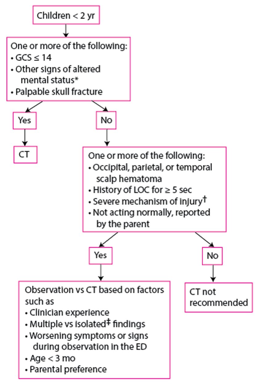
For children with head injury, the Pediatric Emergency Care Applied Research Network (PECARN) has developed an algorithm that may help limit radiation exposure from head CT; clinical observation is used in children who may otherwise have received CT.
Evaluation of Children 0 to 2 Years With a Head Injury
* Include agitation, somnolence, repetitive questioning, and slow response to verbal communication. |
† Include motor vehicle crash involving ejection of patient, death of another passenger, or rollover; collision of a motor vehicle with a pedestrian or bicyclist not wearing a helmet; and a fall of > 0.9 m for children < 2 years; and a blow to the head by a high-impact object. |
‡ No other findings suggesting traumatic brain injury, such as only LOC, headache, vomiting, and certain scalp hematomas in children >3 months. |
ED = emergency department; GCS = Glasgow Coma Scale; LOC = loss of consciousness. |
Adapted from Kupperman N, Holmes JF, Dayan PS, et al for the Pediatric Emergency Care Applied Research Network. Identification of children at very low risk of clinically-important brain injuries after head trauma: A prospective cohort study. Lancet. 2009;374: 1160-1170. doi:10.1016/S0140-6736(09)61558-0 |

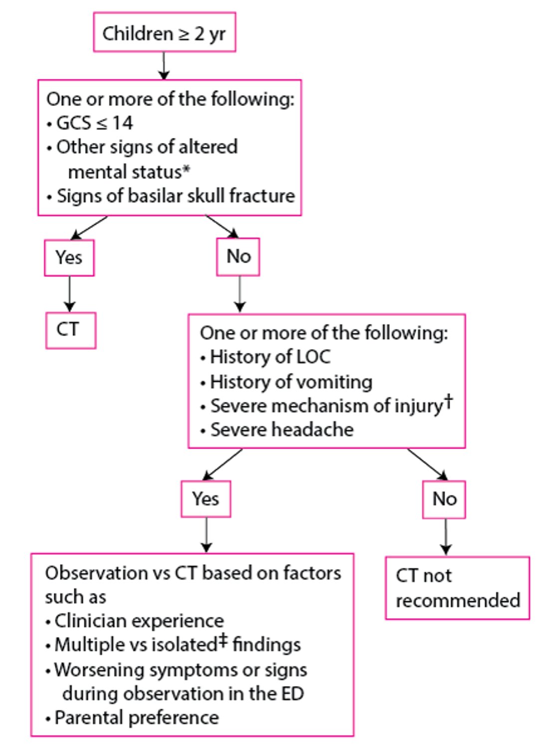
Evaluation of Children ≥ 2 Years With a Head Injury
* Include agitation, somnolence, repetitive questioning, and slow response to verbal communication. |
† Include motor vehicle crash involving ejection of patient, death of another passenger, or rollover; collision of a motor vehicle with a pedestrian or bicyclist not wearing a helmet; and a fall of >1.5 m for children ≥ 2 years; and a blow to the head by a high-impact object. |
‡ No other findings suggesting traumatic brain injury, such as only LOC, headache, vomiting, and certain scalp hematomas in children >3 months. |
ED = emergency department; GCS = Glasgow Coma Scale; LOC = loss of consciousness. |
Adapted from Kupperman N, Holmes JF, Dayan PS, et al for the Pediatric Emergency Care Applied Research Network. Identification of children at very low risk of clinically-important brain injuries after head trauma: A prospective cohort study. Lancet. 2009;374: 1160-1170. doi:10.1016/S0140-6736(09)61558-0 |

Patients with suspected spinal cord injury should be imaged using MRI.
Aortic injury should be considered in patients with severe deceleration chest injury or suggestive signs (eg, pulse deficits or asymmetric blood pressure measurements, end-organ ischemia, suggestive findings on chest radiograph); these patients may require CT angiography or other aortic imaging. Short-acting beta-blockers can be used to control heart rate and blood pressure in patients with traumatic aortic injury.
All patients suspected of having significant blunt chest injury should be placed on a cardiac monitor and have an ECG to detect myocardial injury and arrhythmias. Patients with abnormalities on ECG usually have blood levels of cardiac markers measured and sometimes echocardiography to evaluate the patient for possible cardiac contusion.
Vascular injury to the carotid and vertebral vessels should be considered in patients with trauma to the head and neck, particularly those with unilateral neurologic findings, a neck seat belt sign (linear ecchymosis due to the shoulder strap), or a predisposing injury (eg, fracture of C1, C2, or C3; other C-spine fracture with subluxation; hanging mechanism). Such patients typically should have CT angiography.
Plain radiographs are obtained of any suspected fractures and dislocations of the extremities. Other imaging tests are obtained for specific indications (eg, angiography to diagnose and sometimes embolize vascular injury; CT to better delineate spinal, pelvic, or complex joint fractures).
Laboratory evaluation
Laboratory tests that may be useful include:
Complete blood count to establish a baseline
Serial hemoglobin levels to assess for bleeding
Blood gas determination for partial pressure of oxygen, partial pressure of carbon dioxide, and base deficit
Urine examination for blood
Glucose to evaluate for hypoglycemia
Type and crossmatch for possible blood transfusion
Coagulation studies
Measures of perfusion (serum lactate, base deficit on arterial blood gas measurement, and, in patients with a catheterized central vein, central venous oxygen saturation) are indicated to help identify early or partially treated shock. Other reflexively obtained tests (eg, electrolytes and other chemistries) are unlikely to be helpful unless suggested by relevant medical history (eg, renal insufficiency, diuretic use).
Toxicology screening (eg, blood alcohol, urine toxicology screen) is often performed; results of this testing rarely change immediate management but can help identify substance use disorder as the possible cause of injury, allowing intervention to prevent subsequent trauma.
D-Dimer, fibrinogen, and fibrin degradation products may be measured in pregnant trauma patients. Test results may be abnormal in patients with placental abruption; however, these tests are neither sensitive nor specific and cannot definitively confirm or exclude the diagnosis.
Evaluation and treatment references
1. Del Rios M, Bartos JA, Panchal AR, et al. Part 1: Executive Summary: 2025 American Heart Association Guidelines for Cardiopulmonary Resuscitation and Emergency Cardiovascular Care. Circulation. 2025;152(16_suppl_2):S284-S312. doi:10.1161/CIR.0000000000001372
2. American College of Surgeons/American Congress of Rehabilitation Medicine. Best Practices Guidelines: Spine Injury. March 2022. Accessed March 30, 2026.
3. Bouida W, Marghli S, Souissi S, et al. Prediction value of the Canadian CT head rule and the New Orleans criteria for positive head CT scan and acute neurosurgical procedures in minor head trauma: A multicenter external validation study. Ann Emerg Med. 2013;61(5): 521-527. doi: 10.1016/j.annemergmed.2012.07.016
More Information
The following English-language resources may be useful. Please note that The Manual is not responsible for the content of these resources.


