Common causes of eye injury include physical injury due to accidents, physical assault, sporting injuries (including air-gun, paint-gun, pellet-gun injuries), motor vehicle crashes (including airbag injuries), or exposure to toxic chemicals (eg, some cleaning products). Exposure to strong ultraviolet light, as from a welding arc or bright sunlight reflected off snow, can injure the cornea (see Superficial Punctate Keratitis). Injury may be to the eyeball (globe), surrounding soft tissues (including muscles, nerves, tendons), and/or bones of the orbit.
General evaluation for an eye injury should include the following:
Tests of visual acuity
Pupillary appearance and responses
Range of extraocular motion
Visual fields to confrontation
Location and depth of eyelid and conjunctival lacerations
Location of any foreign bodies
Depth of anterior chamber
Presence of anterior chamber or vitreous hemorrhage, cataract, or absence of a red reflex
Retinal examination
Intraocular pressure determination
Imaging studies as indicated
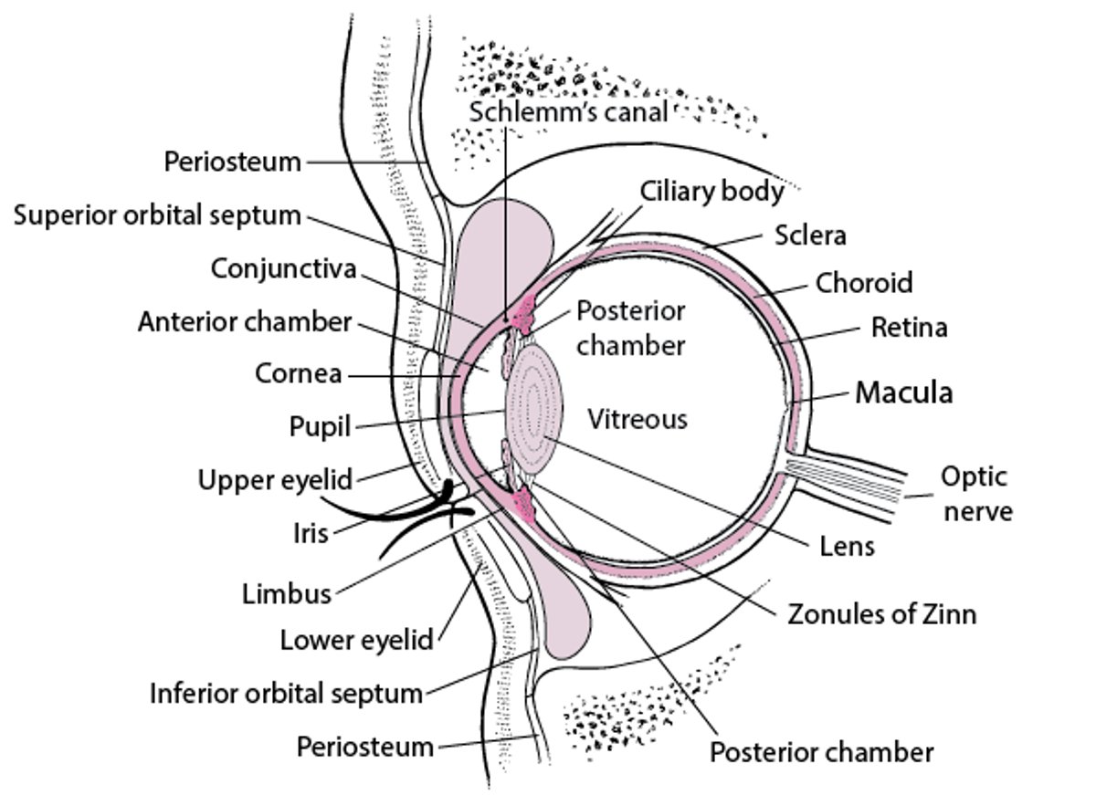
Cross Section of the Eye
The zonules of Zinn keep the lens suspended, and the muscles of the ciliary body focus the lens. The ciliary body also secretes aqueous humor, which fills the anterior and posterior chambers, passes through the pupil into the anterior chamber, and drains primarily via the Schlemm canal. The iris regulates the amount of light entering the eye by adjusting the size of its central opening, the pupil. Visual images are focused on the retina. The conjunctiva covers the eyeball and lines the upper and lower eyelids; it ends at the limbus. The cornea is covered with epithelium that is more sensitive than and differs from the conjunctival epithelium. |

A red reflex is the normal reflection of light from the retina. It is best seen in patients with dilated pupils and in photographs—unless the "red eye reduction" function is chosen on the camera. Detailed examination of the sclera, anterior segment (cornea, conjunctiva, anterior chamber, ciliary body, iris), lens, and anterior vitreous is best performed with a slit lamp (see Slit-lamp examination).
Although direct ophthalmoscopy can be used to examine the lens and some of the posterior structures of the eye, indirect ophthalmoscopy, usually performed by an ophthalmologist, provides a more detailed and binocular view of these structures. Indications for indirect ophthalmoscopy include clinical suspicion of vitreous abnormalities (eg, hemorrhage, foreign body) and retinal abnormalities; clinical suspicion may be based on symptoms, injury mechanism, absence of the red reflex, or retinal abnormalities visible with direct ophthalmoscopy.
Because direct and indirect ophthalmoscopy are best performed through a dilated pupil, approximately 15 to 30 minutes before this examination, mydriatics can be instilled when there is no contraindication to a dilated eye examination (eg, shallow anterior chamber, allergy, ruptured globe). Tropicamide 1% is preferred because of its rapid onset and relatively short duration of action when compared with other medications, such as cyclopentolate. Dose is one drop in each eye.
CT scan is the preferred imaging choice in cases of trauma or acute pathology (such as orbital abscess) that require urgent intervention. CT of the orbits and skull base is highly diagnostic, is easily available, provides excellent images of bony anatomy, and takes less than 60 seconds to perform.
Although CT is the preferred initial imaging method, MRI is also an option. However, MRI should not be used if there is a possibility that a metallic foreign body is present. MRI can cause additional injury to the eye by inducing movement of any metallic foreign bodies in the magnetic field. A screening plain film should be used to definitively rule out a metallic foreign body in suspected cases if an MRI is necessary for diagnostic purposes. MRI provides far better detail than CT of the orbital apex and skull base, and specific sequences can be used for specific pathologic entities. However, MRI of the orbit takes approximately 45 minutes to perform, may not be readily available in an emergency situation, and may suffer from severe degradation artifacts in uncooperative or claustrophobic patients.
In general, plain radiographs are not a helpful diagnostic tool for ocular and orbital pathology. A plain radiograph of the face does not adequately detect all orbital or periorbital foreign bodies. However, if performed for other indications, a plain radiograph may show the presence of a foreign body, especially one composed of metal or other radio-opaque material.
Use of eye guards, goggles, or special eyeglasses, such as those constructed of polycarbonate lenses in a wrap-around polyamide frame, is a simple precaution that greatly reduces the risk of injury.
Drug Information for the Topic