


Eye pain may be severe and seem sharp, aching, or throbbing, or people may feel only mild irritation of the eye surface or the sensation of a foreign object in the eye (foreign body sensation). Many causes of eye pain also cause the eye to look red. Other symptoms may be present depending on the cause of eye pain. For example, people may have blurred vision, a bulging eye, or pain worsened by bright light.
The cornea (the clear layer in front of the iris and pupil) is highly sensitive to pain. Many disorders that affect the cornea also affect the anterior chamber (the fluid-filled space between the iris and the inner part of the cornea) and cause spasm of the muscle that controls the iris (the ciliary muscle). When such spasm is present, bright light causes muscle contraction and worsening pain.
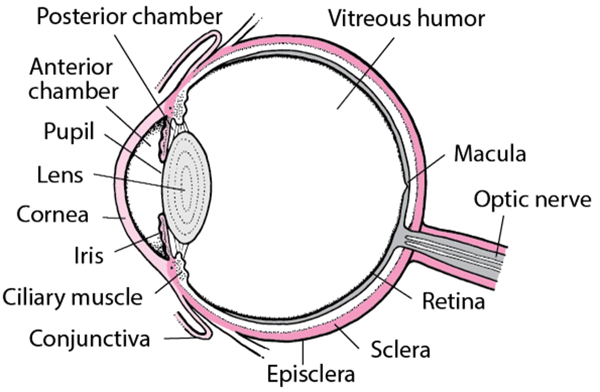
An Inside Look at the Eye

Causes of Eye Pain
Disorders that cause eye pain can be divided into disorders that affect primarily the cornea, disorders of other parts of the eye, and disorders of other areas of the body that cause pain to be felt in the eye.
Common causes
Corneal disorders are the most common causes of eye pain overall, particularly
Corneal scratches (corneal abrasions)
However, most corneal disorders can cause eye pain.
A feeling of scratchiness or a foreign body sensation may be caused by a disorder of the conjunctiva (the thin membrane that lines the eyelid and covers the front of the eye) or of the cornea.
Some Causes and Features of Eye Pain
Cause | Common Features* | Diagnostic Approach |
|---|---|---|
Disorders that affect the cornea primarily | ||
Contact lens keratitis (inflammation of the cornea—the clear layer in front of the iris and pupil—caused by wearing contact lenses for long periods of time) | Usually affecting both eyes Eye ache and a feeling of grittiness in the eye Eye redness, tearing, and sensitivity to light In people who wear contact lenses for long periods of time | A doctor's examination |
Corneal scratch (abrasion) A foreign object (body) | Symptoms that begin after an eye injury or high-risk activity (for example, grinding metal), which may not be noticed in infants and young children Pain in the affected eye when blinking and a foreign body sensation Eye redness, tearing, and usually sensitivity to light | A doctor's examination |
Often a grayish patch on the cornea that later becomes an open, painful sore Eye ache and a foreign body sensation Eye redness, tearing, and sensitivity to light Sometimes in people who have had an eye injury or who have slept with their contact lenses in | A doctor's examination Culture of a sample taken from the ulcer, done by an ophthalmologist | |
Epidemic keratoconjunctivitis (pink eye—inflammation of the conjunctiva, the membrane that lines the eyelids and covers the front of the eye, and the cornea caused by an adenovirus) | Usually in both eyes Eye ache and a feeling of grittiness in the eye Eye redness, tearing, and usually sensitivity to light Often eyelid swelling and swollen, tender lymph nodes in front of the ears Rarely temporary, severe blurring of vision | A doctor's examination |
Herpes simplex keratitis (infection of the cornea caused by the herpes simplex virus) | Usually affecting only one eye Early: Symptoms that begin after an episode of conjunctivitis Blisters on the eyelid, sometimes with crusting Late or recurring: Eye redness and watering, eye pain, impaired vision, and sensitivity to light | Usually only a doctor's examination Sometimes, testing for the virus in scrapings obtained from the surface of the cornea or from blisters around the eye |
Herpes zoster ophthalmicus (shingles that affects the face and eye, caused by the varicella-zoster virus) | Usually affecting only one eye Early: A rash with blisters and/or crusts on one side of the face, around the eye, on the forehead, and sometimes on the tip of the nose Late: Eye redness, tearing, usually sensitivity to light, and eyelid swelling | Usually only a doctor's examination Sometimes, testing for the virus in scrapings obtained from blisters around the eye |
Welder’s (ultraviolet) keratitis (inflammation of the cornea caused by exposure to excessive ultraviolet light) | Usually affects both eyes Symptoms that begin hours after exposure to excessive ultraviolet light (as is produced during arc welding, by a sunlamp, or by bright sun reflecting off snow, particularly at high altitudes) Eye ache and a feeling of grittiness in the eye Eye redness, tearing, and sensitivity to light | A doctor's examination |
Other eye disorders | ||
Severe eye ache and redness Headache, nausea, vomiting, and pain with exposure to light Disturbances in vision such as halos seen around lights and/or decreased vision | Tonometry (measurement of pressure inside the eye)† Examination of the eye's drainage channels with a special lens (gonioscopy), done by an ophthalmologist | |
Anterior uveitis (inflammation of the anterior chamber—the fluid-filled space between the iris and cornea) | Eye ache and sensitivity to light Eye redness (particularly around the cornea) Blurring or loss of vision Often in people who have an autoimmune disorder or who recently had an eye injury | A doctor's examination |
Endophthalmitis (infection inside the eye)‡ | Affecting only one eye Eye ache, intense eye redness, sensitivity to light, and severely decreased vision Often in people who have had recent eye surgery, a serious eye injury, or bloodstream infection | A doctor's examination Cultures of fluids inside of the eye, done by an ophthalmologist |
Optic neuritis (inflammation of the optic nerve), which can be related to multiple sclerosis‡ | Usually mild pain that may worsen when eyes are moved Partial or complete loss of vision Eyelids and corneas that appear normal | A doctor's examination Often, MRI with contrast of the brain and orbit |
Orbital cellulitis (infection of the tissue within the eye socket, or orbit)‡ | Affecting only one eye Bulging of the eye, eye redness, pain deep within the eye, and aches in and around the eye Red and swollen eyelids Inability to fully move the eye in all directions Impaired vision or loss of vision Fever Sometimes preceded by symptoms of sinusitis (see below) | A doctor's examination Usually, CT or MRIs of the orbit |
Orbital pseudotumor (a noncancerous accumulation of inflammatory and fibrous tissue in the eye socket)‡ | Aches in and around the eye, which may be very severe Often bulging of the eye Often inability to fully move the eye in all directions Swelling around the eye | A doctor's examination Usually, CT or MRI of the orbit Sometimes, biopsy |
Scleritis (inflammation of the white of the eye called the sclera) | Very severe pain, often described as boring, and sensitivity to light Watering of the eyes Red or violet patches on the white of the eye Often in people who have an autoimmune disorder | A doctor's examination Sometimes, ultrasound or CT of the orbit Rarely, biopsy |
Other disorders that cause eye pain | ||
In people who have had previous episodes of severe headaches Cluster headaches: Headaches that
Migraines: Headaches that
| A doctor's examination Sometimes, MRI or CT of the brain | |
Sometimes swelling around the eye but no other eye symptoms A yellow or green thick nasal discharge (sometimes with bleeding), headache, or eye or facial pain that varies with head position Fever, tenderness of the face, sometimes a productive cough during the night, and bad breath | A doctor's examination Sometimes, CT of the sinuses | |
* Features include symptoms and the results of the doctor's examination. Features mentioned are typical but not always present. Disorders usually affect only one eye unless otherwise specified. † Doctors almost always do a slit-lamp examination with fluorescein staining and measure the pressure inside the eye (called tonometry).† Doctors almost always do a slit-lamp examination with fluorescein staining and measure the pressure inside the eye (called tonometry). ‡ These causes are uncommon. CT = computed tomography; MRI = magnetic resonance imaging. | ||
Evaluation of Eye Pain
Mild eye irritation or a foreign body sensation is common and not usually serious. However, true pain in the eye can be a sign of a severe, vision-threatening disorder. The following information can help people decide when to see a doctor and help them know what to expect during the evaluation.
Warning signs
In people with eye pain, certain symptoms and characteristics are cause for concern. They include
Vomiting
Seeing halos around lights
Fever, chills, fatigue, or muscle aches
Decreased sharpness of vision (visual acuity)
Bulging of one or both eyes (proptosis)
Inability to move the eye in all directions (such as right, left, up, and down)
When to see a doctor
People who have severe pain, eye redness, or warning signs should see a doctor right away. People with mild pain and no eye redness or warning signs can wait a day or two to see if the discomfort goes away on its own.
What the doctor does
Doctors first ask questions about the person's symptoms and medical history. Doctors then do a physical examination. What they find during the history and physical examination often suggests a cause of the eye pain and the tests that may need to be done (see table Some Causes and Features of Eye Pain).
Doctors ask the person to describe the pain, including when it started, how severe it is, and whether it hurts to look in different directions or blink. They ask about whether the person has ever had eye pain and whether the person is sensitive to light, has blurred vision, or feels as if the eye contains a foreign object.
During the physical examination, doctors check for the presence of fever or a runny nose. They check the face for tenderness.
Most important is the eye examination, including the entire eye, eyelids, and the region around the eye. Doctors check
Whether the eyes are red or swollen
How clearly a person can see using a standard eye chart (visual acuity)
Whether the person can see in each part of the field of vision (visual field testing)
How the pupils react to light
Whether shining a light into the unaffected eye causes pain in the affected eye when the affected eye is closed (called true photophobia)
If doctors suspect a foreign object but do not see one, they turn the eyelids inside out to search for hidden foreign objects.
Doctors usually do a slit-lamp examination. A slit lamp is an instrument that enables a doctor to examine the eye under high magnification. Doctors place a drop of fluorescein stain on the cornea to show scratches or certain kinds of infection, including ulcers. Doctors use . A slit lamp is an instrument that enables a doctor to examine the eye under high magnification. Doctors place a drop of fluorescein stain on the cornea to show scratches or certain kinds of infection, including ulcers. Doctors usetonometry to measure the pressure inside the eye (intraocular pressure). They use a slit-lamp and/or an ophthalmoscope (a light with magnifying lenses that shines into the back of the eye) to examine the lens and use an ophthalmoscope to examine the vitreous humor (the jellylike substance that fills the eyeball), retina (the light-sensing structure at the back of the eye), optic nerve, and the retinal veins and arteries.
Sometimes findings are helpful in making a diagnosis. Particular findings or combinations may point to particular disorders.
Findings may also help suggest or eliminate certain types of disorders.
Corneal disorders, among other disorders, tend to cause eye redness, tearing, and pain. If those symptoms are absent, a corneal disorder is very unlikely.
Pain on the surface of the eye, a foreign body sensation, and pain with blinking suggest a foreign object.
People who wear contact lenses may have a corneal scratch, a corneal ulcer, or contact lens keratitis.
When measuring eye pressure, doctors put a drop of anesthetic into the eye. If pain then disappears, the cause of pain is probably a corneal disorder.
Deep, aching, throbbing pain often indicates a possibly serious disorder, such as acute closed-angle glaucoma, anterior uveitis, scleritis, endophthalmitis, orbital cellulitis, or orbital pseudotumor. If, in addition, there is eyelid swelling, bulging of the eye, or inability to move the eye to look in all directions, the most likely disorders are orbital pseudotumor, orbital cellulitis, or possibly severe endophthalmitis.
Fever, chills, and tenderness suggest infections such as orbital cellulitis or sinusitis.
Testing
The need for tests depends on what doctors find during the history and physical examination. Testing is usually not necessary. However, if doctors find increased intraocular pressure (indicating glaucoma), they may refer the person to an ophthalmologist (a medical doctor who specializes in the evaluation and treatment—surgical and nonsurgical—of eye disorders) for gonioscopy. A gonioscope is a special lens that allows doctors to examine the drainage channels in the eye, which can help determine whether the glaucoma is of the open-angle or closed-angle type.
Bulging of the eye and inability of an eye to move in all directions without moving the head can indicate orbital cellulitis or orbital pseudotumor. Computed tomography (CT) or magnetic resonance imaging (MRI) is then done to check for these disorders. CT may also be done if sinusitis is suspected but the diagnosis is not otherwise clear or if complications are suspected. MRI with dye may be done if optic neuritis is suspected.
Doctors send a sample of fluids from inside the eye (vitreous or aqueous humor) to the laboratory if endophthalmitis seems likely. They may send a sample from the cornea or a blister if herpes simplex keratitis or herpes zoster ophthalmicus seems likely but the diagnosis is not certain. In the laboratory, technicians try to grow bacteria or viruses (culture) to confirm infections and determine the organism causing the infection.
Treatment of Eye Pain
The best way to treat eye pain is to treat the cause of the pain. People may also need to take pain relievers (such as acetaminophen or a nonsteroidal anti-inflammatory drug) until the pain stops. Sometimes people with pain caused by The best way to treat eye pain is to treat the cause of the pain. People may also need to take pain relievers (such as acetaminophen or a nonsteroidal anti-inflammatory drug) until the pain stops. Sometimes people with pain caused byanterior uveitis or corneal disorders also need to use an eye drop that prevents ciliary muscle spasm by dilating the pupil and thus reduces eye pain with light exposure. For example, cyclopentolate may be used.also need to use an eye drop that prevents ciliary muscle spasm by dilating the pupil and thus reduces eye pain with light exposure. For example, cyclopentolate may be used.
Key Points
Usually doctors can determine the cause of eye pain during an examination.
People with severe pain, eye redness, or warning signs (vomiting, halos around lights, fever, decreased visual clarity, bulging eyes, and inability to move the eye in all directions) should see a doctor right away.


