


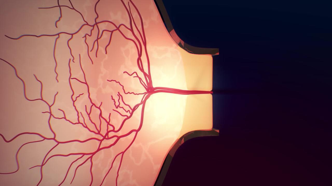
Optic neuritis is inflammation of the optic nerve.
Multiple sclerosis is the most common cause.
Loss of vision may develop, and there may be pain with eye movement.
Magnetic resonance imaging is done.
Steroids (also sometimes called glucocorticoids or corticosteroids) may be given.


In this illustration, the images in the left column show a healthy optic nerve and healthy myelin sheaths.
The images in the right column show an inflamed optic nerve and damaged myelin.
The bottom line graphics show visual evoked potentials (VEP). A VEP measures how quickly and strongly visual signals travel from the eye to the brain. In optic neuritis, these signals are usually slower, and in some cases weaker.
Alila Medical Media/stock.adobe.com
Causes of Optic Neuritis

Optic neuritis is most common optic nerve disorder in adults younger than 50. Optic neuritis is most often caused by multiple sclerosis. Some people with optic neuritis have a known diagnosis of multiple sclerosis, while other people who have optic neuritis are later found to have multiple sclerosis. Optic neuritis may also be caused by the following:
Neuromyelitis optica (NMO)
Myelin oligodendrocyte glycoprotein antibody–associated disease (MOGAD)
Infections such as viral encephalitis (especially in children), meningitis, syphilis, sinusitis, tuberculosis, and human immunodeficiency virus (HIV)
Medications such as tumor necrosis factor (TNF)-alpha inhibitors or checkpoint inhibitors
Autoimmune conditions such as systemic lupus erythematosus
However, the cause of optic neuritis is often unknown.
Symptoms of Optic Neuritis
Optic neuritis causes vision loss, which may be severe and may occur in one or both eyes. Loss of vision may increase over several days. Vision in the involved eye or eyes can range from almost normal to complete blindness. Color vision may be particularly affected, but the person may not realize it. Most people have mild eye pain, which often feels worse with eye movement.
Depending on the cause, vision usually returns within 2 to 3 months but not always completely. Some people have repeat episodes of optic neuritis.
Diagnosis of Optic Neuritis
A doctor's evaluation with eye and neurological exam
Usually magnetic resonance imaging
Diagnosis involves examination of the reactions of the pupils and observing the back of the eyes with a light with magnifying lenses (ophthalmoscope). The head of the optic nerve at the back of the eye (optic disc) may appear swollen. Testing the field of vision usually reveals loss of a portion of the visual field.
Magnetic resonance imaging (MRI) of the brain may show evidence of multiple sclerosis; myelin oligodendrocyte glycoprotein antibody–associated disease (also called MOGAD), a neurologic, immune-mediated disease in which the optic nerve becomes inflamed; or neuromyelitis optica (also called NMO), a rare immunologic disease that damages the spinal cord and optic nerve. MRI of the brain and orbits will usually show abnormality of the optic nerve. Spinal cord imaging may be done in people with neurologic symptoms.
Treatment of Optic Neuritis
Sometimes steroids (also known as glucocorticoids or corticosteroids)
In some instances, steroids are given by vein to treat optic neuritis. After a few days, steroids can be given by mouth. These medications may hasten recovery. If the vision loss is severe and does not start to resolve after steroids, plasma exchange can sometimes be used. If the optic neuritis is related to multiple sclerosis, NMO, MOGAD, or an infection, the underlying disease should also be treated.
Magnifiers, large-print devices, and talking watches (low-vision aids) may help people with loss of vision.


