

Nail dystrophies are changes in nail texture or composition. Approximately 50% of nail dystrophies are due to fungal infection (onychomycosis) (1). The remainder are due to trauma, including mechanical disorders; congenital abnormalities; inflammatory disorders, including psoriasis, atopic dermatitis, and lichen planus; benign tumors; and occasionally cancer.
The diagnosis of onychomycosis as a cause of nail dystrophy requires confirmation of fungi in the nail because it can often clinically resemble noninfectious nail conditions. Confirmatory testing options include examination of subungual scrapings microscopically with a potassium hydroxide (KOH) preparation, histopathologic examination of a nail clipping with periodic acid-Schiff (PAS) or Grocott-Gomori methenamine silver (GMS) stain, fungal culture, or polymerase-chain reaction (PCR) based-assays. A biopsy of the nail matrix and/or nail bed is necessary to diagnose suspected inflammatory disorders or to exclude malignancy (The diagnosis of onychomycosis as a cause of nail dystrophy requires confirmation of fungi in the nail because it can often clinically resemble noninfectious nail conditions. Confirmatory testing options include examination of subungual scrapings microscopically with a potassium hydroxide (KOH) preparation, histopathologic examination of a nail clipping with periodic acid-Schiff (PAS) or Grocott-Gomori methenamine silver (GMS) stain, fungal culture, or polymerase-chain reaction (PCR) based-assays. A biopsy of the nail matrix and/or nail bed is necessary to diagnose suspected inflammatory disorders or to exclude malignancy (2, 3).
(See also Overview of Nail Disorders.)
General references
1. Faergemann J, Baran R. Epidemiology, clinical presentation and diagnosis of onychomycosis. Br J Dermatol. 2003;149 Suppl 65:1-4. doi:10.1046/j.1365-2133.149.s65.4.x
2. Hafirassou AZ, Valero C, Gassem N, et al: Usefulness of techniques based on real time PCR for the identification of onychomycosis-causing species. Mycoses 60(10):638–644, 2017. doi: 10.1111/myc.12629
3. Gupta AK, Nakrieko KA: Onychomycosis infections: Do polymerase chain reaction and culture reports agree? J Am Podiatr Med Assoc 107(4):280–286, 2017. doi: 10.7547/15-136
Congenital Nail Disorders
In some congenital ectodermal dysplasias, patients have no nails (anonychia).
In pachyonychia congenita, the nail beds are thickened, discolored, and transversely hypercurved (pincer nail deformity). A defining feature is painful plantar keratoderma (tender, marked thickening and induration on the palms and soles).
Nail-patella syndrome causes triangular lunulae, absent or small nails, and iliac horns (bony outgrowths in the ilium of the pelvis).
Patients with Darier disease can have nails with red and white streaks (known as "candy-cane" nails) and a distal V-shaped nick.


This image shows the characteristic V-shaped notching of the free edges of the nails and longitudinal red and white bands due to Darier disease.
Image courtesy of Karen McKoy, MD.
Nail Disorders and Dystrophies Associated With Systemic Disease or Exposures
Plummer-Vinson syndrome (esophageal webs caused by severe, untreated iron deficiency) is often associated with koilonychia (concave, spoon-shaped nails). Patients should be followed for the development of aerodigestive malignancies.
Yellow nail syndrome is a rare condition characterized by thickened, hypercurved, and yellow nails. A defining feature is arrest of nail growth. This condition typically occurs as part of a triad along with lymphedema and/or chronic respiratory disorders. Chronic bronchial infections are present in approximately half of reported cases.
Lindsay nails (half-and half nails) and Terry nails are types of leukonychia (white nails). The pathology is not in the nail plate itself but rather in the nail bed, causing the nail to appear white. Lindsay nails present as a the proximal white portion occupying about half of the nail bed with the distal half occupied by a pink or red-brown band (1). Lindsay nails occur in 20 to 50% of patients who have chronic kidney disease (2); however, they are also associated with Crohn disease, cirrhosis, pellagra, and Kawasaki disease. Lindsay nails also occur in healthy people. Terry nails are characterized by whiteness of approximately 80% of the nail bed with a 0.5- to 3.0-mm brown-to-pink distal band (1). Terry nails are associated with chronic liver failure (with hypoalbuminemia) or cirrhosis, chronic kidney failure, chronic heart failure, and adult-onset diabetes mellitus. Differentiation from Lindsay nails can be difficult. In Terry nails, nearly the entire nail is opaque white and the lunula is not visible. There is a thin zone of normal pink nail bed at the distal edge of the nail. Terry nails may sometimes occur as part of normal aging.
Beau lines are horizontal grooves in the nail plate that occur when nail growth temporarily slows, which can occur after infection, trauma, systemic illness, or during cycles of chemotherapy. Onychomadesis similarly results from temporary growth arrest of the nail matrix and causes a proximal separation of the nail plate from the nail bed. Nails affected by Beau lines regrow normally with time.
Onychomadesis differs from and is more severe than Beau lines because the full thickness of the nail is involved. In onychomadesis, the nail plate separates from the nail matrix and bed, often leading to shedding of the nail. This condition most frequently occurs several months after hand-foot-and-mouth disease but can occur after other viral infections, autoimmune disorders (eg, pemphigus vulgaris), chronic medication use (eg, antiepileptics, chemotherapy) or critical illness (3). Nails affected by onychomadesis regrow normally with time. Localized nail shedding may occur in response to trauma and can clinically resemble onychomadesis; however, all nails are affected in onychomadesis.
Chronic use of oral retinoids, such as isotretinoin and etretinate, can cause dryness and brittleness of the nails.Chronic use of oral retinoids, such as isotretinoin and etretinate, can cause dryness and brittleness of the nails.

Characteristic spoon-shaped (concave) nail of a patient with iron-deficiency anemia.
Characteristic spoon-shaped (concave) nail of a patient with iron-deficiency anemia.
DR P. MARAZZI/SCIENCE PHOTO LIBRARY

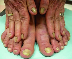
This image shows yellow nails with increased thickening and hypercurvature characteristic of yellow nail syndrome.
This image shows yellow nails with increased thickening and hypercurvature characteristic of yellow nail syndrome.
© Springer Science+Business Media


This image shows Beau lines in a patient undergoing multiple cycles of chemotherapy. Each line corresponds to a cycle of treatment.
This image shows Beau lines in a patient undergoing multiple cycles of chemotherapy. Each line corresponds to a cycle o
© Springer Science+Business Media

The transverse depression of the nail plate is easily appreciated looking at the nail from the side.
The transverse depression of the nail plate is easily appreciated looking at the nail from the side.
© Springer Science+Business Media
Characteristic spoon-shaped (concave) nail of a patient with iron-deficiency anemia.
Characteristic spoon-shaped (concave) nail of a patient with iron-deficiency anemia.
DR P. MARAZZI/SCIENCE PHOTO LIBRARY
This image shows yellow nails with increased thickening and hypercurvature characteristic of yellow nail syndrome.
This image shows yellow nails with increased thickening and hypercurvature characteristic of yellow nail syndrome.
© Springer Science+Business Media
This image shows Beau lines in a patient undergoing multiple cycles of chemotherapy. Each line corresponds to a cycle of treatment.
This image shows Beau lines in a patient undergoing multiple cycles of chemotherapy. Each line corresponds to a cycle o
© Springer Science+Business Media
The transverse depression of the nail plate is easily appreciated looking at the nail from the side.
The transverse depression of the nail plate is easily appreciated looking at the nail from the side.
© Springer Science+Business Media
Systemic nail dystrophies references
1. Pitukweerakul S, Pilla S: Terry's nails and Lindsay's nails: Two nail abnormalities in chronic systemic diseases. J Gen Intern Med 31(8):970, 2016. doi: 10.1007/s11606-016-3628-z
2. Oanță A, Iliescu V, Țărean S. Half and Half Nails in a Healthy Person. Acta Dermatovenerol Croat. 2017;25(4):303-304.
3. Hardin J, Haber RM. Onychomadesis: literature review. Br J Dermatol. 2015;172(3):592-596. doi:10.1111/bjd.13339
Nail Dystrophies Associated With Dermatologic Conditions
In psoriasis, nails may have a number of changes, including irregular pits, oil spots (localized areas of tan-brown discoloration), separation of a part of the nail from the nail bed (onycholysis), thickening and crumbling of the nail plate, red spots in the lunula, and splinter hemorrhages. Nail psoriasis may occur as an isolated phenomenon or as a feature of cutaneous psoriasis. Nail psoriasis is also a risk factor for development of psoriatic arthritis.
Treatment of nail psoriasis is challenging due to poor penetration of the nail plate by topical medications and suboptimal patient adherence (1). Treatment options include topical therapies (topical glucocorticoids, vitamin D analogues), oral small molecules (eg, apremilast, deucravacitinib), immunosuppressants (eg, methotrexate, cyclosporine, azathioprine), and biologic medications (eg, IL-17, IL-12/23, and JAK inhibitors) (). Treatment options include topical therapies (topical glucocorticoids, vitamin D analogues), oral small molecules (eg, apremilast, deucravacitinib), immunosuppressants (eg, methotrexate, cyclosporine, azathioprine), and biologic medications (eg, IL-17, IL-12/23, and JAK inhibitors) (2, 3). Biologic medications also used to treat cutaneous psoriasis have the greatest efficacy in treating nail psoriasis (4). Pulsed-dye laser has demonstrated efficacy and can be recommended for nail psoriasis; however, other device-based therapies (eg, light) need further study to judge their effectiveness (5).
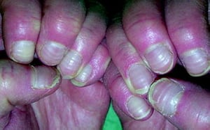
Lichen planus of the nail unit requires immediate clinical attention because it can cause permanent nail changes and nail loss if untreated. Therefore, early diagnosis and treatment is imperative. Nail changes include longitudinal ridging, fissuring, erythema of the lunula, and distal splitting of the nail. Over time, scarring and irreversible changes may occur, including nail atrophy, pterygium formation, and total nail loss. Treatment options include topical (high-potency), intralesional, and systemic glucocorticoids, and immunosuppressant medications (eg, mycophenolate mofetil, azathioprine, methotrexate, and cyclophosphamide). Low-dose naltrexone has also been used successfully in case reports (unit requires immediate clinical attention because it can cause permanent nail changes and nail loss if untreated. Therefore, early diagnosis and treatment is imperative. Nail changes include longitudinal ridging, fissuring, erythema of the lunula, and distal splitting of the nail. Over time, scarring and irreversible changes may occur, including nail atrophy, pterygium formation, and total nail loss. Treatment options include topical (high-potency), intralesional, and systemic glucocorticoids, and immunosuppressant medications (eg, mycophenolate mofetil, azathioprine, methotrexate, and cyclophosphamide). Low-dose naltrexone has also been used successfully in case reports (6). Maintenance therapy is needed to prevent relapse. Pterygium of the nail, which is a complication of lichen planus, is characterized by scarring from the proximal nail outward in a V formation, which ultimately leads to nail loss (7).
Alopecia areata of the nail can be accompanied by regular pits that form a geometric pattern. Pits are small and fine. Alopecia areata may also be associated with severe onychorrhexis (brittleness with nail breakage) (8). Treatment options include intralesional and topical glucocorticoids and topical sensitizers such as squaric acid dibutylester. Biologic therapies and oral small molecules including tofacitinib and apremilast have shown some promise.). Treatment options include intralesional and topical glucocorticoids and topical sensitizers such as squaric acid dibutylester. Biologic therapies and oral small molecules including tofacitinib and apremilast have shown some promise.
Twenty-nail dystrophy, also known as trachyonychia, is a clinical term describing a nail disorder characterized by roughness, excessive longitudinal ridging, and a sandpaper-like texture that affects all twenty fingernails and toenails. It is sometimes associated with alopecia areata and associated conditions like psoriasis, atopic dermatitis, alopecia areata, or lichen planus. It can also be idiopathic.

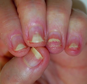
This photo shows irregular pits and areas of yellow-brown discoloration (oil spots) in a person with psoriasis.
This photo shows irregular pits and areas of yellow-brown discoloration (oil spots) in a person with psoriasis.
© Springer Science+Business Media

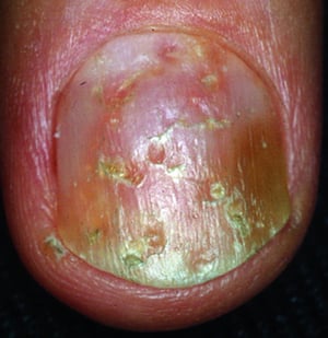
Nail bed psoriasis causes onycholysis (as evidenced by the white discoloration of the distal nails with erythematous border and absence of distal nail on the ring and little fingers). The underlying nail bed is hyperkeratotic.
Nail bed psoriasis causes onycholysis (as evidenced by the white discoloration of the distal nails with erythematous bo
© Springer Science+Business Media

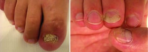
The photo on the left shows thickening and crumbling of the nail of the great toe. Pitting and onycholysis are visible on the hand (right), suggesting nail psoriasis as the diagnosis.
The photo on the left shows thickening and crumbling of the nail of the great toe. Pitting and onycholysis are visible
© Springer Science+Business Media


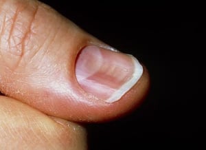
This photo shows longitudinal ridging of the thumbnail of a patient with lichen planus of the nail.
This photo shows longitudinal ridging of the thumbnail of a patient with lichen planus of the nail.
DR P. MARAZZI/SCIENCE PHOTO LIBRARY

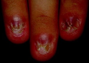
This photo shows dorsal pterygium of several nails accompanied by atrophy of the nails.
This photo shows dorsal pterygium of several nails accompanied by atrophy of the nails.
© Springer Science+Business Media
This photo shows irregular pits and areas of yellow-brown discoloration (oil spots) in a person with psoriasis.
This photo shows irregular pits and areas of yellow-brown discoloration (oil spots) in a person with psoriasis.
© Springer Science+Business Media
Nail bed psoriasis causes onycholysis (as evidenced by the white discoloration of the distal nails with erythematous border and absence of distal nail on the ring and little fingers). The underlying nail bed is hyperkeratotic.
Nail bed psoriasis causes onycholysis (as evidenced by the white discoloration of the distal nails with erythematous bo
© Springer Science+Business Media
The photo on the left shows thickening and crumbling of the nail of the great toe. Pitting and onycholysis are visible on the hand (right), suggesting nail psoriasis as the diagnosis.
The photo on the left shows thickening and crumbling of the nail of the great toe. Pitting and onycholysis are visible
© Springer Science+Business Media
This photo shows longitudinal ridging of the thumbnail of a patient with lichen planus of the nail.
This photo shows longitudinal ridging of the thumbnail of a patient with lichen planus of the nail.
DR P. MARAZZI/SCIENCE PHOTO LIBRARY
This photo shows dorsal pterygium of several nails accompanied by atrophy of the nails.
This photo shows dorsal pterygium of several nails accompanied by atrophy of the nails.
© Springer Science+Business Media
Dermatologic conditions references
1. Hwang JK, Lipner SR. Treatment of Nail Psoriasis. Dermatol Clin. 2024;42(3):387-398. doi:10.1016/j.det.2024.02.004
2. van de Kerkhof P, Guenther L, Gottlieb AB, et al: Ixekizumab treatment improves fingernail psoriasis in patients with moderate-to-severe psoriasis: Results from the randomized, controlled and open-label phases of UNCOVER-3. : Ixekizumab treatment improves fingernail psoriasis in patients with moderate-to-severe psoriasis: Results from the randomized, controlled and open-label phases of UNCOVER-3.J Eur Acad Dermatol Venereol 31(3):477–482, 2017. doi: 10.1111/jdv.14033
3. Merola JF, Elewski B, Tatulych S, et al: Efficacy of tofacitinib for the treatment of nail psoriasis: Two 52-week, randomized, controlled phase 3 studies in patients with moderate-to-severe plaque psoriasis. Efficacy of tofacitinib for the treatment of nail psoriasis: Two 52-week, randomized, controlled phase 3 studies in patients with moderate-to-severe plaque psoriasis.J Am Acad Dermatol 77(1):79–87, 2017. doi: 10.1016/j.jaad.2017.01.053
4. Hwang JK, Ricardo JW, Lipner SR. Efficacy and Safety of Nail Psoriasis Targeted Therapies: A Systematic Review. Am J Clin Dermatol. 2023;24(5):695-720. doi:10.1007/s40257-023-00786-4
5. Elmets CA, Lim HW, Stoff B, et al. Joint American Academy of Dermatology-National Psoriasis Foundation guidelines of care for the management and treatment of psoriasis with phototherapy. J Am Acad Dermatol. 2019;81(3):775-804. doi:10.1016/j.jaad.2019.04.042
6. Bray ER, Morrison BW. Low-Dose Naltrexone Use in Biopsy-Proven Lichen Planus of the Nails. . Low-Dose Naltrexone Use in Biopsy-Proven Lichen Planus of the Nails.JAMA Dermatol. 2024;160(12):1334-1337. doi:10.1001/jamadermatol.2024.4098
7. Hwang JK, Grover C, Iorizzo M, et al. Nail psoriasis and nail lichen planus: Updates on diagnosis and management. J Am Acad Dermatol. 2024;90(3):585-596. doi:10.1016/j.jaad.2023.11.024
8. Chelidze K, Lipner SR. Nail changes in alopecia areata: an update and review. Int J Dermatol. 2018;57(7):776-783. doi:10.1111/ijd.13866
Nail Discoloration
Chemotherapeutic cancer medications (especially the taxanes) can cause melanonychia (nail plate pigmentation), which can be diffuse or may occur in transverse bands. Some medications can cause characteristic changes in nail coloration:
Quinacrine: Nails appear greenish yellow or white under ultraviolet light.
Cyclophosphamide: The onychodermal bands (seal formed at the junction of the nail plate and distal nail bed at the free edge of the nail plate) become slate-gray or bluish. Cyclophosphamide: The onychodermal bands (seal formed at the junction of the nail plate and distal nail bed at the free edge of the nail plate) become slate-gray or bluish.
Arsenic: Nails may turn diffusely brown.
Tetracyclines, ketoconazole, phenothiazines, sulfonamides, and phenindione: Nails may have brownish or blue discoloration.Tetracyclines, ketoconazole, phenothiazines, sulfonamides, and phenindione: Nails may have brownish or blue discoloration.
Gold therapy: Nails may be light or dark brown.
Silver salts (argyria): Nails may be diffusely blue-gray.
Tobacco smoking or nail polish can result in yellow or brownish discoloration of nails and fingertips.

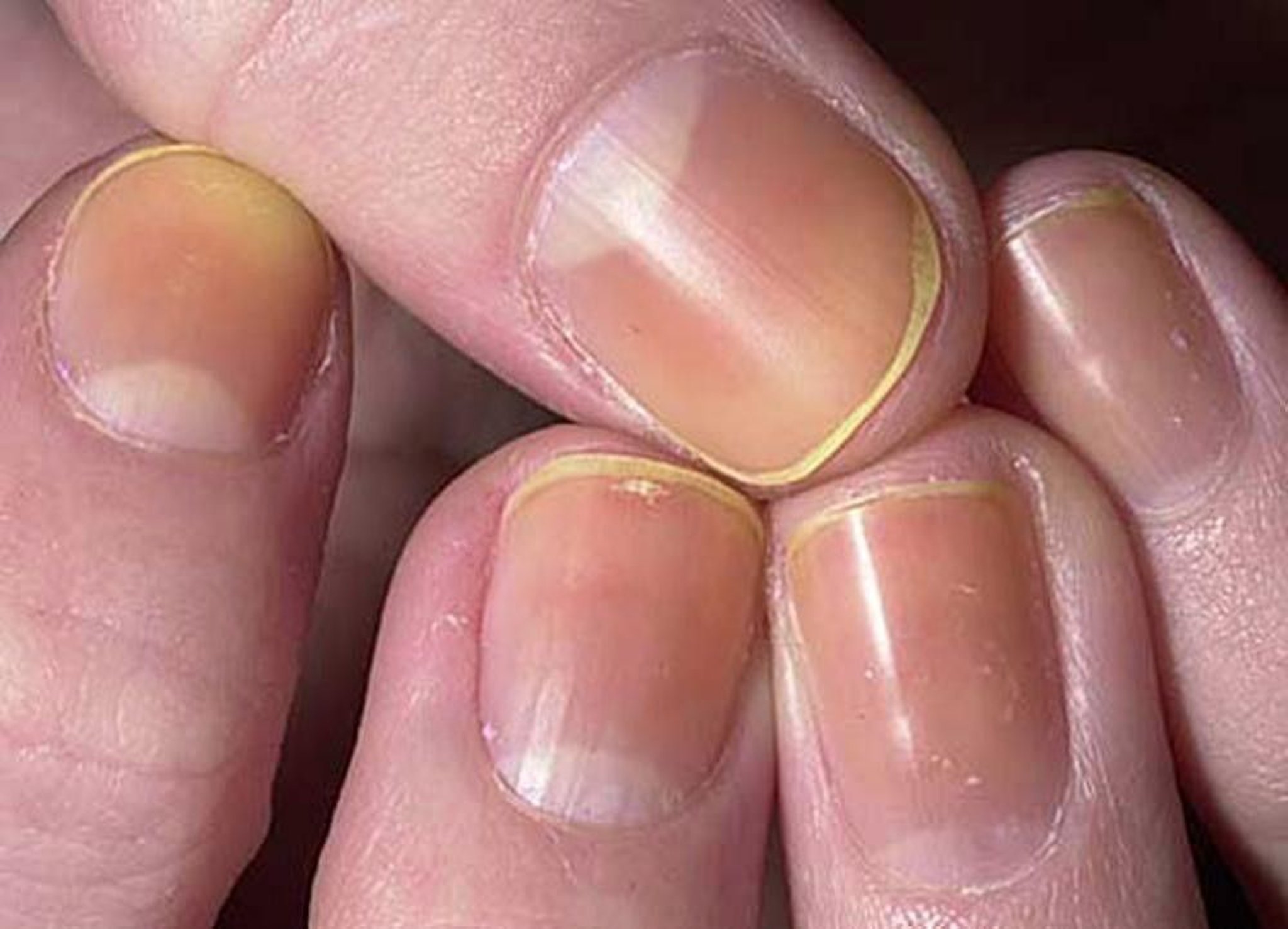
Image provided by Thomas Habif, MD.
White transverse lines of the nails (Mees lines) may occur as a result of chemotherapy, acute arsenic intoxication, malignant tumors, myocardial infarction, thallium and antimony intoxication, fluorosis. Mees lines can also occur during etretinate therapy. These lines are not due to changes in the nail bed, but are a true leukonychia. The nails can grow out normally if the insulting exposure has been removed. Mees lines also develop with trauma to the finger, although traumatic white lines usually do not span the entire nail.


This photo shows white transverse lines on the fingernail of an adult.
SCIENCE PHOTO LIBRARY
Onychomycosis Trichophyton mentagrophytes or T. rubrum causes a chalky white discoloration of the surface of the nail plate.
Green-nail syndrome is caused by colonization with Pseudomonas aeruginosa (1). It most often affects the first digits of the dominant hand, and is noteworthy for its striking blue-green color. It often occurs in patients with onycholysis or chronic paronychia whose nails have been exposed to irritants or have had excessive exposure to water. If the onycholysis or chronic paronychia is treated effectively, the P. aeruginosa infection will resolve. Alternatively, topical gentamicin ointment can be effective in chronic cases. Patients should avoid irritants and excess moisture. Frequent clipping of the nail increases the response to treatment. infection will resolve. Alternatively, topical gentamicin ointment can be effective in chronic cases. Patients should avoid irritants and excess moisture. Frequent clipping of the nail increases the response to treatment.
Red nail discoloration can be due to benign nail tumors (eg, onychopapilloma, glomus tumor), malignant nail tumors (eg, squamous cell carcinoma, amelanotic melanoma), infectious diseases (eg, subungual verruca), genetic disorders (eg, Darier disease), and inflammatory disorders (eg, nail psoriasis, nail lichen planus).
Blue nail discoloration involving a single nail may be due to a glomus tumor; a blue nevus; or less commonly, a nail unit melanoma (2). Blue nail discoloration involving multiple nails is associated with medications (eg, minocycline, zidovudine, and hydroxyurea), toxic and exogenous exposures (eg, silver), and other medical conditions (such as HIV and systemic lupus erythematosus). ). Blue nail discoloration involving multiple nails is associated with medications (eg, minocycline, zidovudine, and hydroxyurea), toxic and exogenous exposures (eg, silver), and other medical conditions (such as HIV and systemic lupus erythematosus).


The photo on the left shows green-nail syndrome with onycholysis of the fourth fingernail and green discoloration due to the presence of pigments produced by Pseudomonas. The photo on the right shows the green discoloration of the nail bed after clipping off the detached nail plate.
© Springer Science+Business Media
Nail discoloration references
1. Geizhals S, Lipner SR. Retrospective Case Series on Risk Factors, Diagnosis and Treatment of Pseudomonas aeruginosa Nail Infections. Am J Clin Dermatol. 2020;21(2):297-302. doi:10.1007/s40257-019-00476-0
2. Hwang JK, Lipner SR. Blue Nail Discoloration: Literature Review and Diagnostic Algorithms. Am J Clin Dermatol. 2023;24(3):419-441. doi:10.1007/s40257-023-00768-6
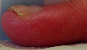
Median Nail Dystrophy (Median Canaliform Dystrophy)
Median nail dystrophy is characterized by small cracks in the nail that extend laterally and look like the branches of an evergreen tree. The cracks and ridges are similar to those seen in habit-tic nail deformity (which is dystrophy of the central nail caused by repetitive trauma to the nail matrix resulting from rubbing or picking with another finger). The cause of median nail dystrophy is unknown in some cases, but trauma is thought to play a role. Frequent use of personal digital devices that subject the nails to repetitive striking has been implicated in several cases. Tacrolimus 0.1% at bedtime without occlusion has been successful when patients stop all activities that might lead to repetitive low-level trauma. Median nail dystrophy is characterized by small cracks in the nail that extend laterally and look like the branches of an evergreen tree. The cracks and ridges are similar to those seen in habit-tic nail deformity (which is dystrophy of the central nail caused by repetitive trauma to the nail matrix resulting from rubbing or picking with another finger). The cause of median nail dystrophy is unknown in some cases, but trauma is thought to play a role. Frequent use of personal digital devices that subject the nails to repetitive striking has been implicated in several cases. Tacrolimus 0.1% at bedtime without occlusion has been successful when patients stop all activities that might lead to repetitive low-level trauma.


This photo of median nail dystrophy shows disrupted growth in the middle of the fingernails.
DR P. MARAZZI/SCIENCE PHOTO LIBRARY
Melanonychia Striata
Melanonychia striata are hyperpigmented bands that are longitudinal and extend from the proximal nail fold and cuticle to the free distal end of the nail plate. Pigmentation results from deposition of melanin by melanocytes in the nail matrix. Deposition of melanin is increased by melanocyte activation (increased production of melanin in the nail cells) or by melanocytic hyperplasia (increased production of melanocytes in the nail matrix).

Melanonychia striata are hyperpigmented bands in the nail plate caused by melanin. In this image, a line is most noticeable in the nail of the left ring finger (arrow).
Image provided by Chris G. Adigun, MD.
Melanocyte activation may be a normal physiologic variant in people with dark skin. This variant, often called ethnic melanonychia, requires no treatment. Other causes of melanocyte activation include nail biting, trauma, pregnancy, Addison disease and other endocrine disorders, infections, postinflammatory hyperpigmentation, and friction. Melanocyte activation can also occur from the use of certain medications, including doxorubicin, 5-fluorouracil, zidovudine, and psoralens. may be a normal physiologic variant in people with dark skin. This variant, often called ethnic melanonychia, requires no treatment. Other causes of melanocyte activation include nail biting, trauma, pregnancy, Addison disease and other endocrine disorders, infections, postinflammatory hyperpigmentation, and friction. Melanocyte activation can also occur from the use of certain medications, including doxorubicin, 5-fluorouracil, zidovudine, and psoralens.
Melanocytic hyperplasia can be caused by benign conditions, such as nail matrix melanocytic nevus or nail lentigo, or by malignant melanoma. Factors more often associated with malignant melanoma of the nail matrix include new onset after middle age, presence of pigmentation on the dominant thumb or hallux, rapid growth or darkening, bandwidth > 3 mm, associated nail plate dystrophy, or Hutchinson sign (extension of hyperpigmentation onto the proximal and/or lateral nail fold). Nail unit biopsy is essential in cases of suspected melanoma (1). Children often present with longitudinal melanonychia that would be concerning in adults, but nail unit melanoma is exceedingly rare in children (2).


Longitudinal pigmentation of the nail (melanonychia striata; blue arrow) and hyperpigmentation extending across lunula to proximal nail fold (Hutchinson sign; red arrow) of the middle finger with rounded pigmented lesion of the thumbnail. This patient was diagnosed with acral-lentiginous melanoma (a form of malignant melanoma).
Image courtesy of Carl Washington, MD, and Mona Saraiya, MD, MPH, via the Public Health Image Library of the Centers for Disease Control and Prevention.
Melanonychia striata references
1. Leung AKC, Lam JM, Leong KF, Sergi CM: Melanonychia striata: Clarifying behind the black curtain. A review on clinical evaluation and management of the 21st century. Int J Dermatol 58(11):1239–1245, 2019. doi: 10.1111/ijd.14464
2. Ricardo JW, Bellet JS, Jellinek N, et al. Evaluation and diagnosis of longitudinal melanonychia: A clinical review by a nail expert group. J Am Acad Dermatol. 2025;93(1):176-187. doi:10.1016/j.jaad.2025.02.075
Onycholysis
Onycholysis is separation of the nail plate from the nail bed or complete nail plate loss. It can occur as a drug reaction in patients treated with tetracyclines (photo-onycholysis), doxorubicin, 5-fluorouracil, cardiovascular medications (particularly practolol and captopril), cloxacillin and cephaloridine (rarely), trimethoprim/sulfamethoxazole, diflunisal, etretinate, indomethacin, isoniazid, griseofulvin, and isotretinoin. Simple (ie, not associated with another nail or skin disorder) onycholysis may also result from exposure to irritants, such as frequent exposure to water, citrus fruits, or chemicals. Onycholysis is separation of the nail plate from the nail bed or complete nail plate loss. It can occur as a drug reaction in patients treated with tetracyclines (photo-onycholysis), doxorubicin, 5-fluorouracil, cardiovascular medications (particularly practolol and captopril), cloxacillin and cephaloridine (rarely), trimethoprim/sulfamethoxazole, diflunisal, etretinate, indomethacin, isoniazid, griseofulvin, and isotretinoin. Simple (ie, not associated with another nail or skin disorder) onycholysis may also result from exposure to irritants, such as frequent exposure to water, citrus fruits, or chemicals.Irritant contact dermatitis of the hands and fingers may also lead to onycholysis (1). Colonization of the nail bed with Candida albicans may occur, but treating the underlying irritant exposure leads to resolution of the onycholysis, with or without treating the Candida.
Onycholysis is common in patients with psoriasis or may occur with thyroid disease or thyrotoxicosis.


Onycholysis (as evidenced by the white discoloration of the distal nail), as well as pitting, is visible in this patient with psoriasis.
© Springer Science+Business Media
Onycholysis reference
1. Vélez NF, Jellinek NJ: Simple onycholysis: A diagnosis of exclusion. J Am Acad Dermatol 70(4):793–794, 2014. doi: 10.1016/j.jaad.2013.09.061
Pincer Nail Deformity
Pincer nail deformity is a transverse overcurvature of the nail plate. It is most often caused by onychomycosis, psoriasis, tumors of the nail apparatus, osteoarthritis, and poorly fitting shoes. It has also been reported in patients with systemic lupus erythematosus, Kawasaki disease, end-stage renal disease, and some genetic syndromes (eg, paronychia congenita). Patients often have pain at the borders of the nail where the nail plate curves into the tips of the fingers or toes. There is no established treatment of choice, but a number of surgical techniques have been successful (1, 2, 3).


In this patient with hereditary pincer nails, the edges of the nails curve inwardly into the skin of the toes.
© Springer Science+Business Media
Pincer nail deformity references
1. Demirkiran ND: Suture treatment for pincer nail deformity: An inexpensive and simple technique. Dermatol Surg 46(4):573–576, 2019. doi: 10.1097/DSS.0000000000001818
2. Shin WJ, Chang BK, Shim JW, et al: Nail plate and bed reconstruction for pincer nail deformity. Clin Orthop Surg 10(3):385–388, 2018. doi: 10.4055/cios.2018.10.3.385
3. Won JH, Chun JS, Park YH, et al: Treatment of pincer nail deformity using dental correction principles. J Am Acad Dermatol 78(5):1002–1004, 2018. doi: 10.1016/j.jaad.2017.08.014
Trachyonychia
Trachyonychia (rough, opaque nails) may occur with alopecia areata, lichen planus, atopic dermatitis, and psoriasis. It is most common among children. When present in all nails, trachyonychia is often called 20-nail dystrophy. When it occurs in children, it tends to resolve spontaneously. When it occurs in adults, treatment is aimed at the underlying disorder. A clue to the diagnosis is the nails feel like sandpaper.


In this photo, the nails are opaque and rough.
© Springer Science+Business Media
Nail Tumors
Benign and malignant tumors can affect the nail unit, causing onychodystrophy. Benign tumors include myxoid cysts, pyogenic granulomas, and glomus tumors. Malignant tumors include Bowen disease, squamous cell carcinoma, and malignant melanoma. When malignancy is suspected, expeditious biopsy or referral for biopsy is strongly advised.

© Springer Science+Business Media
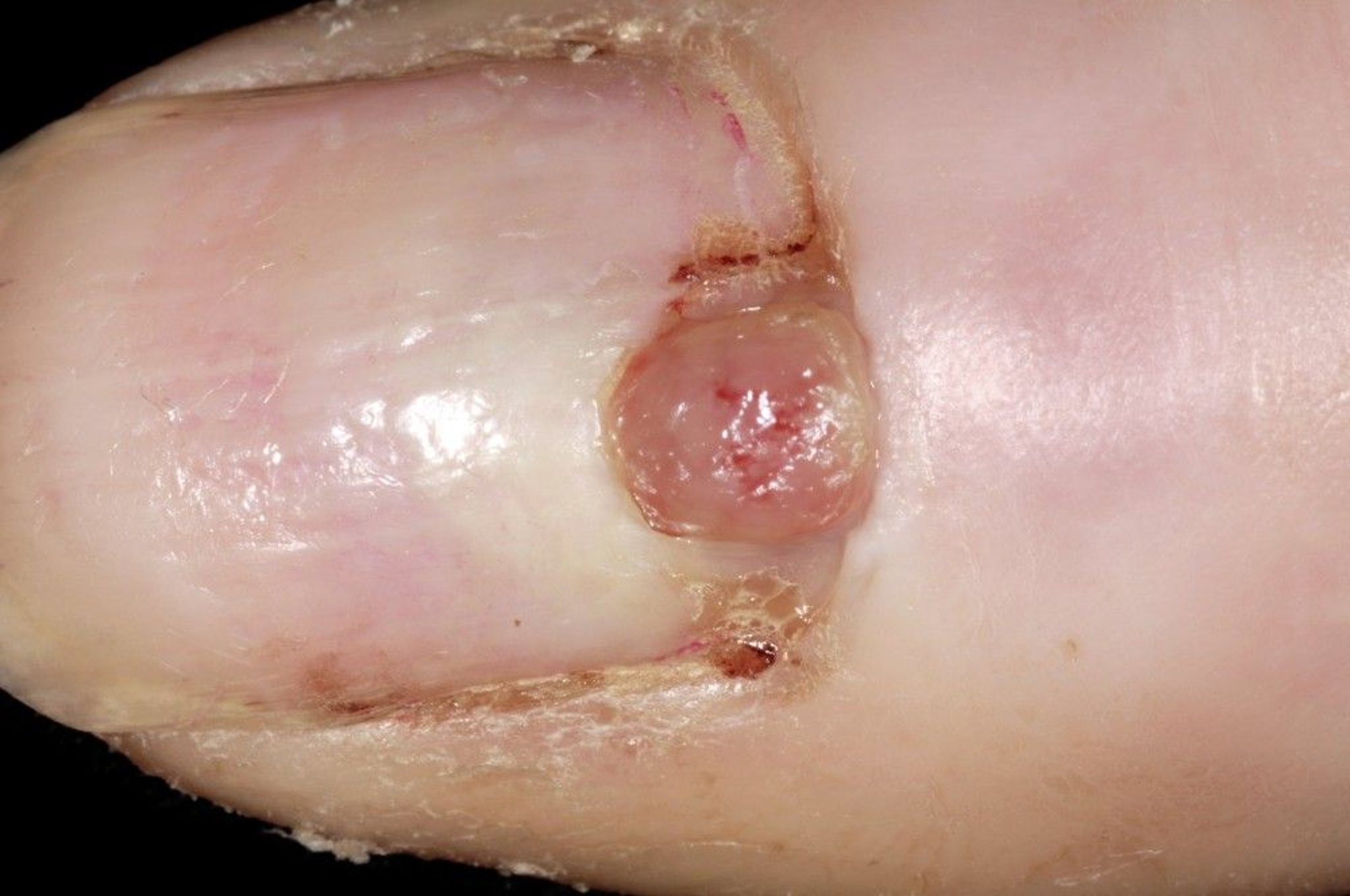
Pyogenic granuloma is a benign growth.
DR P. MARAZZI/SCIENCE PHOTO LIBRARY


