

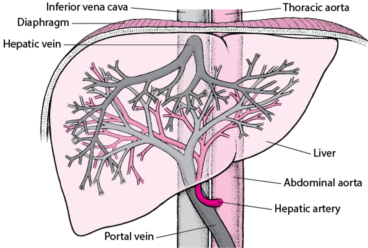
The liver has a dual blood supply. The portal vein (which is rich in nutrients and relatively high in oxygen) provides two-thirds of blood flow to the liver. The hepatic artery (which is oxygen-rich) supplies the rest. The hepatic veins drain the liver into the inferior vena cava. When portal vein blood flow increases, hepatic artery flow decreases and vice versa (the hepatic arterial buffer response). This dual, reciprocally compensatory blood supply provides some protection from hepatic ischemia in healthy people.
Blood Supply of the Liver

Despite its dual blood supply, the liver, a metabolically active organ, can be injured by
Ischemia
Insufficient venous drainage
Specific vascular lesions
Ischemia results from reduced blood flow, reduced oxygen delivery, increased metabolic activity, or all 3. Diffuse ischemia can cause ischemic hepatitis; focal ischemia can cause hepatic infarction or ischemic cholangiopathy. Hepatic infarction results from hepatic artery disorders.
Insufficient venous drainage may result from focal or diffuse obstruction or from right-sided heart failure, as in congestive hepatopathy. Obstruction can occur in the intrahepatic or extrahepatic veins (Budd-Chiari syndrome) or in the intrahepatic terminal hepatic venules and hepatic sinusoids (sinusoidal obstruction syndrome, previously called veno-occlusive disease) but often occurs in both. Cirrhosis is the most common cause of diffuse intrahepatic venous outflow obstruction (1). Diffuse obstruction results in congestion of the sinusoids, hepatomegaly, portal hypertension, reduced portal blood flow, ascites, and splenomegaly. Manifestations of focal venous obstruction depend on the location.
Specific vascular lesions may occur in the hepatic artery, hepatic vein, or portal vein. The hepatic artery may be occluded. Uncommonly, aneurysms develop. In peliosis hepatis, blood-filled cystic spaces develop in the sinusoids (microvascular anastomoses between the portal and hepatic veins).
Hepatic vein disorders can result in focal or diffuse venous obstruction.
Nearly all portal vein disorders obstruct portal vein blood flow and cause portal hypertension. Obstruction can be
Extrahepatic—portal vein thrombosis due to a hypercoagulable state, a vessel wall lesion (eg, pylephlebitis, omphalitis), an adjacent lesion (eg, pancreatitis, tumor), or congenital atresia of the portal vein
Intrahepatic—eg, microvascular portal vein obstruction as occurs in schistosomiasis, primary biliary cholangitis (PBC, previously called primary biliary cirrhosis), sarcoidosis, and noncirrhotic portal hypertension
Reference
1. Northup PG, Garcia-Pagan JC, Garcia-Tsao G, et al: Vascular Liver Disorders, Portal Vein Thrombosis, and Procedural Bleeding in Patients With Liver Disease: 2020 Practice Guidance by the American Association for the Study of Liver Diseases. Hepatology 73(1):366-413, 2021. doi: 10.1002/hep.31646


