

Tinea corporis is a dermatophyte infection of the face, trunk, and extremities. Diagnosis is based on clinical appearance and by examination of skin scrapings on potassium hydroxide wet mount. Treatment involves topical or oral antifungals.
Tinea corporis is a dermatophytosis that causes pink-to-red annular (ring-shaped) patches and plaques with raised scaly borders that expand peripherally and tend to clear centrally. Postinflammatory hyperpigmentation can make the centers appear less clear on dark skin.
A rare variant form appears as nummular (circle- or round-shaped) scaling patches studded with small papules or pustules that have no central clearing.
Common causes are Trichophyton mentagrophytes, Trichophyton rubrum, and Microsporum canis.


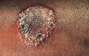
This photo shows a pink-to-red, round patch of body ringworm. The patch has raised borders, some scaling, and some central clearing towards the bottom border.
This photo shows a pink-to-red, round patch of body ringworm. The patch has raised borders, some scaling, and some cent
Image provided by Thomas Habif, MD.

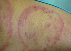
The border is raised and scaly and in this example has visible tiny pustules.
The border is raised and scaly and in this example has visible tiny pustules.
© Springer Science+Business Media

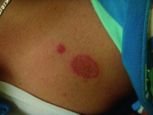
Both lesions are due to tinea corporis. The lesion on the right shows typical peripheral scale and slight central lesion clearing.
Both lesions are due to tinea corporis. The lesion on the right shows typical peripheral scale and slight central lesio
© Springer Science+Business Media

© Springer Science+Business Media

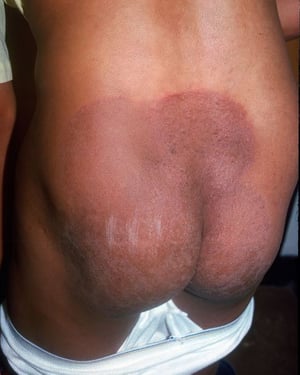
This photo shows a well-demarcated, scaly, erythematous plaque characteristic of tinea corporis. Postinflammatory hyperpigmentation makes the center appear less clear than on light skin.
This photo shows a well-demarcated, scaly, erythematous plaque characteristic of tinea corporis. Postinflammatory hyper
Image courtesy of Karen McKoy, MD.
This photo shows a pink-to-red, round patch of body ringworm. The patch has raised borders, some scaling, and some central clearing towards the bottom border.
This photo shows a pink-to-red, round patch of body ringworm. The patch has raised borders, some scaling, and some cent
Image provided by Thomas Habif, MD.
The border is raised and scaly and in this example has visible tiny pustules.
The border is raised and scaly and in this example has visible tiny pustules.
© Springer Science+Business Media
Both lesions are due to tinea corporis. The lesion on the right shows typical peripheral scale and slight central lesion clearing.
Both lesions are due to tinea corporis. The lesion on the right shows typical peripheral scale and slight central lesio
© Springer Science+Business Media
© Springer Science+Business Media
This photo shows a well-demarcated, scaly, erythematous plaque characteristic of tinea corporis. Postinflammatory hyperpigmentation makes the center appear less clear than on light skin.
This photo shows a well-demarcated, scaly, erythematous plaque characteristic of tinea corporis. Postinflammatory hyper
Image courtesy of Karen McKoy, MD.
Diagnosis of Tinea Corporis
Primarily physical examination
Potassium hydroxide (KOH) wet mount
Tinea corporis is diagnosed by clinical appearance and by potassium hydroxide wet mount of skin scrapings.
Differential diagnosis of tinea corporis includes:
Allergic or irritant contact dermatitis
Erythema migrans (Lyme disease)
Treatment of Tinea Corporis
Topical or oral antifungals
Treatment of mild-to-moderate lesions is with terbinafine, naftifine, and imidazoles (eg, clotrimazole, oxiconazole) in cream, lotion, or gel formulations (Treatment of mild-to-moderate lesions is with terbinafine, naftifine, and imidazoles (eg, clotrimazole, oxiconazole) in cream, lotion, or gel formulations (1). The medication should be rubbed in 2 times a day continuing at least 7 to 10 days after lesions disappear, typically at approximately 2 to 3 weeks. (See table .)
Extensive and resistant lesions occur in patients infected with Trichophyton rubrum and in people with debilitating systemic diseases. For such cases, prolonged (eg, 3 to 4 weeks) oral antifungal therapy with either terbinafine or itraconazole is recommended (and in people with debilitating systemic diseases. For such cases, prolonged (eg, 3 to 4 weeks) oral antifungal therapy with either terbinafine or itraconazole is recommended (2). Fluconazole is a reasonable alternative(). Fluconazole is a reasonable alternative(3).
Treatment references
1. El-Gohary M, van Zuuren EJ, Fedorowicz Z, et al. Topical antifungal treatments for tinea cruris and tinea corporis. Cochrane Database Syst Rev. 2014;2014(8):CD009992. Published 2014 Aug 4. doi:10.1002/14651858.CD009992.pub2
2. Barac A, Stjepanovic M, Krajisnik S, et al. Dermatophytes: Update on Clinical Epidemiology and Treatment. Mycopathologia. 2024;189(6):101. Published 2024 Nov 21. doi:10.1007/s11046-024-00909-3
3. Faergemann J, Mörk NJ, Haglund A, Odegård T. A multicentre (double-blind) comparative study to assess the safety and efficacy of fluconazole and griseofulvin in the treatment of tinea corporis and tinea cruris. Br J Dermatol. 1997;136(4):575-577.. A multicentre (double-blind) comparative study to assess the safety and efficacy of fluconazole and griseofulvin in the treatment of tinea corporis and tinea cruris. Br J Dermatol. 1997;136(4):575-577.
Key Points
Tinea corporis typically causes pink-to-red annular (ring-shaped) patches and plaques with raised scaly borders that expand peripherally and clear centrally.
Diagnosis is based on appearance and potassium hydroxide wet mount.
If mild to moderate, treat using an imidazole, ciclopirox, naftifine, or terbinafine cream, lotion, or gel applied 2 times a day for at least 7 to 10 days after lesions disappear.If mild to moderate, treat using an imidazole, ciclopirox, naftifine, or terbinafine cream, lotion, or gel applied 2 times a day for at least 7 to 10 days after lesions disappear.
If severe, oral terbinafine or itraconazole is recommended; oral fluconazole is another alternative. If severe, oral terbinafine or itraconazole is recommended; oral fluconazole is another alternative.
Drug Information for the Topic


