


Angiography is a type of medical imaging that uses x-rays and a contrast agent to produce images of blood vessels.
In angiography, x-rays are used to produce detailed images of blood vessels. It is sometimes called conventional angiography to distinguish it from computed tomography (CT) angiography (CTA) and magnetic resonance angiography (MRA). During angiography, doctors can also treat disorders of blood vessels. Angiography, although invasive, is relatively safe.
Angiography can provide still images or motion pictures (called cineangiography). Cineangiography can show how fast blood travels through blood vessels. (See also Coronary Angiography and Overview of Imaging Tests.)
Procedure for Angiography
Before the angiography procedure, people are usually asked to refrain from eating and drinking for 12 hours.
For the procedure, people lie on an x-ray table (one that x-rays can easily pass through). Because the table may be tilted, straps may be applied across the chest and legs. X-ray cameras can be positioned as needed. Electrodes are placed on the chest to monitor the heart. Blood pressure and oxygen levels are also monitored.
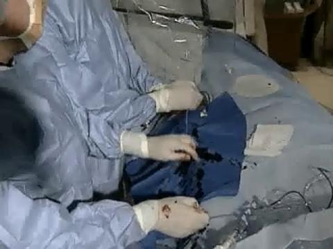
After injecting a local anesthetic, a doctor makes a small incision, typically in the groin or sometimes in the arm. Then a thin, flexible tube (catheter) is inserted, usually into an artery, and is threaded through blood vessels to the area being evaluated. When the catheter is in place, a radiopaque contrast agent (a liquid that contains iodine and can be seen on x-rays) is injected. The contrast agent flows through the blood vessels and outlines them. The images appear on a video screen and are recorded. Thus, doctors can assess the structure of blood vessels and identify any abnormalities present.
Before angiography, people are often given a sedative intravenously to help them relax and remain calm, but they remain conscious during the procedure. During the procedure, people may be asked to take deep breaths, hold their breath, or cough. People should report any discomfort they feel.
Angiography may take less than an hour up to several hours, depending on the area of the body being evaluated and the type of the examination or procedures being done. It is usually done as an outpatient procedure, meaning that the person can return home within a short time after the procedure is finished.
If the catheter is inserted into an artery, the insertion site must be steadily compressed for 10 to 20 minutes after all the instruments are removed. Compression reduces bleeding and bruises.

Uses of Angiography
Angiography is used to check for abnormalities in blood vessels, usually arteries. Abnormalities may include:
Blockages
Narrowing
Abnormal connections between arteries and veins (arteriovenous malformations)
Inflammation (vasculitis)
Bulges (aneurysms) in a weakened blood vessel wall
Tears (dissection) in a blood vessel wall
During angiography, procedures to treat the abnormalities detected can sometimes be done:
Narrowed arteries can be widened.
Blockages can be removed.
A tube made of wire mesh (stent) can be placed to keep an artery open.
Tears or weakened areas in a blood vessel can be repaired.
Blood flow to tumors or arteriovenous malformations can be blocked.
Types of Conventional Angiography
Arteriography
Arteriography produces an image of arteries and is the most common type of angiography.
Venography
Venography produces an image of veins. Ultrasound has largely replaced venography in the diagnosis of clots in veins (deep vein thrombosis).
Digital subtraction angiography
Digital subtraction angiography provides images of the brain's blood vessels. During this procedure, x-ray images of blood vessels are taken before and after a radiopaque contrast agent is injected. Then a computer subtracts one image from the other. Images of structures other than arteries (such as bones) are thus eliminated. As a result, the arteries can be seen more clearly.
Common Types of Angiography
Type | Area to Be Evaluated | Uses |
|---|---|---|
Blood vessels of the heart With cardiac catheterization, the heart itself | To diagnose coronary artery disease and other heart disorders To determine whether angioplasty or coronary artery bypass surgery is feasible To determine the severity of a heart disorder To identify the cause of chest pain, shortness of breath, or certain other symptoms To clarify the specific structure of a person’s heart before heart valve replacement surgery | |
Aortography | Aorta | To check for the following:
|
Cerebral angiography | Blood vessels of the brain | To check for the following:
|
Blood vessels of the eye | To evaluate damage to the retina due to diabetes (diabetic retinopathy) or macular degeneration To evaluate the retina before laser therapy | |
Peripheral arteriography | Arteries of the arms, legs, and trunk, except the aorta and arteries of the heart | To check for the following:
|
Pulmonary angiography* | Blood vessels of the lungs | To diagnose pulmonary embolism (blockage by blood clots in the pulmonary arteries, which lead from the heart to the lungs) and identify abnormalities of pulmonary arteries and veins |
* CT pulmonary angiography has largely replaced conventional pulmonary angiography because it is less invasive. | ||
CT = computed tomography. | ||
Disadvantages of Angiography
For some people, angiography is uncomfortable. In a few people, allergic-type reactions to the contrast agent occur. The injection site may bleed, become infected, or be painful. Rarely, the catheter damages a blood vessel.
Serious complications, such as shock, seizures, kidney damage, and sudden stopping of the heart’s pumping (cardiac arrest), are very rare. Sometimes during cardiac catheterization, the heart skips beats or slows briefly.
The risk of complications is higher in older adults, although it is still low.
The dose of radiation used in angiography varies depending on the procedure but typically is higher than in x-ray tests. For example, the radiation dose in coronary angiography is 350 to 750 times as much as that used in a single-view x-ray of the chest.
Angiography is not always readily available. It must be done by highly skilled doctors.


