


Magnetic resonance imaging (MRI) is a type of medical imaging that uses a strong magnetic field and very high frequency radio waves to produce highly detailed images.
During an MRI, a computer records changes in the magnetic field around a person's body to create cross-sectional, detailed images. Unlike computed tomography (CT) scans and positron emission tomography (PET) scans, an MRI does not use x-rays (radiation). MRI (See also Overview of Imaging Tests.)

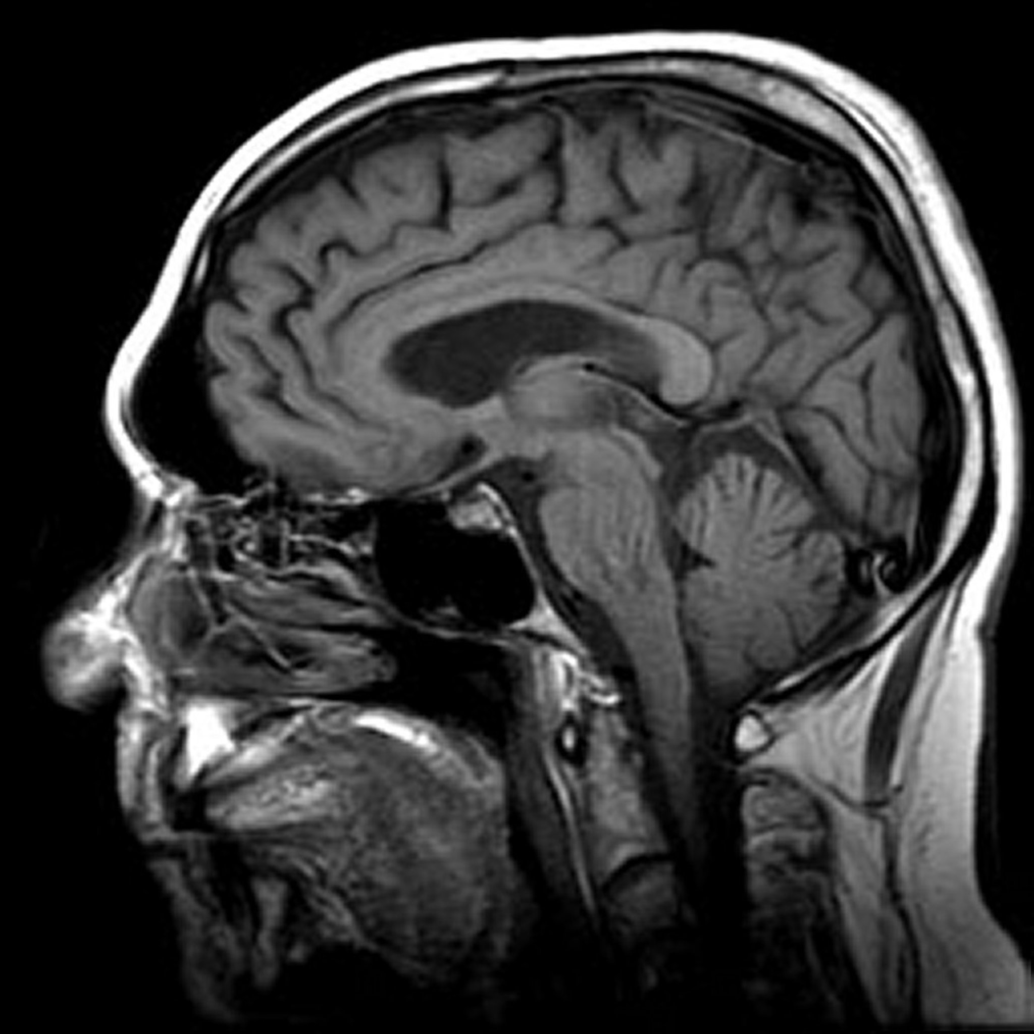
Image provided by Jon A. Jacobson, MD.
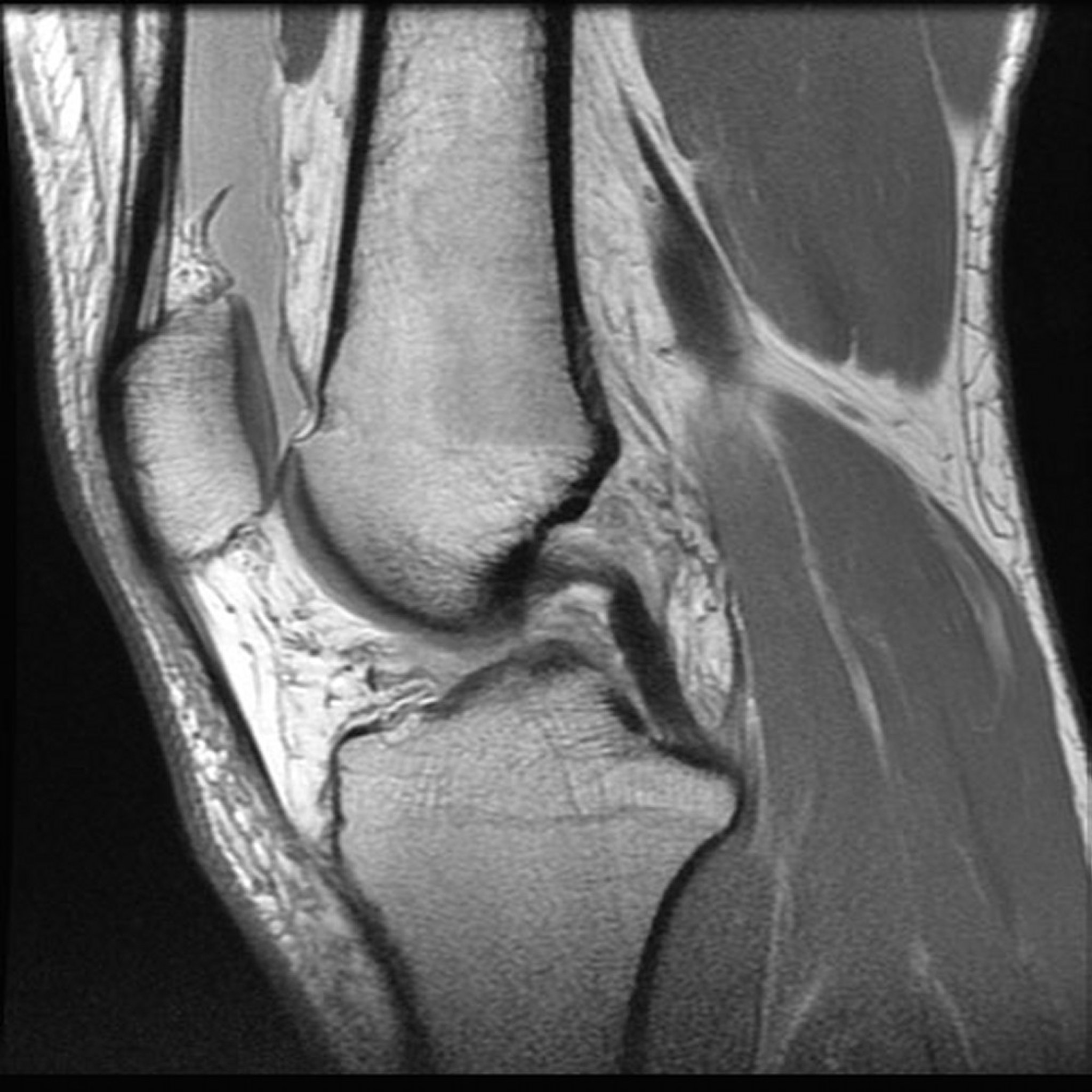
Image provided by Jon A Jacobson, MD.
Procedure for MRI
For MRI, a person lies on a motorized table that is moved into the narrow interior of a large tubular scanner, which produces a strong magnetic field. Normally, protons (positively charged parts of an atom) in tissues are in no particular arrangement. But when protons are surrounded by a strong magnetic field, as in an MRI scanner, they line up with the magnetic field. Then, the scanner emits a pulse of radio waves, which momentarily knocks the protons out of line. As the protons line up with the magnetic field again, they release energy (called signals). The strength of the signal varies depending on the type of tissue it passes through. The MRI scanner records these signals. A computer is used to analyze the signals and produce images.
Examiners can change how various tissues appear on a scan by varying the radio wave pulses, the strength and direction of the magnetic field, and other factors. For example, fat tissue appears dark on one type of scan and bright on another. These different scans provide complementary information, so more than one is often obtained.
A contrast agent containing gadolinium (a paramagnetic contrast agent) may be injected into a vein or a joint. Gadolinium agents change the magnetic field in a way that makes images clearer.
Before the test, people remove most or all of their clothing and are given a gown that has no buttons, snaps, zippers, or other metal on it to wear. All metal objects (such as keys, jewelry, and cell phones) and other objects that could be affected by the magnetic field (such as credit cards and watches) should be left outside the MRI scanning room. People must lie still when images are taken and may have to hold their breath at times. Because the scanner makes loud banging noises, people may be given headphones or ear plugs to wear. A scan may take 20 to 60 minutes. After the test, people can resume their usual activities immediately.
Uses of MRI
MRI is preferred to computed tomography (CT) when doctors need more detail about soft tissues—for example, to image abnormalities in the brain, spinal cord, muscles, and liver. MRI is particularly useful for identifying tumors in these tissues.
MRI is also used to do the following:
Measure certain molecules in the brain that distinguish a brain tumor from a brain abscess
Identify abnormalities in female reproductive organs and fractures in the hip and pelvis
Help doctors evaluate joint abnormalities (such as tears in ligaments or cartilage in the knee) and sprains
Help doctors evaluate bleeding and infection
MRI is also used when the risks of CT are high. For example, MRI may be preferred for people who have had a reaction to the iodinated contrast agents used in CT and for pregnant women (because radiation can cause problems in the fetus).
Gadolinium contrast agent, injected into a vein before an MRI, helps doctors evaluate inflammation, tumors, and blood vessels. Injecting this agent into a joint helps doctors get a clearer picture of joint abnormalities, particularly if they are complex (as in injuries or degeneration of ligaments and cartilages in the knee).
Variations of MRI
Functional MRI
Functional MRI detects metabolic changes that occur when the brain is active. Thus, it can show which areas of the brain are active when a person does a specific task, such as reading, writing, remembering, calculating, or moving a limb. Functional MRI can be used in research and clinical settings, for example, to plan brain surgery for epilepsy, tumors, or abnormal connections between arteries and veins (arteriovenous malformations).
Perfusion MRI
With perfusion MRI, doctors can estimate blood flow in a particular area. This information can be useful during a stroke to determine whether blood flow to parts of the brain is decreased. It can also be used to identify areas where blood flow is increased—for example, in tumors.
Diffusion-weighted MRI
Diffusion-weighted MRI detects changes in water movement in cells that are not functioning normally. It is used primarily to identify early stroke. It is also used to detect certain brain disorders and to determine whether tumors have spread to the brain, or to differentiate a brain abscess from a tumor. Use of this technique to image areas other than the brain is limited. Diffusion-weighted MRI is often combined with other techniques to evaluate tumors, mostly in the brain.
Magnetic resonance spectroscopy
Magnetic resonance spectroscopy uses radio waves that are emitted almost continuously rather than in pulses as in conventional MRI. Magnetic resonance spectroscopy is used to detect brain disorders, such as seizure disorders, Alzheimer disease, brain tumors, and brain abscesses. It can distinguish between the dead debris inside an abscess and multiplying cells inside a tumor.
This technique is also used to evaluate metabolic disorders in muscles and the nervous system.
Magnetic resonance angiography (MRA)
MRA, like conventional angiography and CT angiography, can provide detailed images of blood vessels. MRA is safer and easier to do, and often can be done without injection of a contrast agent.
MRA can show blood flow through arteries and veins or blood flow only in one direction and thus show only arteries or only veins. As in CT angiography, a computer is used to remove all tissues except the blood vessels from the image.
Often, a gadolinium contrast agent is injected into a vein to outline blood vessels. The examiner carefully times the scanning so that the images are taken when gadolinium is concentrated in the blood vessels being evaluated.
MRA is used to evaluate blood vessels of the brain, heart, abdominal organs, arms, and legs. It is used to detect the following:
Narrowed arteries in the limbs
Blood clots in veins of the limbs and pelvis
Blood flow to tumors
Tumors that are affecting blood vessels
Magnetic resonance venography
Magnetic resonance venography is an MRA of veins. It is often used to detect a blood clot in a vein that carries blood away from the brain (cerebral venous thrombosis) and to monitor the effect of treatment on this disorder.
Echo planar imaging
Echo planar imaging produces sequences of images in only seconds. It can be used to image the brain, heart, and abdomen. Because it is fast, movement by the person being examined does not blur the images as much. Also, the technique can provide information about how tissues are functioning.
However, it requires special equipment and is more likely to misrepresent certain structures than conventional MRI because of the nature of the technique.
Disadvantages of MRI
The time needed for MRI is longer than that needed for CT. Also, MRI is usually less likely to be immediately available than CT. Thus, CT may be better in emergencies, such as serious injuries and stroke. MRI is also more expensive than CT.
Other disadvantages include:
Claustrophobia and sometimes difficulty fitting within the MRI scanner because it is a small, enclosed space
The effects of the magnetic field on metal devices implanted in the body
Reactions to the contrast agent
Problems related to the small enclosed space
Space in the MRI scanner is small and enclosed, making some people feel claustrophobic, even people who usually are not anxious about confined spaces. Some people with obesity have difficulty fitting within the scanner.
Some MRI scanners (called open MRI scanners) have an open side and a larger interior. In them, people may feel less claustrophobic, and people with obesity may fit more easily. The images produced in open MRI scanners may be inferior to those produced by enclosed scanners depending on the magnet strength, but they can still be used to make a diagnosis.
People who are anxious about MRI can be given an antianxiety medication, such as alprazolam or lorazepam, 15 to 30 minutes before scanning.
Magnetic field effects
Usually, MRI is not used if people have:
Certain materials (such as shrapnel) in specific parts of their body, especially in the eye
Implanted devices that can be affected by powerful magnetic fields
These devices include some cardiac pacemakers, defibrillators, cochlear implants, and magnetic metallic clips used to treat aneurysms. The magnetic field used in MRI can cause an implanted device to move, overheat, or malfunction. The device is more likely to be affected if it was implanted within the previous 6 weeks (because scar tissue, which can help hold the device in place, has not yet formed). These devices can also distort MRI images.
Some devices, such as common dental implants, an artificial hip, or rods used to straighten the spine, are not affected by MRI.
Before MRI is done, people who have any implanted devices should tell their doctor, who can determine whether imaging is safe.
The MRI magnetic field is very strong and always on. Thus, if a metal object (such as an oxygen tank or an IV pole) is near the entrance of the scanning room, the object may be pulled into the scanner at high speed. The person being evaluated may be injured, and separating the object from the magnet may be difficult.
Reactions to the MRI contrast agent
Gadolinium contrast agents can cause headache, nausea, pain and a sensation of cold at the injection site, distortion of taste, and dizziness.
These agents are much less likely to cause severe reactions than the iodinated contrast agents used in conventional and CT angiography.
However, nephrogenic systemic fibrosis—a severe, life-threatening disorder—has occurred in a small number of people with advanced chronic kidney disease. Most cases of nephrogenic systemic fibrosis are linked to a type of contrast agent called group I gadolinium-based contrast media (GBCM), which is no longer administered in the United States.
Drug Information for the Topic


