

The long QT interval syndromes are disorders of the heart's electrical activity that predispose people to dangerous heart rhythms and sudden death.
People may be born with an abnormality that causes long QT interval or they may develop it as a complication of other disorders.
People are at risk for torsades de pointes ventricular tachycardia, which may stop on its own or degenerate into ventricular fibrillation.
Diagnosis is by electrocardiography (ECG).
Treatment is avoidance of triggers, use of beta-blockers, and sometimes an implantable cardioverter-defibrillator (ICD).
(See also Overview of Abnormal Heart Rhythms and Overview of Cardiac Channelopathies.)
The QT interval refers to the time between 2 events on the electrocardiogram (ECG)—from the beginning of the QRS complex to the end of the T wave. Any abnormality that prolongs the QT interval increases the risk of a rapid, dangerous heart rhythm called torsades de pointes ventricular tachycardia (torsades). Ventricular tachycardia is a heart rhythm that originates in the ventricles (lower chambers of the heart) and produces a rapid heart rate. Torsades often turns into ventricular fibrillation, in which the heart stops beating, which rapidly causes death.
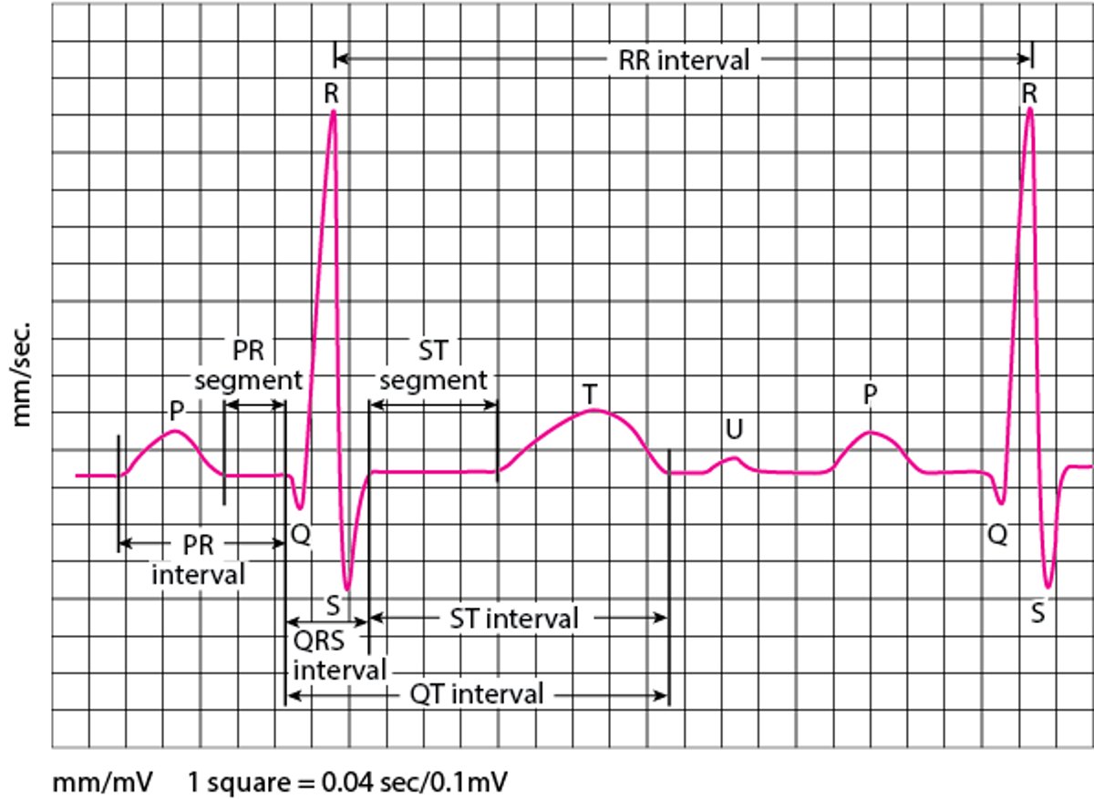
ECG: Reading the Waves
An electrocardiogram (ECG) represents the electrical current moving through the heart during a heartbeat. The current's movement is divided into parts, and each part is given an alphabetic designation in the ECG. Each heartbeat begins with an impulse from the heart's pacemaker (sinus or sinoatrial node). This impulse activates the upper chambers of the heart (atria). The P wave represents activation of the atria. Next, the electrical current flows down to the lower chambers of the heart (ventricles). The QRS complex represents activation of the ventricles. The ventricles must then undergo an electrical change to get ready for the next heart beat. This electrical activity is called the recovery wave, which is represented by the T wave. Many kinds of abnormalities can often be seen on an ECG. They include a previous heart attack (myocardial infarction), an abnormal heart rhythm (arrhythmia), an inadequate supply of blood and oxygen to the heart (ischemia), and excessive thickening (hypertrophy) of the heart's muscular walls. Certain abnormalities seen on an ECG can also suggest bulges (aneurysms) that develop in weaker areas of the heart's walls. Aneurysms may result from a heart attack. If the rhythm is abnormal (too fast, too slow, or irregular), the ECG may also indicate where in the heart the abnormal rhythm starts. Such information helps doctors begin to determine the cause. |

Did You Know...
|
Causes of Long QT Interval Syndromes
Some people are born with a genetic abnormality that causes a long QT interval (called long QT syndrome). These inherited disorders are one type of heart disorder called a channelopathy.
In other people, the long QT interval is not inherited but results from disorders such as low serum levels of potassium (hypokalemia), a very slow heart rhythm, or a medication. Often, medications used to treat abnormal heart rhythms cause a long QT interval, but certain antidepressants and certain antiviral and antifungal medications also can cause it.
The inherited long QT interval syndromes are classified based on the specific gene that has mutated. More than 15 forms of long QT syndrome have been identified. Many of these are inherited in an autosomal dominant pattern, in which a person with the disorder has a 50% chance of passing it on to each child they have. However, a specific genetic abnormality is found in only 50 to 75% of people. The likelihood of detecting an abnormality varies depending on the person's symptoms and ECG findings.
Triggers
Sometimes, exercise brings on the ventricular tachycardia (see Sudden Cardiac Death in Athletes). Other factors that increase the risk of torsades include female sex, older age, an underactive thyroid gland (hypothyroidism), brain disorders such as a stroke, and certain types of heart disease such as a heart attack or inflammation of the heart (myocarditis).
Symptoms of Long QT Interval Syndromes
The long QT interval syndromes do not cause symptoms unless torsades de pointes ventricular tachycardia occurs, which can cause palpitations (awareness of heart beats), light-headedness, or fainting. Torsades de pointes ventricular tachycardia may turn into ventricular fibrillation and cause cardiac arrest and sudden death.
Diagnosis of Long QT Interval Syndromes
Electrocardiography (ECG)
Usually exercise testing and ambulatory ECG monitoring
Sometimes genetic testing
A long QT interval may be found when an ECG is done for other reasons. People with unexplained cardiac arrest or fainting or a family history of such when the affected people do not have other heart disease are evaluated for long QT interval syndromes. Doctors typically do an ECG along with an exercise test and also have the person wear a heart monitor at home for several days or longer.
Not all people with a long QT interval have a congenital long QT syndrome, and not all people with a congenital long QT syndrome have a long QT interval on any given ECG. Thus, doctors use a scoring system based on the results of the tests and the person's symptoms to predict how likely the diagnosis is. The score helps determine which people need genetic testing or provocative testing with exercise, sudden standing, or IV epinephrine (Not all people with a long QT interval have a congenital long QT syndrome, and not all people with a congenital long QT syndrome have a long QT interval on any given ECG. Thus, doctors use a scoring system based on the results of the tests and the person's symptoms to predict how likely the diagnosis is. The score helps determine which people need genetic testing or provocative testing with exercise, sudden standing, or IV epinephrine (adrenaline).
With provocative testing using intravenous epinephrine, people are given the medication in to try to cause the characteristic long QT interval on the ECG. People with a low likelihood of having a congenital long QT interval syndrome do not need genetic testing, but when the likelihood is intermediate or high, genetic testing is needed. With provocative testing using intravenous epinephrine, people are given the medication in to try to cause the characteristic long QT interval on the ECG. People with a low likelihood of having a congenital long QT interval syndrome do not need genetic testing, but when the likelihood is intermediate or high, genetic testing is needed.
Close family members of a person with a congenital long QT syndrome should be examined by a doctor to detect symptoms that may suggest an abnormal heart rhythm and should undergo ECG. Genetic testing and exercise testing also are sometimes needed.
Treatment of Long QT Interval Syndromes
Converting dangerous abnormal heartbeats to normal rhythm by giving an electric shock (defibrillation)
Eliminating predisposing causes and triggers, especially electrolyte abnormalities and use of certain medications
Usually beta-blockers
Sometimes an implantable cardioverter-defibrillator (ICD)
If people are experiencing episodes of ventricular tachycardia or ventricular fibrillation, treatment includes
Converting heartbeat to normal rhythm by applying an electric shock (defibrillation)
Measures to prevent sudden death include avoiding specific triggers, including strenuous exercise. When possible, any medications that are likely to cause abnormal heart rhythms are stopped.
Beta-blockers are given to some people, such as those who have had episodes of fainting, cardiac arrest, or ventricular fibrillation or ventricular tachycardia.
A permanent pacemaker may be needed to reduce the likelihood of recurrent torsades de pointes ventricular tachycardia. People who have been resuscitated after cardiac arrest and certain others need an implantable cardioverter-defibrillator (ICD).
More Information
The following English-language resource may be useful. Please note that The Manual is not responsible for the content of this resource.
American Heart Association: Arrhythmia: Information to help people understand their risks of arrhythmias as well as information on diagnosis and treatment


