


Ventricular tachycardia is a heart rhythm that originates in the ventricles (lower chambers of the heart) and produces a heart rate of at least 120 beats per minute (the normal heart rate is usually between 60 and 100 beats per minute).
People almost always have awareness of heartbeats (palpitations) and other symptoms of heart failure (for example, shortness of breath, chest discomfort, and/or fainting).
Electrocardiography is used to make the diagnosis.
Medications and procedures to destroy abnormal areas of the ventricles may be used, but usually an automatic implantable cardioverter-defibrillator is required.
(See also Overview of Abnormal Heart Rhythms.)
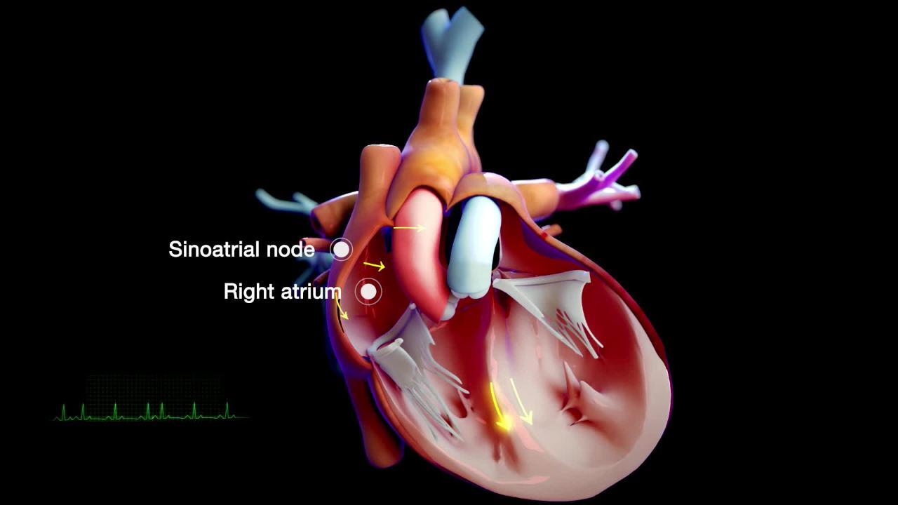
The electrical current that begins each heartbeat originates in the heart’s pacemaker (called the sinus node or sinoatrial node), located in the top of the upper right heart chamber (right atrium). However, a heartbeat is sometimes triggered from one of the lower chambers of the heart, the ventricles. People may have only a few heartbeats triggered by the ventricles (ventricular premature beats), but sometimes people have a sequence of consecutive ventricular premature beats. Sometimes only a few such beats occur together, and then the heart returns to a normal rhythm. Ventricular tachycardia that lasts more than 30 seconds is called sustained ventricular tachycardia.
Sustained ventricular tachycardia usually occurs in people with a structural heart disorder such as a heart attack, heart failure, or a cardiomyopathy. It is more common among older people. However, rarely, ventricular tachycardia develops in young people who do not have a structural heart disorder. Such young people may have a heart disorder called long QT syndrome, which can be inherited or caused by certain medications. It can also be due to other rare inherited disorders such as cardiac channelopathies.

Symptoms of Ventricular Tachycardia
People with ventricular tachycardia almost always have awareness of heart beats (palpitations). They may have weakness, light-headedness, and/or chest discomfort.
Sustained ventricular tachycardia can be dangerous because the ventricles cannot fill adequately or pump blood normally. Blood pressure tends to fall, and heart failure follows. Sustained ventricular tachycardia is also dangerous because it can turn into ventricular fibrillation—a form of cardiac arrest. Because of this, even though short periods of ventricular tachycardia may cause few symptoms, even at rates of up to 200 beats per minute, is still extremely dangerous.
Diagnosis of Ventricular Tachycardia
Electrocardiography
Electrocardiography (ECG) is used to diagnose ventricular tachycardia and to help determine whether treatment is required.
Treatment of Ventricular Tachycardia
Converting heartbeat to normal rhythm
Preventing further episodes
Immediate treatment
Ventricular tachycardia is treated when it causes symptoms or when episodes last more than 30 seconds even without causing symptoms.
People who have symptoms, particularly if blood pressure is too low, require immediate cardioversion (an electrical shock to convert the heart to normal rhythm).
People who have no symptoms but who have had ventricular tachycardia for more than 30 seconds should be treated either with cardioversion or intravenous medications.
Cardioversion is painful, so sedation is required, but cardioversion is almost always effective and aside from the discomfort has few side effects.
Medications are not uncomfortable but are not as effective or as rapid as cardioversion in stopping the abnormal heart rhythm (arrhythmia) and are more likely to cause side effects. The most commonly used medications are procainamide, amiodarone, and lidocaine (see table Medications are not uncomfortable but are not as effective or as rapid as cardioversion in stopping the abnormal heart rhythm (arrhythmia) and are more likely to cause side effects. The most commonly used medications are procainamide, amiodarone, and lidocaine (see table).
Long-term treatment
The long-term goal is to prevent sudden death, rather than simply stopping the abnormal rhythm. In people with ventricular tachycardia who have an underlying heart disorder, particularly if their heart does not pump well, an implantable cardioverter-defibrillator (ICD, a small device that can detect an arrhythmia and deliver a shock to correct it) is often used. This procedure is similar to implantation of an artificial pacemaker.
Certain procedures may be used to destroy the small abnormal area in the ventricles, identified by ECG, that is usually responsible for sustained ventricular tachycardia. They include catheter ablation (delivery of energy using radiowaves, laser pulses, or high-voltage electrical current or freezing with cold through a catheter inserted in the heart) and open-heart surgery.
More Information
The following English-language resource may be useful. Please note that The Manual is not responsible for the content of this resource.
American Heart Association: Arrhythmia: Information to help people understand their risks of arrhythmias as well as information on diagnosis and treatment
Drug Information for the Topic


