

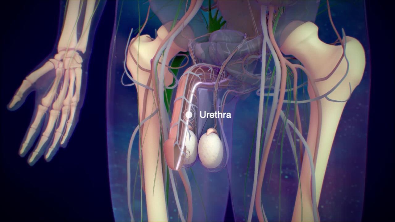
The urethra is the tube that drains urine from the bladder out of the body.
(See also Overview of Kidney and Urinary Tract Birth Defects.)
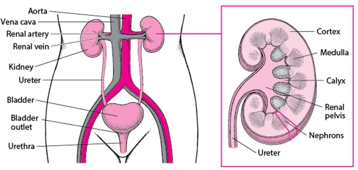
A Look Inside the Urinary Tract

Types of Birth Defects of the Urethra
There are several types of birth defects of the urethra. The urethra may be
Partially blocked
In the wrong place
Bulging out from its opening (prolapsed urethra)
Duplicated (2 or more urethras instead of just 1)
These defects may
Block the flow of urine
Cause urine to come out from the wrong location
Any urethra defect that blocks or slows the flow of urine can cause urine to become stagnant, which can result in urinary tract infections (UTIs). Blockage of urine flow also can raise the pressure inside the bladder and/or kidneys and damage them over time. Frequent infections also can damage the kidneys. Kidney damage can cause high blood pressure and rarely kidney failure.

Partially blocked urethra
Several birth defects partially block the urethra.
Posterior urethral valves occur only in boys. In posterior urethral valves, folds of abnormal tissue in the urethra block the flow of urine from the bladder. Posterior urethral valves almost always occur only in boys. The blockage increases the pressure in the bladder and can cause difficulty urinating and a weak urine stream. In more severe forms, the blockage affects the developing fetus. The increased urine pressure from the blockage can interfere with development of the bladder and kidneys. The blockage also may reduce the amount of urine the fetus releases into the amniotic fluid (the fluid that surrounds the fetus in the uterus). If the fetus does not release enough urine into the amniotic fluid, the amount of amniotic fluid is reduced. If there is too little amniotic fluid, problems occur with the development of the fetus's lungs, heart, and limbs. Poor lung development can be fatal shortly before or after birth. After birth, affected infants have symptoms of poor bladder drainage or poor kidney function.
A urethral stricture is a narrowing of the urethra that is usually caused by an injury, most commonly a crush injury that occurs when boys fall straddling a hard object. Sometimes urethral stricture is a birth defect or occurs after surgical repair is done to correct a defect of the penis called hypospadias. It is more common among boys.
In urethral meatal stenosis, the outside opening of the urethra (meatus) is narrow and decreases and misdirects the flow of urine. This occurs mostly in boys who previously had surgery on their penis or were circumcised as infants.
Urethra in the wrong place
The opening of the urethra may be in the wrong place.
In boys, the opening of the urethra may be on the underside of the penis (called hypospadias) or on the topside of the penis (called epispadias), instead of at the tip. Boys who have hypospadias often have another defect called chordee (a downward bend of the penis) and a hooded foreskin (where the foreskin has not grown together on the underside of the penis). Children who have epispadias may have involuntary release of urine (urinary incontinence).
In girls, the opening of the urethra may be misplaced between the clitoris and the labia, inside the opening of the vagina, or rarely on the abdomen.
Urethral prolapse
Urethral prolapse occurs mainly in girls, more commonly in Black girls than in White girls.
In this disorder, the inner lining of the urethra sticks out through the opening of the urethra. When urethral prolapse occurs, the opening of the urethra looks like a small, red and swollen donut.
Urethral prolapse typically does not cause symptoms, but the prolapsed tissue may bleed, causing blood spots on the girl's diaper or underwear.
Duplicated urethras (extra urethras)
Rarely, children are born with 2 or more urethras. Usually only 1 of them is connected to the bladder, but sometimes they are all connected to the bladder or to each other.

Diagnosis of Urethra Birth Defects
Physical examination
Sometimes voiding cystourethrography
Before birth, doctors sometimes discover defects of the urethra that cause significant urinary blockage during routine prenatal ultrasound.
After birth, doctors often find defects of the urethra during a physical examination or a well-child examination. If doctors suspect a newborn boy has posterior urethral valves, they do a test called voiding cystourethrography (VCUG) before the boy is discharged from the hospital. For voiding cystourethrography, a catheter is passed through the urethra into the bladder, a liquid that shows up on x-rays (contrast agent) is put in through the catheter, and x-rays are taken before, during, and after the child urinates.
Treatment of Urethra Birth Defects
Usually surgical repair
Defects of the urethra that cause symptoms, such as a blockage, usually need to be surgically corrected.
Children who have a blockage in the urethra have surgery to open the blockage as soon as possible. Children whose urethra is abnormal, narrow, or missing may need surgery to correct these defects.
Boys who have posterior urethral valves have surgery when they are diagnosed. Surgery is done to relieve the blockage and prevent further kidney damage. The surgical procedure is usually done with a cystoscope (a small tube with a camera on the end that is inserted into the urethra) to cut the extra tissue that is causing the obstruction. Even after surgery, the bladder may not function normally and boys may need catheterization or additional surgery. Catheterization is draining the bladder by inserting a thin, flexible tube (catheter) through the urethral opening into the bladder.
Boys who have hypospadias may have surgery to repair the defect and correct any other existing defects, such as chordee, depending on the degree of severity.
Girls who have urethral prolapse may be given a cream that contains estrogen to lessen symptoms. Urethral prolapse usually goes away with time and rarely requires surgery.


