

Torsades de pointes ventricular tachycardia is a specific type of ventricular tachycardia that occurs in people who have a particular disorder of the heart's electrical activity called a long QT interval.
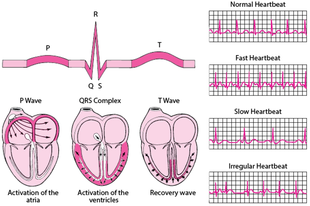
The QT interval refers to the time between 2 events on the electrocardiogram (ECG)—from the beginning of the QRS complex to the end of the T wave.
(See also Overview of Abnormal Heart Rhythms and Ventricular Tachycardia.)
ECG: Reading the Waves
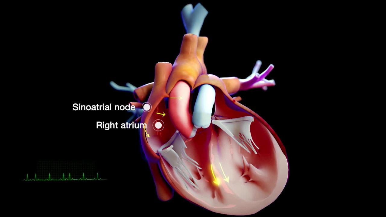
An electrocardiogram (ECG) represents the electrical current moving through the heart during a heartbeat. The current's movement is divided into parts, and each part is given an alphabetic designation in the ECG. Each heartbeat begins with an impulse from the heart's pacemaker (sinus or sinoatrial node). This impulse activates the upper chambers of the heart (atria). The P wave represents activation of the atria. Next, the electrical current flows down to the lower chambers of the heart (ventricles). The QRS complex represents activation of the ventricles. The ventricles must then undergo an electrical change to get ready for the next heart beat. This electrical activity is called the recovery wave, which is represented by the T wave. Many kinds of abnormalities can often be seen on an ECG. They include a previous heart attack (myocardial infarction), an abnormal heart rhythm (arrhythmia), an inadequate supply of blood and oxygen to the heart (ischemia), and excessive thickening (hypertrophy) of the heart's muscular walls. Certain abnormalities seen on an ECG can also suggest bulges (aneurysms) that develop in weaker areas of the heart's walls. Aneurysms may result from a heart attack. If the rhythm is abnormal (too fast, too slow, or irregular), the ECG may also indicate where in the heart the abnormal rhythm starts. Such information helps doctors begin to determine the cause. |

Any abnormality that prolongs the QT interval increases the risk of a dangerous heart rhythm called torsades de pointes ventricular tachycardia. Some people are born with a genetic abnormality that causes a long QT interval (called long QT syndrome). In other people, the long QT interval results from low serum levels of potassium (hypokalemia), a very slow heart rhythm, or a medication. Often, medications used to treat abnormal heart rhythms cause a long QT interval, but certain antidepressants and certain antiviral and antifungal medications can also cause it.
Ventricular tachycardia is a heart rhythm that originates in the ventricles (lower chambers of the heart) and produces a rapid heart rate. People with a long QT interval may develop a particular form of ventricular tachycardia called torsades de pointes ventricular tachycardia (torsades). Torsades has a characteristic appearance on the electrocardiogram (ECG) and often turns into ventricular fibrillation, in which the heart stops beating, which rapidly causes death.
Sometimes, exercise brings on the ventricular tachycardia (see Sudden Cardiac Death in Athletes). Other factors that increase the risk of torsades include female sex, older age, an underactive thyroid gland (hypothyroidism), brain disorders such as a stroke, and certain types of heart disease such as a heart attack or inflammation of the heart (myocarditis).

Symptoms of Torsades de Pointes Ventricular Tachycardia
People who develop torsades de pointes ventricular tachycardia may have palpitations (awareness of heartbeats) and feel light-headed or faint. Torsades de pointes runs of ventricular tachycardia usually stop on their own but frequently recur. Ventricular fibrillation causes cardiac arrest and sudden collapse.
Diagnosis of Torsades de Pointes Ventricular Tachycardia
Electrocardiography
Electrocardiography (ECG) is used to detect torsades.
If doctors diagnose torsades in a person, they ask whether there is a family history of the disorder or if relatives have died unexpectedly because of a heart problem. Such people may then have genetic testing for congenital long QT syndrome, and their close family members should be evaluated.
Treatment of Torsades de Pointes Ventricular Tachycardia
Converting heartbeat to normal rhythm by applying an electric shock (defibrillation)
Preventing further episodes
Sometimes doctors give magnesium sulfate. Defibrillation is needed if ventricular fibrillation develops.
If a medication is the cause, it is stopped.
People may need to limit their physical activity to prevent a recurrence. They may also need to take beta-blockers (see table ) or have an artificial pacemaker or cardioverter-defibrillator implanted.
More Information
The following English-language resource may be useful. Please note that The Manual is not responsible for the content of this resource.
American Heart Association: Arrhythmia: Information to help people understand their risks of arrhythmias as well as information on diagnosis and treatment


