Globe injury can occur from blunt or penetrating trauma to the eye.
Trauma to the globe may cause the following:
Globe rupture (laceration)
Traumatic optic neuropathy
Retinal detachment
Loss of visual function
(See also Overview of Eye Trauma.)


Paul Whitten/SCIENCE PHOTO LIBRARY
The following injuries, which are covered elsewhere in The Manual, can have a variety of causes, including a precipitating traumatic injury:
Other common globe injuries include conjunctival, subconjunctival, and scleral hemorrhages, which typically do not require treatment; retinal hemorrhage, retinal edema, or retinal detachment; vitreous hemorrhage; laceration of the iris; and a dislocated lens, all of which typically require urgent evaluation by an ophthalmologist.
Evaluation can be difficult when massive eyelid edema, ecchymosis, or laceration is present. Even so, unless the need for immediate eye surgery is obvious (necessitating urgent evaluation by an ophthalmologist), the lid is opened, taking care not to exert pressure on the globe, and as complete an examination as possible is conducted. At a minimum, the following are noted:
Pupil shape and pupillary responses
Extraocular movements
Anterior chamber depth or hemorrhage
Presence of red reflex
Intraocular pressure
An analgesic or an anxiolytic may be given to facilitate examination. Gentle and careful use of eyelid retractors or an eyelid speculum makes it possible to open the lids if the patient requires assistance. If a commercial instrument is not available, the eyelids can be separated with makeshift retractors fashioned by opening a paperclip to an S shape, then bending the U-shaped ends to 180°. Instruments should not place pressure on the globe. Globe laceration should be suspected with any of the following:
A corneal or scleral laceration is visible.
Aqueous humor is leaking (positive Seidel sign).
The anterior chamber is very shallow (eg, making the cornea appear to have folds) or very deep (due to rupture posterior to the lens).
The pupil is irregular or misshapen (possibly indicating globe perforation or herniation of the iris).
The red reflex is absent (possibly indicating vitreous hemorrhage or retinal injury).
If globe laceration is suspected, the following measures should take place before an ophthalmologist is available (1):
Administer intravenous antibiotics (eg, ceftazidime and vancomycin)
A CT scan should be performed to look for a foreign body and other injuries, such as fractures; MRI is avoided because of the possibility of an occult metallic foreign body. Topical antibiotics are avoided. Vomiting, which can increase intraocular pressure (IOP) and contribute to extravasation of ocular contents, is suppressed using antiemetics as needed. Because fungal contamination of open wounds is dangerous, corticosteroids are contraindicated until after wounds are closed surgically. Tetanus prophylaxis is indicated for open globe injuries. The patient should not be allowed any oral liquids or food and should be cleared medically for general anesthesia.
Operative repair under general anesthesia typically requires globe exploration, removal of any foreign bodies, layered closures of the sclera and cornea, and injection of intravitreal antibiotics and/or antifungal agents. Postoperatively, the eye is shielded. Mydriatics and topical antibiotics/antifungals are applied depending on the specific etiology of the initial injury and the intraoperative findings. The intraocular pressure is followed and any increase is treated with topical medications. The patient is closely followed for several days because of the risk of posttraumatic endophthalmitis. Rarely, after laceration of the globe, the uninjured contralateral eye becomes inflamed (sympathetic ophthalmia) and, untreated, may lose vision to the point of blindness. The mechanism is an autoimmune reaction, and the time course is variable (2).
Intraocular Foreign Bodies
Penetrating trauma to the eye may also result in a retained intraocular foreign body (eg, broken pencil, fragment of metal from a rotary tool). Intraocular foreign bodies require immediate surgical removal by an ophthalmologist. Intravitreal, systemic, and topical antimicrobials with effectiveness against Bacillus cereus are indicated for any injury possibly contaminated with soil or vegetation; these antimicrobials include intravitreal ceftazidime in combination with vancomycin followed by intravenous doses of vancomycin and ceftazidime or a topical fluoroquinolone such as moxifloxacin (1, 3). Ointment should be avoided if the globe is lacerated.
A protective shield (eg, plastic or aluminum eye shield or the bottom third of a paper cup) is placed and taped over the bony orbital rim to avoid inadvertent pressure on the globe that could extrude ocular contents through the penetration site. Patches must be avoided because they can also inadvertently apply pressure to the globe. Tetanus prophylaxis is indicated after open globe injuries.
As with any laceration of the globe, vomiting (eg, due to pain) can increase intraocular pressure and should be prevented. If nausea occurs, antiemetics should be prescribed.
The patient should not be allowed any oral liquids or food (eg, be made NPO) and should be cleared medically for general anesthesia.
Hyphema (Anterior Chamber Hemorrhage)
Hyphema usually results from injury to the anterior segment of the eye, specifically the iris and its insertion onto the sclera, resulting in hemorrhage into the anterior chamber. Hyphema may be followed by recurrent bleeding, increased intraocular pressure (IOP), and/or blood staining of the cornea, any of which may result in permanent vision loss. Symptoms are those of associated injuries unless the hyphema is large enough to obstruct vision. Direct inspection typically reveals blood cells in the anterior chamber, with or without layering of blood, and/or the presence of a clot in the anterior chamber. Layering is visible as a meniscus-like blood level in the dependent (usually inferior) part of the anterior chamber. Microhyphema, a less severe form of hyphema, may only be detectable upon direct inspection of the anterior chamber by slit-lamp examination, which allows for visualization of suspended red blood cells.
An ophthalmologist should attend to the patient as soon as possible. The patient is placed on bed rest with the head elevated 30 to 45° and an eye shield is placed to protect the eye from further trauma (see Corneal Abrasions and Foreign Bodies). Hospitalization may be indicated for patients who are at high risk of recurrent bleeding (ie, those who have large hyphemas, bleeding diatheses, sickle cell disease, or who use anticoagulants) who have IOP that is difficult to control, or who may not be able to administer recommended treatment. Testing for bleeding abnormalities should be considered if the cause of the hyphema is not known. Oral and topical nonsteroidal anti-inflammatory drugs (NSAIDs) are contraindicated because they may contribute to recurrent bleeding. Antifibrinolytics, such as tranexamic acid, are not routinely used but may be helpful in patients at high risk of rebleeding or other complications (4).
Treatment of hyphema is mainly focused on management of IOP. Intraocular pressure can rise acutely (within hours, usually in patients with sickle cell disease or trait) or months to years later. Thus, IOP is monitored daily for several days and then regularly over subsequent weeks and months and if symptoms develop (eg, development of eye ache, decreased vision, nausea, all of which are similar to the symptoms of acute angle-closure glaucoma). If pressure rises, timolol 0.5% twice a day, brimonidine 0.2% or 0.15% twice a day, or both are given. Response to treatment is determined by IOP, often checked every 1 or 2 hours until controlled or until a significant rate of reduction is demonstrated; thereafter, it is usually checked once or twice daily. Mydriatic drops (eg, scopolamine 0.25% or atropine 1%) and topical corticosteroids (eg, prednisolone acetate 1%) are often given to reduce inflammation and scarring. However, the evidence to support the use of mydriatics and corticosteroids for hyphema is not definitive (5).
Rebleeding from retraction of the clot is reported to occur in up to 38% of patients (4). An ophthalmologist should be consulted for management, as the rebleed may be more severe than the initial bleeding. Administration of antifibrinolytics may be considered, and miotic or mydriatic medications are viable options. IOP should be monitored. Rarely, recurrent bleeding with secondary glaucoma requires surgical evacuation of the blood.

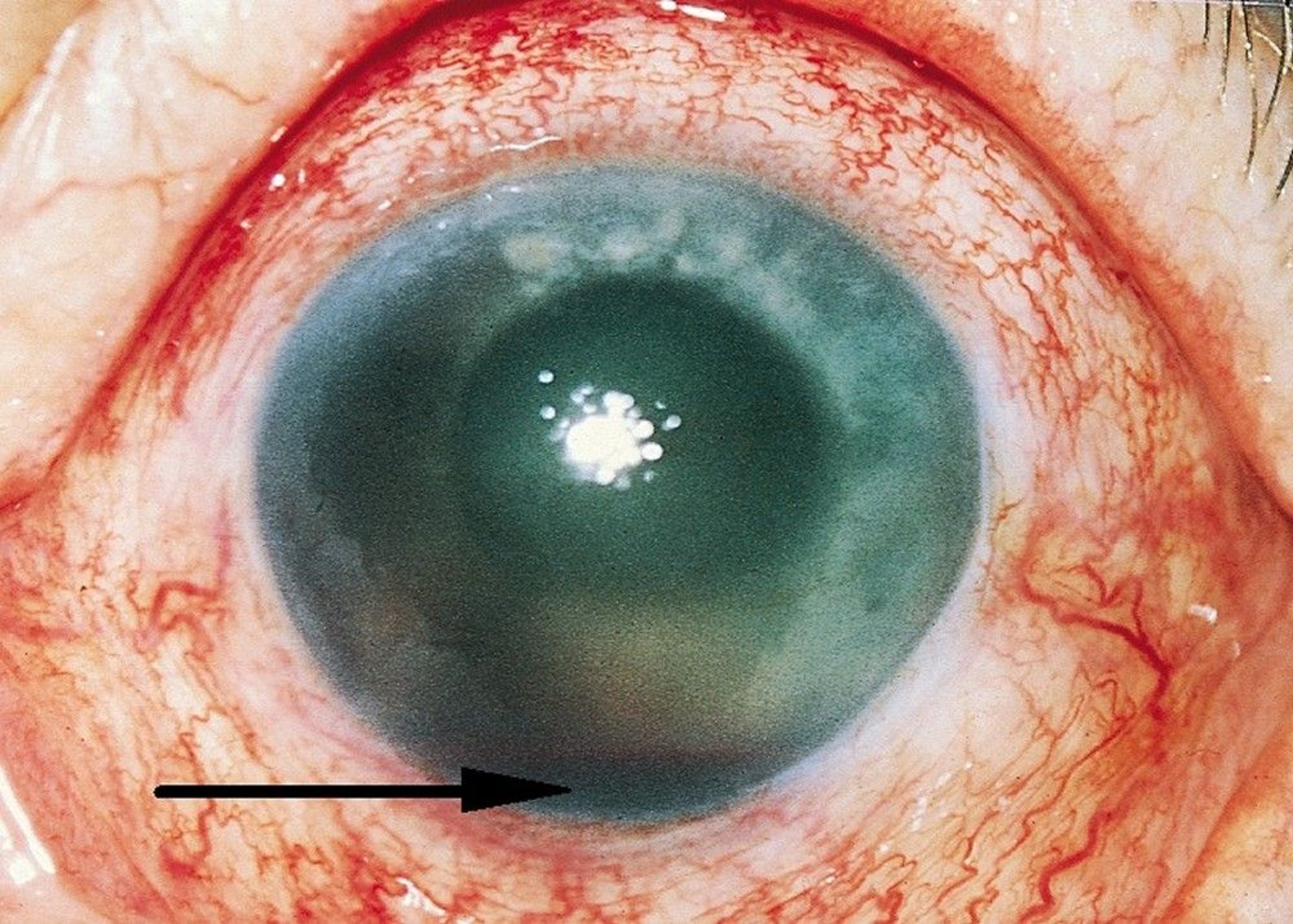
This patient has a traumatic hyphema (arrow) and conjunctival injection after a traumatic injury to the eye.
© Springer Science+Business Media
Blowout Fracture
Blowout fracture occurs when blunt trauma forces the orbital contents through one of the more fragile portions of the orbital wall, typically the floor (6). Medial, lateral, and roof fractures also can occur. Patients may have facial or orbital pain, diplopia, enophthalmos, hypesthesia of the cheek and upper lip (due to infraorbital nerve injury), epistaxis, and/or subcutaneous emphysema. Other facial fractures or injuries must also be ruled out. Globe, optic nerve, and brain injuries must be considered in all cases of orbital fracture and evaluated as indicated.
Diagnosis is best made using CT with thin cuts through the facial bones. If ocular motility is impaired (eg, causing diplopia), extraocular muscles should be assessed for signs of entrapment.
If diplopia or clinically significant enophthalmos are present, surgical repair may be indicated. However, many, if not most, patients with orbital floor fractures can be followed conservatively without surgery. Patients should be told to avoid blowing the nose to prevent orbital compartment syndrome from air reflux (7). Using a topical intranasal vasoconstrictor for 2 to 3 days may alleviate epistaxis. Oral antibiotics can be used if patients have sinusitis.
One exception to conservative management of blowout fracture is the so-called white-eyed blowout fracture, which typically occurs in children, teenagers, and young adults. In such cases, the patient has few if any external signs of injury (thus the term "white-eyed"), but presents with severe extraocular muscle restriction from direct muscle entrapment within the fracture. Patients may also have severe nausea and vomiting, often misdiagnosed as concussion, and bradycardia because the muscle entrapment causes a vasovagal response. In contradistinction to adult blowout fractures, the orbital bone in white-eyed blowout fractures bends or "greensticks" rather than cleanly breaking, resulting in a trapdoor phenomenon. Such fractures require urgent repair with in 24 to 48 hours to minimize the risk of ischemic contracture of the entrapped extraocular muscle, as well as to relieve symptoms of the vasovagal reflex. Of note, because of the trapdoor nature of white-eyed blowout fractures, the fractures may be missed on CT reading unless a full history is provided to the radiologist.
Orbital Compartment Syndrome
Orbital compartment syndrome (OCS) is an ophthalmic emergency. OCS occurs when orbital pressure increases suddenly, usually due to trauma that causes orbital hemorrhage. Anything that increases orbital volume (air, blood, or pus in the orbit) can lead to an OCS. Symptoms can include sudden vision loss, as well as diplopia, eye pain, and lid swelling (8).
Physical examination findings may include decreased vision, chemosis, ecchymosis, limited and/or painful eye motility, afferent pupillary defect, proptosis, ophthalmoplegia, and elevated intraocular pressure (IOP). Diagnosis is clinical and treatment should not be delayed for imaging (9).
Emergent consultation with ophthalmology is indicated for consideration of surgery; medical therapy should not delay this consultation. Treatment is immediate lateral canthotomy and cantholysis (surgical exposure of the lateral canthal tendon with incision of its inferior branch) followed by:
Elevation of the head of the bed to 45°
Monitoring of IOP
Treatment of elevated IOP; in the acute phase, intravenous mannitol may be administered short-term (as long as no systemic or neurosurgical contraindications are present) (10)
Reversal of any coagulopathy
Prevention of further increasing intraorbital pressure (preventing or minimizing pain, nausea, cough, straining, severe hypertension)
Application of ice or cool compresses


References
1. Bhagat N, Nagori S, Zarbin M. Post-traumatic infectious endophthalmitis. Surv Ophthalmol. 2011;6(3):214-251. doi: 10.1016/j.survophthal.2010.09.002
2. Parchand S, Agrawal D, Ayyadurai N, et al. Sympathetic ophthalmia: A comprehensive update. Indian J Ophthalmol. 2022;70(6):1931-1944. doi: 10.4103/ijo.IJO_2363_21
3. Knox FA, Best RM, Kinsella F, et al. Management of endophthalmitis with retained intraocular foreign body. Eye (Lond). 2004;18(2):179-182. doi: 10.1038/sj.eye.6700567
4. Deans R, Noël LP, Clarke WN. Oral administration of tranexamic acid in the management of traumatic hyphema in children. Can J Ophthalmol. 1992;27(4):181-183.
5. Woreta FA, Lindsley KB, Gharaibeh A, Ng SM, Scherer RW, Goldberg MF. Medical interventions for traumatic hyphema. Cochrane Database Syst Rev. 2023;3(3):CD005431. doi: 10.1002/14651858.CD005431.pub5
6. Iftikhar M, Canner JK, Hall L, Ahmad M, Srikumaran D, Woreta FA. Characteristics of orbital floor fractures in the United States from 2006 to 2017. Ophthalmology. 2021;128(3):463-470. doi: 10.1016/j.ophtha.2020.06.065
7. Kersten RC, Vagefi MR, Bartley GB. Orbital "blowout" fractures: Time for a new paradigm. Ophthalmology. 2018;125(6):796-798. doi: 10.1016/j.ophtha.2018.02.014
8. Papadiochos I, Petsinis V, Sarivalasis S-E. Acute orbital compartment syndrome due to traumatic hemorrhage: 4-year case series and relevant literature review with emphasis on its management. Oral Maxillofac Surg. 2023;27(1):101-116. doi: 10.1007/s10006-021-01036-9
9. Hatton MP, Rubin PA. Management of orbital compartment syndrome. Arch Ophthalmol. 2007;125(3):433-434; author reply 434. doi: 10.1001/archopht.125.3.433-b
10. Johnson D, Winterborn A, Kratky V. Efficacy of intravenous mannitol in the management of orbital compartment syndrome: A nonhuman primate model. Ophthalmic Plast Reconstr Surg. 2016;32(3):187-190. doi: 10.1097/IOP.0000000000000463
Key Points
Globe trauma may cause hyphema, blowout fracture, globe laceration, orbital compartment syndrome, cataract, lens dislocation, glaucoma, vitreous hemorrhage, or retinal damage (hemorrhage, detachment, or edema).
Treat corneal foreign bodies by removing foreign material, prescribing a topical antibiotic, and sometimes instilling a cycloplegic.
Suspect an intraocular foreign body if fluorescein streams away from a corneal defect, if the pupil is irregular, or if the mechanism of injury involves a high-speed machine (eg, drill, saw, anything with a metal-on-metal mechanism), hammering, or explosion.
For intraocular foreign bodies, give systemic and topical antibiotics, apply a shield, control pain and nausea, and consult an ophthalmologist for surgical removal.
Never patch an eye with suspected penetrating injury. Always use only an eye shield.
Hyphema, best diagnosed by slit-lamp examination, requires bed rest with head elevation at 30 to 45° and close monitoring of intraocular pressure.
Refer patients for surgical repair of blowout fractures that cause diplopia or enophthalmos.
Obtain immediate ophthalmologic consultation; do immediate lateral canthotomy and inferior cantholysis if orbital compartment syndrome, is present.
Drug Information for the Topic