


A cataract is a congenital or degenerative opacity of the lens. The main symptom is gradual, painless vision blurring. Diagnosis is by ophthalmoscopy and slit-lamp examination. Treatment is surgical removal and placement of an intraocular lens.
Cataracts are the leading cause of blindness worldwide (1). In the United States, almost 20% of people aged 65 to 74 have cataracts that interfere with vision. Almost one in two people older than 75 has cataracts.
Lens opacity can develop in several locations:
Central lens nucleus (nuclear cataract)
Beneath the posterior lens capsule (posterior subcapsular cataract)
On the side of the lens (cortical cataract)—these usually do not interfere with central vision
(For developmental or congenital cataracts, see Congenital Cataract.)
Reference
1. Hashemi H. Pakzad R, Yekta A, et al: Global and regional prevalence of age-related cataract: A comprehensive systematic review and meta-analysis. Eye 34:1357–1370, 2020. do: 10.1038/s41433-020-0806-3
Etiology of Cataract
Cataracts occur with aging. Other risk factors may include the following:
Trauma (sometimes causing cataracts years later)
Alcohol use
Exposure to x-rays
Heat from infrared exposure
Systemic disease (eg, diabetes)
Systemic drugs (eg, corticosteroids)
Chronic ultraviolet light exposure
Many people have no risk factors other than age. Some cataracts are congenital with a genetic etiology, or associated with a systemic syndrome or diseases.
Estrogen use by women after menopause may be protective, but estrogen should not be used solely for this purpose.
Symptoms and Signs of Cataract
Cataracts usually develop slowly over years. Early symptoms may be loss of contrast, glare (ie, halos and starbursts around lights, not photophobia), needing more light to see well, and problems distinguishing dark blue from black. Painless blurring eventually occurs. The degree of blurring depends on the location and extent of the opacity. Monocular double vision or ghost images occur rarely.
With a nuclear cataract, distance vision worsens. Near vision may improve in the early stages because of changes in the refractive index of the lens; presbyopic patients may be temporarily able to read without glasses (second sight).
A posterior subcapsular cataract disproportionately affects vision because the opacity is located at the crossing point of incoming light rays. Such cataracts reduce visual acuity more when the pupil constricts (eg, in bright light, during reading). They are also the type most likely to cause loss of contrast as well as glare (halos and starbursts around lights), especially from bright lights or from car headlights while driving at night.

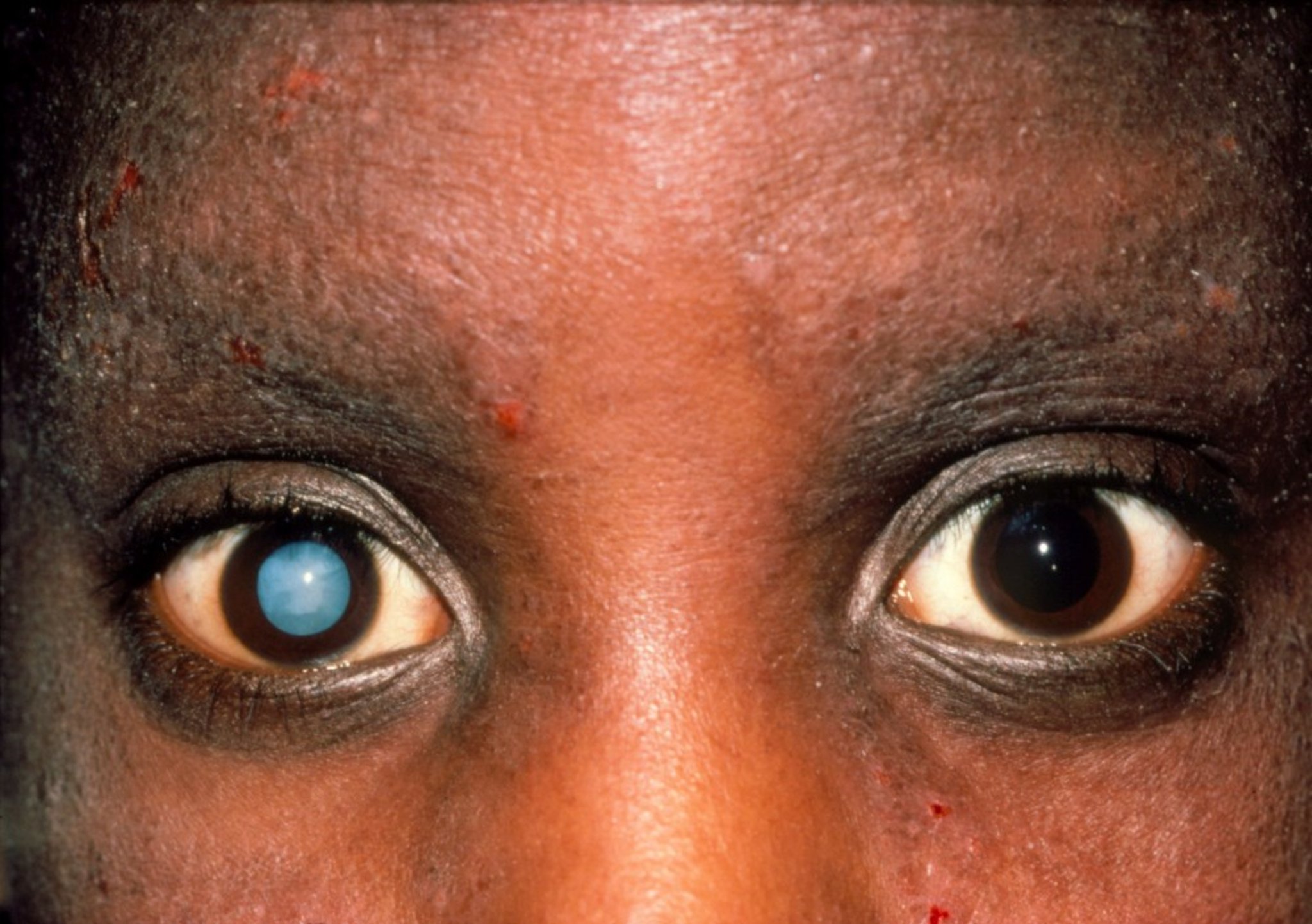
WESTERN OPHTHALMIC HOSPITAL/SCIENCE PHOTO LIBRARY
Rarely, the cataract swells, pushing the iris over the trabecular drainage meshwork and causing its occlusion and thus secondary closed-angle glaucoma and pain.
Diagnosis of Cataract
Ophthalmoscopy followed by slit-lamp examination
Diagnosis is best made with the pupil dilated. Well-developed cataracts appear as gray, white, or yellow-brown opacities in the lens. Examination of the red reflex through the dilated pupil with the ophthalmoscope held about 30 cm away usually discloses subtle opacities. Small cataracts stand out as dark defects in the red reflex. A large cataract may obliterate the red reflex. Slit-lamp examination provides more details about the character, location, and extent of the opacity.
Pearls & Pitfalls
|
Treatment of Cataract
Surgical removal of the cataract
Placement of an intraocular lens
Usual indications for surgery include the following:
Best vision obtained with glasses is worse than 20/40 (< 6/12), or vision is significantly decreased under glare conditions (eg, oblique lighting while trying to read a chart) in a patient with bothersome halos or starbursts.
Patients sense that vision is limiting (eg, by preventing activities of daily living such as driving, reading, hobbies, and occupational activities).
Vision could potentially be meaningfully improved if the cataract is removed (ie, a significant portion of the vision loss must be caused by the cataract).
Far less common indications include cataracts that cause glaucoma or that obscure the fundus in patients who need periodic fundus examinations for management of diseases such as diabetic retinopathy and macular degeneration. There is no advantage to removing a cataract early.
Cataract extraction and lens implant procedures
Cataract extraction is usually done using a topical or local anesthetic and IV sedation. There are 3 extraction techniques:
In intracapsular cataract extraction, the cataract and lens capsule are removed in one piece; this technique is rarely used.
In extracapsular cataract extraction, the hard central nucleus is removed in one piece and then the soft cortex is removed in multiple small pieces.
In phacoemulsification (a type of extracapsular cataract extraction), the hard central nucleus is dissolved by ultrasound and then the soft cortex is removed in multiple small pieces.
Phacoemulsification uses the smallest incision, thus enabling the fastest healing, and is usually the preferred procedure. Femtosecond lasers can be used in refractive laser-assisted cataract surgery prior to phacoemulsification to achieve a more precise, accurate incision and decrease risk of infection. In extracapsular extraction (including phacoemulsification), the lens capsule is not removed.
A plastic or silicone lens is almost always implanted intraocularly to replace the optical focusing power of the removed crystalline lens. The lens implant is usually placed on or within the lens capsule (posterior chamber lens). The lens can also be placed in front of the iris (anterior chamber lens) or attached to the iris and within the pupil (iris plane lens). Iris plane lenses are rarely used in the United States because many designs led to a high frequency of postoperative complications. Multifocal intraocular lenses are newer and have different focusing zones that may reduce dependence on glasses after surgery. Patients occasionally experience glare with these lenses, especially under low-light conditions, and also have problems with reduced contrast sensitivity.
Postsurgical care and complications
1, 23). Patients often wear an eye shield while sleeping and should avoid the Valsalva maneuver, heavy lifting, excessive forward bending, and eye rubbing for several weeks.
Major complications of cataract surgery are rare (4). Complications include the following (5):
Intraoperative: Bleeding beneath the retina, causing the intraocular contents to extrude through the incision (choroidal hemorrhage—very rare and could result in irreversible blindness), vitreous prolapsing out of the incision (vitreous loss), fragments of the cataract dislocating into the vitreous, incisional burn, and detachment of corneal endothelium and its basement membrane (Descemet membrane)
Within the first week: Endophthalmitis (infection within the eye—very rare and could result in irreversible blindness) and glaucoma
Within the first month: Cystoid macular edema
Months later: Bullous keratopathy (ie, swelling of the cornea due to damage to the corneal pump cells during cataract surgery), retinal detachment, and posterior capsular opacification (common but treatable with laser)
After surgery, vision returns to 20/40 (6/12) or better in 95% of eyes if there are no preexisting disorders such as amblyopia, retinopathy, macular degeneration, and glaucoma (4). If an intraocular lens is not implanted, contact lenses or thick glasses are needed to correct the resulting hyperopia.
Treatment references
1. Melega MV, Alves M, Cavalcanti Lira RP, et alJ Cataract Refract Surg 45(3):343-350, 2019. doi: 10.1016/j.jcrs.2018.10.044
2. Shorstein NH, Winthrop KL, Herrinton LJ: Decreased postoperative endophthalmitis rate after institution of intracameral antibiotics in a Northern California eye department. J Cataract Refract Surg 39(1):8-14, 2013. doi: 10.1016/j.jcrs.2012.07.031
3. Walters T, Bafna S, Vold S, et alJ Clin Exp Ophthalmol 7.4 (2016):1-11.
4. Powe NR, Schein OD, Gieser SC, et al: Synthesis of the literature on visual acuity and complications following cataract extraction with intraocular lens implantation. Cataract Patient Outcome Research Team. Arch Ophthalmol. 112(2):239-252, 1994. doi: 10.1001/archopht.1994.01090140115033. Erratum in: Arch Ophthalmol 112(7):889, 1994.
5. Greenberg PB, Liu J, Wu WC, et al: Predictors of mortality within 90 days of cataract surgery. Ophthalmology 117(10):1894-9, 1899.e1, 2010. doi: 10.1016/j.ophtha.2010.02.009
Prevention of Cataract
Many ophthalmologists recommend ultraviolet-coated eyeglasses or sunglasses as a preventive measure. Reducing risk factors such as alcohol, tobacco, and corticosteroids and controlling blood glucose in diabetes1), the effects of supplementation have been mixed (2, 3).
Prevention references
1. Mares JA, Voland R, Adler R, et al: Healthy diets and the subsequent prevalence of nuclear cataract in women. Arch Ophthalmol 128(6):738-749, 2010. doi: 10.1001/archophthalmol.2010.84
2. Mathew MC, Ervin AM, Tao J, et al: Antioxidant vitamin supplementation for preventing and slowing the progression of age-related cataract. Cochrane Database Syst Rev6(6):CD004567, 2012. doi: 10.1002/14651858.CD004567.pub2
3. Christen WG, Glynn RJ, Manson JE, et al: Effects of multivitamin supplement on cataract and age-related macular degeneration in a randomized trial of male physicians. Ophthalmology 121(2):525-534, 2014. doi: 10.1016/j.ophtha.2013.09.038.
Key Points
Modifiable risk factors for cataract include exposure to ultraviolet light; use of alcohol, tobacco, and systemic corticosteroids; and poor control of blood glucose.
Symptoms include loss of contrast, glare (halos and starbursts around lights), and eventually visual blurring.
Diagnosis is by examination with the eye dilated.
Surgical removal and placement of an intraocular lens are usually indicated if the cataract contributes to visual loss that interferes with activities of daily living, causes bothersome glare, or reaches certain degrees of severity (eg, best-corrected visual acuity worse than 20/40).


