Multiple myeloma is a cancer of plasma cells that produce monoclonal immunoglobulin and invade and destroy adjacent bone tissue. Common manifestations include lytic lesions in bones that cause pain and/or fractures, renal insufficiency, hypercalcemia, anemia, and recurrent infections. Diagnosis typically requires demonstration of M-protein (sometimes present in urine and not serum but rarely absent entirely), and/or light-chain proteinuria, and excessive plasma cells in the bone marrow. Specific treatment most often includes some combination of conventional chemotherapy, glucocorticoids, and one or more additional medications such as proteasome inhibitors (eg, bortezomib, carfilzomib, ixazomib), immunomodulatory agents (eg, lenalidomide, thalidomide, pomalidomide), or monoclonal antibodies (eg, daratumumab, isatuximab, elotuzumab). Antibody- and T-cell-based approaches to targeting of B-cell maturation antigen have shown efficacy. High-dose melphalan followed by autologous peripheral blood stem cell transplantation may also be used.
In the United States, the lifetime risk of getting multiple myeloma is less than 1% (1). The average age at diagnosis is approximately 69 years. Prevalence in Black people is twice that in White people. There is a slight male predominance. The etiology is unknown, although chromosomal and genetic factors, radiation, and chemicals, including pesticides, have been suggested.
The American Cancer Society estimates that in 2026 in the United States there will be about 36,000 new cases of multiple myeloma diagnosed and about 10,850 deaths (1). Survival continues to improve with recent reports showing a median survival of over 10 years (2).
General references
1. American Cancer Society. Key statistics about multiple myeloma. Atlanta, Ga., American Cancer Society; 2026. Updated January 13, 2026. Accessed August 3, 2026.
2. Jew S, Bujarski S, Regidor B, et al. Clinical outcomes and serum B-cell maturation antigen levels in a real-world unselected population of newly diagnosed multiple myeloma patients. Target Oncol. 2023;18(5):735-747. doi:10.1007/s11523-023-00990-6
Pathophysiology of Multiple Myeloma
The M-protein (monoclonal immunoglobulin protein) produced by the malignant plasma cells is IgG in about 50 to 55% of myeloma patients and IgA in about 20% (1). Of patients producing either IgG or IgA, 40% also have Bence Jones proteinuria, which is free monoclonal kappa (κ) or lambda (λ) light chains in the urine. In 15 to 20% of patients, plasma cells secrete only Bence Jones protein. IgM and IgD myeloma account for approximately 1 to 2% of cases each; IgD myeloma is more frequent among patients of Asian descent. IgE myeloma is exceedingly rare.
Rarely, patients have no M-protein in blood and urine. However, the serum free light chain assay demonstrates monoclonal light chains in many of the patients who were formerly considered to have nonsecretory myeloma.
Diffuse osteoporosis or discrete osteolytic lesions are often present, with the most common sites being the pelvis, spine, ribs, femur, humerus, and skull. Lesions are caused by bone replacement by expanding plasmacytomas or by cytokines that are secreted by malignant plasma cells that activate osteoclasts and suppress osteoblasts, leading to bone loss. The osteolytic lesions are usually multiple; occasionally, they are solitary intramedullary masses. Increased bone loss may also lead to hypercalcemia.
Extraosseous solitary plasmacytomas are unusual but may occur in any tissue, especially in the upper respiratory tract.
In many patients, kidney failure is present at diagnosis or develops during the course of the disorder. Kidney has many causes, and most commonly, it results from deposition of light chains in the distal tubules (myeloma-related kidney disease) or hypercalcemia.
Patients often develop anemia usually due to kidney disease or suppression of erythropoiesis by cancer cells. Anemia in patients with multiple myeloma also may be due to other unrelated causes, including iron deficiency or vitamin B12 deficiency.
Because of lack of normal antibodies and other immune dysfunction, some patients have increased susceptibility to bacterial infection. Viral infections, especially herpes zoster infections, also may occur as a result of treatment modalities such as proteasome inhibitors (eg, bortezomib, ixazomib, carfilzomib) and monoclonal antibodies (eg, daratumumab, elotuzumab, isatuximab). Newer bispecific antibodies and chimeric antigen receptor (CAR) T-cell therapies are associated with a high risk of severe infections.
Amyloidosis occurs in 10% of patients who have myeloma, most often in patients with lambda-type M-proteins (2).
Variant expressions of multiple myeloma occur (see table ).
Variant Expressions of Multiple Myeloma
Form | Characteristics |
|---|---|
Extramedullary plasmacytoma | Plasmacytomas that occur outside of the bone marrow |
Solitary plasmacytoma of bone | Single bone plasmacytomas, which usually produce no M-protein |
Osteosclerotic myeloma (POEMS syndrome) | Polyneuropathy (chronic inflammatory polyneuropathy) Organomegaly (hepatomegaly, splenomegaly, or lymphadenopathy) Endocrinopathy (eg, gynecomastia, testicular atrophy) M-protein Skin changes (eg, hyperpigmentation, excess hair) |
Nonsecretory myeloma | Absence of M-protein in serum and urine Absence of elevated serum free light chains |
Pathophysiology references
1. Kyle RA, Gertz MA, Witzig TE, et al. Review of 1027 patients with newly diagnosed multiple myeloma. Mayo Clin Proc. 2003;78(1):21-33. doi:10.4065/78.1.21
2. Ríos-Tamayo R, Krsnik I, Gómez-Bueno M, et al. AL Amyloidosis and multiple myeloma: A complex scenario in which cardiac involvement remains the key prognostic factor. Life (Basel). 2023;13(7):1518. doi:10.3390/life13071518
Symptoms and Signs of Multiple Myeloma
Persistent bone pain (especially in the back or thorax), kidney failure, and recurrent bacterial infections are the most common problems on presentation, but many patients are identified when routine laboratory tests show an elevated total protein level in the blood, proteinuria, or unexplained anemia or kidney failure.
Fatigue related to anemia may be the predominate symptom and may be the sole reason for evaluation for myeloma in some patients. Rarely, patients with very high monoclonal protein levels in the blood have manifestations of the hyperviscosity syndrome.
Pathologic (fragility) fractures (ie, fractures that occur with minimal or no trauma) are common, and vertebral collapse may lead to spinal cord compression and paraplegia.
Peripheral neuropathy, carpal tunnel syndrome (especially in patients with amyloidosis), abnormal bleeding, and symptoms of hypercalcemia (eg, polydipsia, dehydration) may occur in some patients.
Lymphadenopathy and hepatosplenomegaly are unusual.
Diagnosis of Multiple Myeloma
Complete blood count (CBC) with platelets, peripheral blood smear, and chemistry panel (blood urea nitrogen [BUN], creatinine, calcium, uric acid, lactate dehydrogenase [LDH])
Serum protein electrophoresis (SPEP) with immunofixation; quantitative immunoglobulins; serum free light chains, and urine protein electrophoresis (UPEP) from a 24-hour urine collection
Radiographs (skeletal survey) and either a positron emission tomography (PET)-CT scan or whole-body MRI
Bone marrow examination, including conventional cytogenetics and fluorescent in situ hybridization studies (FISH)
Multiple myeloma is suspected in patients > 40 years with persistent unexplained bone pain, particularly at night or at rest, other typical symptoms, unexplained laboratory abnormalities (eg, elevated blood protein or urinary protein, hypercalcemia, renal insufficiency, anemia), or radiographs showing a pathologic fracture or lytic lesions.
Laboratory evaluation includes routine blood tests (CBC,chemistry panel), LDH, serum and urine immunofixation and protein electrophoresis, and serum free light chains (1). Patients should also undergo a skeletal survey and either a PET-CT scan or whole-body MRI because these tests are more sensitive to bone disease than radiographs. A bone marrow examination is also required along with conventional cytogenetics and FISH studies (2, 3).
Anemia is present during the course of the disease in 80% of patients, usually a normocytic, normochromic anemia with formation of rouleaux (clusters of 3 to 12 red blood cells that occur in stacks) (3). White blood cell and platelet counts are usually normal. BUN, serum creatinine, LDH, and serum uric acid may be elevated. The anion gap is sometimes low. Hypercalcemia is present at diagnosis in about 10% of patients.
Protein electrophoresis and immunofixation electrophoresis are carried out first on a serum sample. Serum protein electrophoresis identifies M-protein in about 80 to 90% of patients. Most of the remaining 10 to 20% are patients with only free monoclonal light chains (Bence Jones protein), which can be detected in blood using a free light chain assay or in urine using urine protein and immunofixation electrophoresis. In practice, the 24-hour urine collection for urine protein electrophoresis and immunofixation is typically obtained if initial serum testing (serum protein electrophoresis, immunofixation, or an abnormal free light chain ratio) is abnormal. However, when multiple myeloma is suspected, it is part of the complete diagnostic workup and is used to identify and qualify urinary M-protein and to document baseline proteinuria. Monoclonal protein is not evident at diagnosis in a small proportion of patients (nonsecretory disease), and during the course of the disease, some patients no longer produce monoclonal protein.
Immunofixation electrophoresis is performed to identify the immunoglobulin class of the M-protein (IgG, IgA, or uncommonly IgD, IgM, or IgE) and can often detect light-chain protein if serum immunoelectrophoresis is negative; if multiple myeloma is strongly suspected, immunofixation electrophoresis is performed even when the electrophoresis is negative.
Serum free light-chain analysis with delineation of kappa and lambda ratios or differences between the involved and uninvolved light chains helps confirm the diagnosis and can also be used to monitor efficacy of therapy and provide prognostic data.
Serum level of beta-2 microglobulin is generally measured once the diagnosis is confirmed or considered very likely, because its main role is in staging and prognosis; together with serum albumin, it is used as part of the international staging system (see table ). Beta-2 microglobulin is a small protein on the membrane of all cells, and its concentration varies directly with tumor mass and severity of kidney dysfunction. Because kidney impairment from any cause raises the level independently from tumor burden, its reliability as a predictive marker in multiple myeloma is reduced in patients with concurrent kidney disease.
Revised International Staging System for Multiple Myeloma
Stage | Criteria |
|---|---|
I | Beta-2 microglobulin < 3.5 mcg/mL (< 297 nmol/L) and Serum albumin ≥ 3.5 g/dL (≥ 35 g/L) Normal LDH Standard-risk cytogenetic abnormalities by FISH* |
II | Not stage I or III |
III | Beta-2 microglobulin ≥ 5.5 mcg/mL (≥ 466 nmol/L) and High-risk cytogenetic abnormalities by FISH† and/or high LDH |
* Standard-risk cytogenetic abnormalities include t(11;14), t(6;14), and trisomies. | |
† High-risk cytogenetic abnormalities consist of t(4;14), t(14;16), t(14;20), or 17p- and gain/amplification of 1q, along with 1p deletions and TP53. | |
FISH = fluorescent in situ hybridization; LDH = lactate dehydrogenase. | |
Data from Palumbo A, Avet-Loiseau H, Oliva S, et al. Revised International Staging System for Multiple Myeloma: A Report From International Myeloma Working Group. J Clin Oncol. 2015;33(26):2863-2869. doi:10.1200/JCO.2015.61.2267 | |
Radiographs include a skeletal survey (ie, plain radiographs of skull, long bones, spine, pelvis, and ribs). Punched-out lytic lesions or diffuse osteoporosis is present in approximately 80% of cases (4). Radionuclide bone scans usually are not helpful. Whole-body MRI can provide more detail and is obtained if specific sites of pain or neurologic symptoms are present. PET-CT scans provide prognostic information and can help determine whether patients have solitary plasmacytoma or multiple myeloma.
The International Myeloma Working Group (IMWG) and International Myeloma Society (IMS) have developed a category known as high-risk multiple myeloma based on the number and type of specific genetic abnormalities in the myeloma cells and serum beta2 microglobulin levels. This includes patients with (5):
17p- and/or TP53 mutation
IgH translocation including t(4;14), t(14;16), or t(14;20) along with 1q+ and/or del(1p32)
Monoallelic del(1p32) along with 1q+ or biallelic del(1p32)
Beta2 microglobulin > 5.5 mg/L (466 nmol/L) with a serum creatinine < 1.2 mg/dL (106.1 μmol/L)

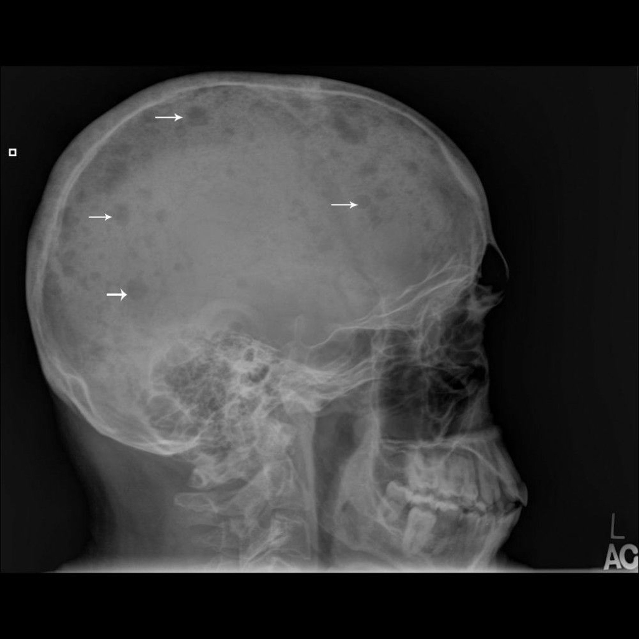
This skull radiograph shows multiple, punched-out lesions in the bone (arrows), which are typical of multiple myeloma.
Image courtesy of Michael J. Joyce, MD, and Hakan Ilaslan, MD.
Bone marrow aspiration and biopsy are done and reveal sheets or clusters of plasma cells; myeloma is diagnosed when ≥ 10% of the cells are of this type. However, bone marrow involvement is patchy; therefore, some samples from patients with myeloma may show < 10% plasma cells. Still, the percentage of plasma cells in bone marrow is rarely normal. Plasma cell morphology does not correlate with the class of immunoglobulin synthesized. Chromosomal studies on bone marrow (eg, using cytogenetic testing methods such as FISH and immunohistochemistry) may reveal specific karyotypic abnormalities in plasma cells that can influence treatment choices and are associated with differences in survival.
Diagnosis and differentiation from other malignancies (eg, metastatic carcinoma, lymphoma, leukemia) and from monoclonal gammopathy of undetermined significance typically require multiple criteria:
Clonal bone marrow plasma cells or plasmacytoma
M-protein in plasma and/or urine
Organ impairment (hypercalcemia, renal insufficiency, anemia, or bony lesions)
In patients without serum M protein, myeloma is indicated by Bence Jones proteinuria > 200 mg/24 hour or abnormal serum free light chain ratio, osteolytic lesions (without evidence of metastatic cancer or granulomatous disease), and sheets or clusters of plasma cells in the bone marrow. Rarely, patients present with the absence of both an M protein and abnormal serum free light chains, so-called nonsecretory myeloma.
Diagnosis references
1. Kumar SK, Callander NS, Adekola K, et al. Multiple Myeloma, Version 5.2026, NCCN Clinical Practice Guidelines In Oncology. J Natl Compr Canc Netw. 2026;24(1):e260001. doi:10.6004/jnccn.2026.0001
2. Sive J, Cuthill K, Hunter H, Kazmi M, Pratt G, Smith D and on behalf of British Society of Haematology. Guidelines on the diagnosis, investigation and initial treatment of myeloma: a British Society for Haematology/UK Myeloma Forum Guideline. Brit J Haematol. 2021;193(2):245-268. doi:10.1111/bjh.17410
3. Rajkumar SV. Multiple myeloma: 2022 update on diagnosis, risk-stratification and management. Am J Hematol. 2022;97(8):1086-1107. doi:10.1002/ajh.26590
4. Kyle RA, Gertz MA, Witzig TE, et al. Review of 1027 patients with newly diagnosed multiple myeloma. Mayo Clin Proc. 2003;78(1):21-33. doi:10.4065/78.1.2
5. Avet-Loiseau H, Davies F, Samur MK, et al. International Myeloma Society/International Myeloma Working Group consensus recommendations on the definition of high-risk multiple myeloma. J Clin Oncol. 2025;43(24):2739-2751. doi:10.1200/JCO-24-01893
Treatment of Multiple Myeloma
Conventional chemotherapy and glucocorticoids for patients with symptoms
Additional therapy with immunomodulatory agents and monoclonal antibodies
Possibly autologous stem cell transplantation
Possibly radiation therapy to specific symptomatic areas that do not respond to systemic therapy
Treatment of complications, eg, anemia, hypercalcemia, kidney dysfunction, infections, and bone disease
Treatment of relapsed or refractory disease with immunomodulatory agents, monoclonal antibodies, proteasome inhibitors, antibody-drug conjugates, bispecific antibodies, and CAR T-cell therapies
Therapy involves direct treatment of malignant cells in patients with symptoms and those with myeloma-related organ dysfunction (anemia, renal dysfunction, hypercalcemia, or bone disease).
Among patients initially presenting without organ dysfunction, the presence of the following findings also requiring rapid treatment of myeloma:
> 60% plasma cells in bone marrow
> 1 lesion on MRI
Ratio of involved to uninvolved serum free light chain ≥ 100
Patients with these risk factors are considered to have active myeloma and require immediate treatment although early treatment of these patients has not been consistently shown to improve their overall survival. Patients without these risk factors or end-organ dysfunction probably do not benefit from immediate treatment, which is usually withheld until symptoms or complications develop.
Treatment of malignant cells
Conventional chemotherapy, consisting of oral melphalan and prednisone given in cycles of 4 to 6 weeks for 8 to 12 cycles with monthly evaluation of response, was the initial treatment of multiple myeloma. However, melphalan-based regimens have fallen out of favor with the improved efficacy of combination therapies that typically include either a proteasome inhibitor (eg, bortezomib, carfilzomib, or ixazomib) or an immunomodulatory agent (lenalidomide or thalidomide).
The preferred standard therapy for most patients is a four-drug regimen combining an anti-CD38 monoclonal antibody ( daratumumab or isatuximab), a proteasome inhibitor (e.g., bortezomib, carfilzomib, or ixazomib), an immunomodulatory agent (lenalidomide or thalidomide), and a glucocorticoid (dexamethasone) (1).
Response to treatment (see table ) is indicated by:
Decreases in serum and urine M-protein
Decreases in levels of the involved serum free light chain
Increases in numbers of red blood cells
Improvement in renal function among patients presenting with kidney failure
Normalization of calcium levels among those presenting with elevated levels
Decrease in bone pain
Decrease in fatigue
Autologous hematopoietic cell transplantation may be considered for patients who have adequate cardiac, hepatic, pulmonary, and renal function, particularly those whose disease is stable or responsive after several cycles of initial therapy. However, pharmacotherapy options are highly effective and may make transplantation less often necessary or unnecessary altogether. Clinical trials show longer progression-free survival but no improvement in overall survival when patients undergo stem cell transplantation as part of initial therapy (2).
Allogeneic stem cell transplantation after nonmyeloablative chemotherapy (eg, low-dose cyclophosphamide and fludarabine) or low-dose radiation therapy can produce myeloma-free survival of 5 to 10 years in some patients via a graft-versus-myeloma effect. However, allogeneic stem cell transplantation with myeloablative or nonmyeloablative chemotherapy remains experimental because of the high morbidity and mortality resulting from graft versus host disease (3).
Treatment of relapsed or refractory myeloma
For patients with relapsed or refractory myeloma, effective regimens are generally built by combining a glucocorticoid (usually dexamethasone) with agents from drug classes the patient has not become refractory to. Available classes include:
Proteasome inhibitors (bortezomib, ixazomib, carfilzomib)
Immunomodulatory agents (thalidomide, lenalidomide, pomalidomide)
Anti CD38 monoclonal antibodies (daratumumab, isatuximab) and an anti-SLAMF7 antibody (elotuzumab)
An antibody-drug conjugate (belantamab mafodotin)
Bispecific antibodies (teclistamab, elranatamab, linvoseltamab, talquetamab)
Chimeric antigen receptor (CAR) T-cell therapies (ciltacabtagene autoleucel, idecabtagene vicleucel)
A GPRC5D-directed bispecific antibody (talquetamab)
A selective inhibitor of nuclear export (selinexor)
A histone deacetylase inhibitor (panobinostat)
A BCL-2 inhibitor (venetoclax) for patients with an 11;14 translocation
Patients with prolonged remission (relapse more than approximately 6 months after discontinuation of therapy) may respond to retreatment with the same regimen that led to the initial remission. Patients who fail to respond to a given combination of medications may respond when another medication in the same class (eg, proteasome inhibitors, immunomodulatory agents, chemotherapeutic agents) is substituted.
Monoclonal antibodies targeting proteins on myeloma cells are also highly effective in relapsed or refractory myeloma. Available monoclonal antibodies include those that target:
CD38 (daratumumab, isatuximab)
Signaling lymphocytic activation molecule F7 (SLAMF7—elotuzumab)
These monoclonal antibodies improve the efficacy of other medications used to treat multiple myeloma, including the immunomodulatory agents thalidomide, lenalidomide, and pomalidomide, and the proteasome inhibitors bortezomib and carfilzomib (4, 5). However, treatment with any of these monoclonal antibodies and only dexamethasone is effective in many patients, especially those whose disease progression is marked by only a rise in measured tumor markers. This approach avoids the adverse effects of additional medications and may allow treatment of other conditions, including malignancies, without the concern of additional adverse effects from multidrug treatment of myeloma.
When combined with other active myeloma medications, especially bortezomib, the following medications are especially effective:
Selective inhibitor of nuclear export: selinexor
Histone deacetylase inhibitor: panobinostat
Effective immune treatments that target B-cell maturation antigen (BCMA) are available. These treatments include:
Cellular therapies for myeloma include the CAR T-cell therapies idecabtagene vicleucel and ciltacabtagene autoleucel.
Bispecific antibodies that also target CD3 on T cells (teclistamab, linvoseltamab, elranatamab ) (6, 7). The combination of the anti-CD38 antibody daratumumab and teclistamab has been shown to be highly effective but is associated with a high risk of serious infections (8). Use of ongoing gammaglobulin may reduce this risk.
The antibody-drug conjugate belantamab mafodotin has shown efficacy for treatment of multiple myeloma (9, 10). It has significant ocular toxicity, and patients must be monitored carefully for potential eye complications.
The bispecific antibody talquetamab targets another protein on myeloma cells, G protein-coupled receptor, class C, group 5, member D (GPRC5D) on multiple myeloma cells and also CD3 (7). The antibody can cause significant nail, skin, and mucosal problems that may be permanent.
Although these treatments are effective, they can cause significant acute adverse effects (cytokine release syndrome (CRS), neurologic problems) and a high risk of ongoing severe infections. Studies show that the use of the anti-IL-6 antibody tocilizumab can greatly reduced the risk of CRS. CAR T-cell treatments may lead to Parkinson-like syndromes as well as severe gastrointestinal problems.
The oral BCL-2 inhibitor venetoclax has been shown to be effective for treating patients whose myeloma cells show the genetic marker t(11;14) and can be effectively combined with steroids and proteasome inhibitors or monoclonal antibodies (11).
Maintenance therapy
Maintenance therapy has been tried with nonchemotherapeutic medications, including interferon alfa, which prolongs remission but does not improve survival and is associated with significant adverse effects. Following a response to glucocorticoid-based regimens, glucocorticoids alone are effective as a maintenance treatment.
Thalidomide may also be effective as a maintenance treatment (12), and lenalidomide alone or with glucocorticoids is effective as maintenance treatment (13). However, data from a randomized trial suggests that continuing lenalidomide beyond two years does not improve overall survival in standard-risk patients (14). The oral proteasome inhibitor ixazomib is also effective as a single agent maintenance therapy (15). Adding lenalidomide to ixazomib has not been shown to improve outcomes over lenalidomide alone (12). There is some concern about secondary cancers among patients receiving long-term lenalidomide therapy, especially after autologous stem cell transplantation. Thus, the risk of developing secondary cancers must be weighed against improved survival.
The role of monoclonal antibodies as maintenance therapy remains to be defined.
Similarly, the optimal duration of biclonal antibody therapy is unknown.
The use of agents from drug classes the patient has not previously used as maintenance therapy has not been evaluated.
Treatment of complications
In addition to direct treatment of malignant cells, therapy must also be directed at complications, which include:
Anemia
Hypercalcemia
Hyperuricemia
Hyperviscosity
Infections
Renal insufficiency
Skeletal lesions
Anemia
Anemia that is inadequately relieved by chemotherapy can be treated with recombinant erythropoietin. If anemia causes cardiovascular symptoms or significant systemic symptoms, packed red blood cells are transfused. Often patients are iron deficient for reasons unrelated to their myeloma and require intravenous iron; other patients sometimes require vitamin B12 supplements. Patients with anemia should have periodic measurement of serum iron, transferrin, and ferritin levels to monitor iron stores as well as vitamin B12 levels.
Hypercalcemia
Hypercalcemia is treated with vigorous saluresis, IV bisphosphonates (preferably zoledronic acid) after rehydration, and sometimes with calcitonin or prednisone. Denosumab can also be used to treat hypercalcemia especially among patients with severe kidney failure. Patients should avoid calcium-containing foods, calcium supplements, and vitamin D.
Hyperuricemia
Hyperuricemia may occur in some patients with high tumor burden and underlying metabolic problems. However, most patients do not require allopurinol. Allopurinol or rasburicase is indicated for patients with high levels of serum uric acid or high tumor burden and a high risk of tumor lysis syndrome with treatment.
Hyperviscosity
If hyperviscosity develops, which rarely occurs in patients with myeloma, plasma exchange is indicated.
Infection
Infection is more likely during treatment-induced neutropenia. In addition, infections with the herpes zoster virus occur frequently among patients treated with certain antimyeloma drugs, especially the proteasome inhibitors (bortezomib, carfilzomib, ixazomib) and the monoclonal antibodies (daratumumab, isatuximab, elotuzumab). The BCMA-directed treatments, both the bispecific antibodies and CAR T-cell therapies, are associated with a very high risk of severe infections, including opportunistic types that can be difficult to treat effectively.
Prophylactic use of antiviral drugs (eg, acyclovir, valganciclovir, famciclovir) is indicated for patients receiving a proteasome inhibitor (bortezomib, carfilzomib, ixazomib), a monoclonal antibody (daratumumab, isatuximab, elotuzumab), a bispecific antibody (teclistamab, elranatamab, talquetamab), or CAR T-cell therapy (idecabtagene vicleucel, ciltacabtagene autoleucel).
Documented bacterial infections should be treated with antibiotics. Prophylactic use of antibiotics is not routinely recommended.
Prophylactic IV immune globulin may reduce the risk of infection but is generally reserved for patients with low uninvolved immunoglobulin levels (< 400 mg/dL IgG) and frequent recurrent infections.
Pneumococcal vaccine and influenza vaccine are indicated to prevent infection but are not effective in most patients because of disease-related and treatment-related immune deficiency. Use of live vaccines is not recommended in patients with a compromised immune system. To prevent herpes zoster infection, the nonviable recombinant zoster vaccine may be given but is of limited effectiveness.
Renal compromise
Renal compromise can often be ameliorated with adequate hydration. Even patients with prolonged, massive Bence Jones proteinuria (≥ 10 to 30 g/day) may have intact kidney function if they maintain a urine output > 2000 mL/day. Dehydration combined with high-osmolar IV contrast may precipitate acute oliguric kidney failure in patients with Bence Jones proteinuria. Plasma exchange may be effective in some cases. Nephrotoxic medications should be avoided. Rapid and aggressive treatment of the underlying myeloma to reduce the levels of the nephrotoxic monoclonal immunoglobulin and treatment of hypercalcemia are both important to reverse this condition.
Skeletal lesions
Skeletal lesions require multiple supportive measures. Maintenance of ambulation and supplemental calcium and vitamin D, as indicated, help preserve bone density in patients without disease-related hypercalcemia. Vitamin D levels should be measured at diagnosis and periodically, and dosing of vitamin D adjusted accordingly. Analgesics and palliative doses of radiation therapy (18 to 24 gray) can relieve bone pain. However, radiation therapy may cause significant toxicity and, because it suppresses bone marrow function, may impair the patient’s ability to receive cytotoxic doses of systemic chemotherapy.
Most patients, especially those with lytic lesions and generalized osteoporosis or osteopenia, should receive a monthly IV bisphosphonate (either pamidronate or zoledronic acid). Bisphosphonates reduce skeletal complications and lessen bone pain and may have an antitumor effect.
For patients with potentially reversible kidney failure resulting from myeloma but unrelated to hypercalcemia or with ongoing infusion reactions after bisphosphonate infusion, an option is monthly denosumab (given subcutaneously), which, unlike bisphosphonates, is not cleared by the kidneys and does not cause infusion reactions.
Both bisphosphonates and denosumab may uncommonly cause osteonecrosis of the jaw. Maintaining excellent dental health and avoiding dental explants and implants are important to minimize the risk of this complication.
Prognosis for Multiple Myeloma
Although multiple myeloma is progressive and remains incurable, the median overall survival has improved to nearly 13 years among unselected patients in a recent study (1).
Unfavorable prognostic signs at diagnosis are higher stage, lower serum albumin levels, higher beta-2 microglobulin levels, elevated LDH levels, specific cytogenetic abnormalities in the tumor cells, and higher levels of malignant cells in the blood. Patients initially presenting with kidney failure also do poorly unless kidney function improves with therapy, which typically happens with current treatment options.
Because multiple myeloma is ultimately fatal, patients are likely to benefit from discussions of end-of-life care that involve clinicians and appropriate family and friends. Points for discussion may include advance directives, the use of feeding tubes, and pain relief.
Prognosis reference
1. Moore, JT, Mettias SM, Cheung J, et al.. Outcomes for unselected, newly diagnosed multiple myeloma patients. Haematologica. 2025;110(11):2823-2826. doi:10.3324/haematol.2025.287458
Key Points
Malignant plasma cells produce monoclonal immunoglobulin and invade and destroy bone.
Expanding plasmacytomas and cytokine secretion cause multiple, discrete, osteolytic lesions (usually in the pelvis, spine, ribs, and skull) and diffuse osteoporosis; pain, fractures, and hypercalcemia are common.
Anemia and kidney failure are common.
Amyloidosis develops in about 10%, typically patients who produce excess lambda light chains.
Perform serum and urine protein electrophoresis followed by immunofixation, quantitative immunoglobulins, and measurement of serum free light chains.
Perform bone marrow aspiration and biopsy.
Patients with symptoms and those with organ dysfunction should be treated with pharmacotherapy, which may include glucocorticoids, chemotherapy agents, proteasome inhibitors, immunomodulatory agents, monoclonal antibodies, selective inhibitors of nuclear export, histone deacetylase inhibitors, and cellular and antibody-based bispecific and antibody-drug conjugate immune therapies targeting the B-cell maturation antigen.
Stem cell transplantation is an option for some patients, but highly effective pharmacotherapy options may make it unnecessary in others.
Drug Information for the Topic