

Photosensitivity is a cutaneous overreaction to sunlight. It can be related to photoallergy or phototoxicity and may be idiopathic or occur after exposure to certain toxic or allergenic substances or chemicals. It can also sometimes be a feature of systemic disorders (eg, systemic lupus erythematosus, porphyria, pellagra, xeroderma pigmentosum). Diagnosis is clinical. Treatment varies by type.
Photosensitivity encompasses a spectrum of symptoms and disorders. In addition to the acute effects of sunlight and the chronic effects of sunlight, a variety of less common reactions may occur after sun exposure. Unless the cause is obvious, patients with pronounced photosensitivity should be evaluated for systemic or cutaneous disorders associated with light sensitivity such as systemic lupus erythematosus (SLE), dermatomyositis, and porphyria.
Photosensitivity can be triggered by:
Idiopathic photodermatoses (eg, polymorphous light eruption, solar urticaria)
Medication- or chemical-induced photosensitivity (presenting as either phototoxicity or photoallergy)
Genetic disorders (eg, xeroderma pigmentosum)
Underlying diseases (eg, systemic lupus erythematosus)
Exposure to certain plants followed by UV radiation (phytophotodermatitis)
Some environmental factors can increase the likelihood of photosensitivity. (See also Overview of Effects of Sunlight.)
Solar urticaria
In certain patients, urticaria develops at a site of sun exposure within a few minutes. Lesions generally resolve within 24 hours. Rarely, if large areas are involved, syncope, dizziness, wheezing, and other systemic symptoms may develop. The etiology is unclear but may involve endogenous skin constituents functioning as photoallergens, leading to mast cell degranulation as in other types of urticaria. Solar urticaria can be distinguished from other types of urticaria in that wheals in solar urticaria occur only on exposed skin after ultraviolet (UV) light exposure.

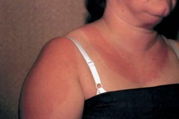
This photo shows solar urticaria in a woman who had been wearing a tank top. These hives appear within minutes of sun exposure.
Solar urticaria can be classified based on the component of the UV spectrum (UVA, UVB, and visible light) that causes lesions. If necessary, patients can be tested by exposing part of the skin to natural light or artificial light at particular wavelengths (phototesting).
Treatment of solar urticaria can be difficult and may include H1 blockers, topical glucocorticoids, and sunscreens (1). If standard treatment fails, desensitization with narrow-band UVB or PUVA (psoralen plus ultraviolet A), may be tried. Omalizumab (anti-IgE therapy) has been successful in a small number of patients. The disorder is chronic and can wax and wane over years.). If standard treatment fails, desensitization with narrow-band UVB or PUVA (psoralen plus ultraviolet A), may be tried. Omalizumab (anti-IgE therapy) has been successful in a small number of patients. The disorder is chronic and can wax and wane over years.
Polymorphous light eruption
Polymorphous light eruption is the most common photosensitive reaction to UV radiation and sometimes visible light (2). It does not seem to be associated with systemic disease or medications. It is considered an immunologically mediated photodermatosis, representing a delayed-type hypersensitivity (type IV hypersensitivity) reaction to unidentified skin antigens that are activated by sunlight (3). Pathogenesis likely involves a combination of failure of normal UV-induced immunosuppression, altered immune cell infiltration, and abnormal cytokine responses such as cutaneous IL-36 upregulation. Genetic factors (eg, polymorphisms in NOD-2, TLR-5) may contribute to susceptibility (4), A positive family history in some patients suggests a genetic risk factor.
Eruptions appear on sun-exposed areas, usually 30 minutes to several hours after exposure; however, sometimes eruptions may not appear for up to several days. Lesions are pruritic, erythematous, and often papular but may be papulovesicular or plaquelike. They are more common among women and people from northern climates when first exposed to spring or summer sun than among those exposed to sun year-round.

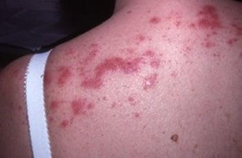
This photo shows erythematous papules and plaques on the upper trunk.
The diagnosis of polymorphous light eruption is made by history, skin findings on physical examination, and exclusion of other photosensitivity disorders. Diagnosis sometimes requires reproduction of the lesions with phototesting when the patient is not using any potentially photosensitizing medications.
The treatment of polymorphous light eruption is guided by the severity of clinical manifestations. Often, lesions are self-limited and spontaneously improve over the course of a few days to weeks as summer progresses. Mild to moderate eruptions are treated with topical glucocorticoids. More severely affected patients may benefit from desensitization in early spring by graduated exposure to UV radiation with low-dose narrowband UVB (311 to 313 nm) phototherapy (see Phototherapy). Patients with disabling disease may require a course of oral immunosuppressive therapy such as prednisone, azathioprine, cyclosporine, or hydroxychloroquine. ). Patients with disabling disease may require a course of oral immunosuppressive therapy such as prednisone, azathioprine, cyclosporine, or hydroxychloroquine.
Preventive measures include using a broad-spectrum sunscreen and avoiding sun exposure. Some evidence suggests that antioxidants such as the dietary supplement Polypodium leucotomos, a natural tropical fern extract, may help prevent polymorphous light eruption, but further studies are needed (5).
Medication or chemical-induced photosensitivity
Over 100 substances, ingested or applied topically, are known to predispose to cutaneous reactions after sun exposure. A limited number are responsible for most reactions (see table ).
Reactions are divided into phototoxicity and photoallergy. Phototesting can help confirm the diagnosis. Treatment for chemical photosensitivity involves topical glucocorticoids and avoidance of the causative substance.

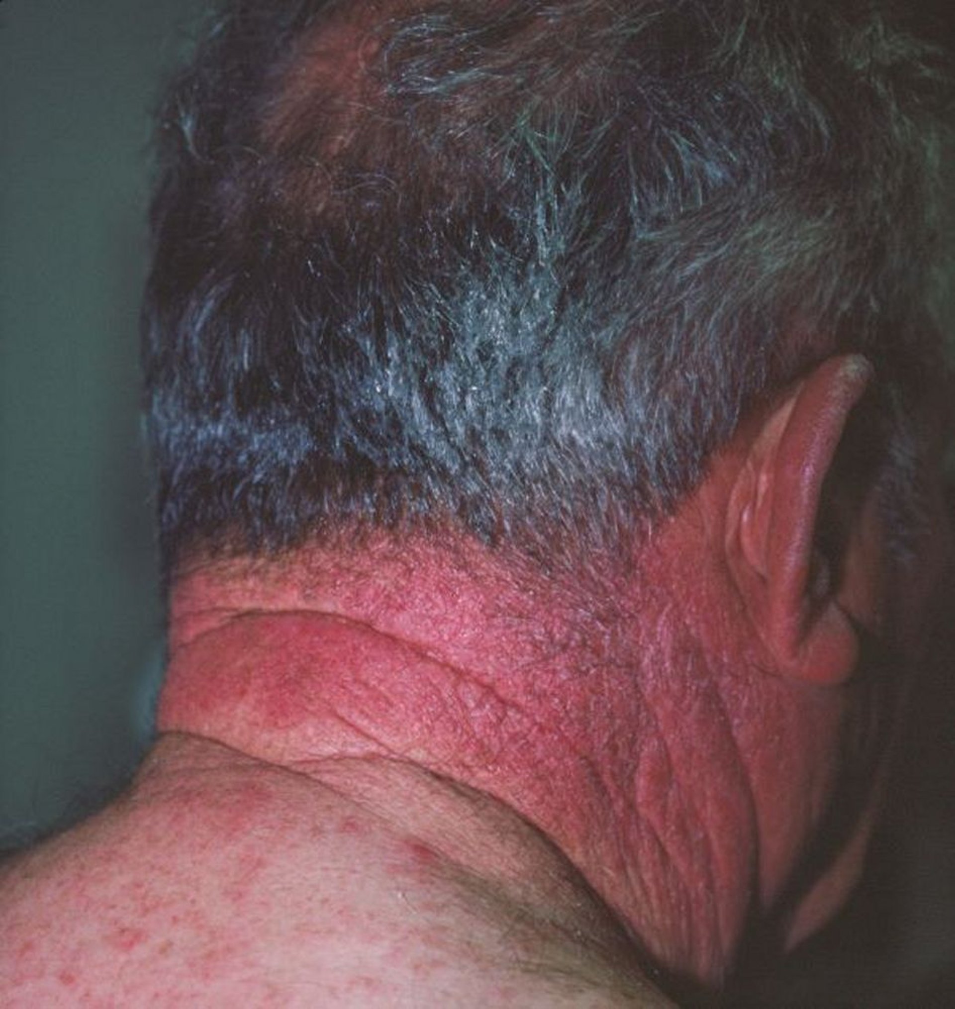
This image shows erythema caused by chemical photosensitivity to hydrochlorothiazide. The skin changes are similar to a sunburn.
Image courtesy of Karen McKoy, MD.
In phototoxicity, light-absorbing compounds generate free radicals and inflammatory mediators, directly causing tissue damage that manifests as pain and erythema (like sunburn) (6). It is not immune mediated. This reaction does not require prior sun exposure and can appear in any person, although reaction severity is highly variable. Typical causes of phototoxic reactions include topical (eg, perfumes, coal tar, furocoumarin-containing plants [such as limes, celery, and parsley], 5-fluorouracil, medications used for photodynamic therapy) or ingested (eg, tetracyclines, thiazides) agents. Phototoxic reactions do not involve non–sun-exposed skin.). It is not immune mediated. This reaction does not require prior sun exposure and can appear in any person, although reaction severity is highly variable. Typical causes of phototoxic reactions include topical (eg, perfumes, coal tar, furocoumarin-containing plants [such as limes, celery, and parsley], 5-fluorouracil, medications used for photodynamic therapy) or ingested (eg, tetracyclines, thiazides) agents. Phototoxic reactions do not involve non–sun-exposed skin.
Photoallergy is a type IV (cell-mediated) immune response (6). Light absorption causes structural changes in the medication or substance, allowing it to bind to tissue protein and function as a hapten, making the complex allergenic. Prior exposure to the allergen is required. The reaction is usually eczematous, with erythema, scaling, pruritus, and sometimes vesicles. Typical causes of photoallergic reactions include aftershave lotions, sunscreens, and sulfonamides. The reaction may extend to non–sun-exposed skin.
Photoallergy occurs less frequently than phototoxicity.
Some Medications or Chemicals That Cause Cutaneous Photosensitivity
Category | Specific Medication or Chemical |
|---|---|
Acne medications | IsotretinoinIsotretinoin |
Analgesics | Nonsteroidal anti-inflammatory drugs (especially piroxicam and ketoprofen)Nonsteroidal anti-inflammatory drugs (especially piroxicam and ketoprofen) |
Antibiotics | Quinolones Sulfonamides Tetracyclines (eg, doxycycline)Tetracyclines (eg, doxycycline) TrimethoprimTrimethoprim |
Antidepressants | Tricyclics |
Antifungals | GriseofulvinGriseofulvin VoriconazoleVoriconazole |
Antihyperglycemics | Sulfonylureas |
Antimalarials | Chloroquine and hydroxychloroquineChloroquine and hydroxychloroquine QuinineQuinine |
Antipsychotics | Phenothiazines (eg, chlorpromazine)Phenothiazines (eg, chlorpromazine) |
Anxiolytics | AlprazolamAlprazolam |
ChlordiazepoxideChlordiazepoxide | |
Chemotherapy medications | DacarbazineDacarbazine Fluorouracil (systemic or topical)Fluorouracil (systemic or topical) MethotrexateMethotrexate VinblastineVinblastine VemurafenibVemurafenib |
Diuretics | FurosemideFurosemide Thiazides |
Cardiac medications | AmiodaroneAmiodarone |
QuinidineQuinidine | |
Topical preparations* | Antibacterials (eg, chlorhexidine, hexachlorophene)Antibacterials (eg, chlorhexidine, hexachlorophene) Coal tarCoal tar Fragrances Furocoumarin-containing plants (eg, limes, celery, parsley) Sunscreens (by photoallergic contact dermatitis) |
* There are many topical preparations that trigger photosensitivity. The specific substances listed are examples. | |
Photosensitivity due to genetic disorders
Several genetic disorders are associated with photosensitive phenotypes. These disorders result in extreme photosensitivity due to impaired ability to repair UV-induced DNA damage. One key example is xeroderma pigmentosum (XP), which is a rare autosomal recessive disorder caused by defects in DNA (specifically nucleotide excision) repair genes, resulting in extreme photosensitivity and dramatically increased risk of UV radiation-induced skin cancers. Several genes have been implicated. The defective DNA repair leads to accumulation of phototoxic DNA damage, causing patients to have a 10,000-fold increased risk of nonmelanoma skin cancer and up to a 2000-fold increased risk of melanoma compared to the general population (7). Defects in DNA repair may also lead to progressive neurologic degeneration and ocular defects in some patients.
Patients with XP typically develop severe sunburns even after minimal sun exposure in infancy or early childhood, followed by freckling in sun-exposed areas (7). Most patients develop their first skin cancer before age 10.
Other genetic disorders causing photosensitivity include Cockayne syndrome (premature aging; photosensitivity; growth failure; progressive neurological, visual, and hearing deterioration) (8) and trichothiodystrophy (brittle, sulfur-deficient hair; extreme photosensitivity; intellectual disability; growth faltering; recurrent infections) (9).
Photosensitivity in systemic lupus erythematosus (SLE)
Photosensitivity in systemic lupus erythematosus (SLE) results from aberrant immune responses to exposure to UV radiation, which triggers inflammatory skin lesions and potentially systemic disease flares. UV radiation induces excessive apoptosis in the keratinocytes of SLE patients. When these apoptotic cells cannot be adequately cleared, they undergo secondary necrosis and release proinflammatory compounds and autoantigens that perpetuate inflammation (10).
Clinical features of UV-induced photosensitivity in SLE include (11):
Acute cutaneous lupus erythematosus with a photosensitive malar or "butterfly" rash (up to 50% of patients)
Subacute cutaneous lupus erythematosus (SCLE) with a photosensitive annular or papulosquamous rashes (10 to 15% of patients)
Chronic cutaneous lupus erythematosus includes discoid lupus erythematosus (DLE), which causes annular scarring plaques, often on sun-exposed areas
For more information, see also Cutaneous Lupus Erythematosus and Diagnosis of SLE.
Phytophotodermatitis
Phytophotodermatitis refers to phototoxic cutaneous reactions that occur when skin is exposed to furocoumarins (psoralens) from certain plants and subsequently exposed to ultraviolet A (UVA) radiation (12). Common sources include citrus fruits (particularly limes), kiwi, celery, and giant hogweed, although several others have been implicated (13). Phytophotodermatitis classically presents with erythematous macules or patches, followed by the formation of vesicles or bullae resembling severe burns. These lesions may sometimes be followed by characteristic linear or bizarre-patterned hyperpigmentation that can persist for weeks to months. Diagnosis is primarily clinical, through careful elicitation of history of relevant plant contact and sun exposure.


This photo shows an erythematous eruption with blistering on a person's forearm resulting from exposure to phytochemicals from a fig tree followed by exposure to sunlight (phytophotodermatitis).
Berto/stock.adobe.com
General references
1. Engler Markowitz M, Noyman Y, Khanimov I, et al. Efficacy of Therapies for Solar Urticaria: A Systematic Review and Meta-Analysis. J Clin Med. 2025;14(16):5736. Published 2025 Aug 13. doi:10.3390/jcm14165736
2. Morison WL. Clinical practice. Photosensitivity. N Engl J Med. 2004 Mar 11;350(11):1111-7. doi: 10.1056/NEJMcp022558
3. Kadurina M, Kazandjieva J, Bocheva G. Immunopathogenesis and management of polymorphic light eruption. Dermatol Ther. 2021 Nov;34(6):e15167. doi: 10.1111/dth.15167
4. Kurz B, Arndt S, Unger P, et al. Association of polymorphous light eruption with NOD-2 and TLR-5 gene polymorphisms. J Eur Acad Dermatol Venereol. 2022 Nov;36(11):2172-2180. doi: 10.1111/jdv.18364
5. Nestor MS, Berman B, Swenson N: Safety and efficacy of oral Polypodium leucotomos extract in healthy adult subjects. J Clin Aesthet Dermatol 8(2):19–23, 2015
6. Epstein JH. Phototoxicity and photoallergy. Semin Cutan Med Surg. 1999 Dec;18(4):274-84. doi: 10.1016/s1085-5629(99)80026-
7. Medline Plus. Xeroderma Pigmentosum. Accessed December 3, 2025.
8. Medline Plus. Cockayne Syndrome. Accessed December 3, 2025.
9. Medline Plus. Trichothiodystrophy. Accessed December 3, 2025.
10. Kuhn A, Wenzel J, Weyd H. Photosensitivity, apoptosis, and cytokines in the pathogenesis of lupus erythematosus: a critical review. Clin Rev Allergy Immunol. 2014 Oct;47(2):148-62. doi: 10.1007/s12016-013-8403-x
11. Siegel CH, Sammaritano LR. Systemic Lupus Erythematosus: A Review. JAMA. 2024 May 7;331(17):1480-1491. doi: 10.1001/jama.2024.2315. Erratum in: JAMA. 2024 Jun 25;331(24):2136. doi: 10.1001/jama.2024.10468
12. Maniam G, Light KM, Wilson J. Margarita Burn: Recognition and Treatment of Phytophotodermatitis. J Am Board Fam Med. 2021 Mar-Apr;34(2):398-401. doi: 10.3122/jabfm.2021.02.20038
13. Petit R, Izambart J, Guillou M, et al. A Review of Phototoxic Plants, Their Phototoxic Metabolites, and Possible Developments as Photosensitizers. Chem Biodivers. 2024 Feb;21(2):e202300494. doi: 10.1002/cbdv.202300494
Drug Information for the Topic


