


Growths can originate in any type of tissue in and around the mouth, including connective tissues, bone, muscle, and nerve. Most commonly, growths form on the lips, the sides of the tongue, the floor of the mouth, and the soft palate. Some growths cause pain or irritation. Growths may be noticed by the patient or discovered during examination.
(See also Evaluation of the Dental Patient, Oral Squamous Cell Carcinoma, Oropharyngeal Squamous Cell Carcinoma, and Candidiasis [Mucocutaneous].)
Etiology of Oral Growths
Oral growths can be
Benign
Premalignant (dysplastic)
Malignant
Benign oral growths
Most oral growths are benign (1); there are numerous types.

This photo shows an oral growth caused by chronic irritation. In this case, a fibroma on the inside of the cheek formed due to rubbing from orthodontic braces.
This photo shows an oral growth caused by chronic irritation. In this case, a fibroma on the inside of the cheek formed
DR P. MARAZZI/SCIENCE PHOTO LIBRARY
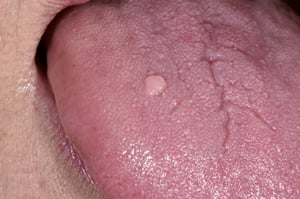
This example is a common oral wart (verruca vulgaris), appearing as a raised, rounded, flesh-colored lesion on the surface of the tongue. Common warts often arise in children but can develop later. It is often difficult to distinguish squamous papilloma from verruca vulgaris.
This example is a common oral wart (verruca vulgaris), appearing as a raised, rounded, flesh-colored lesion on the surf
DR P. MARAZZI/SCIENCE PHOTO LIBRARY
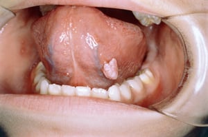
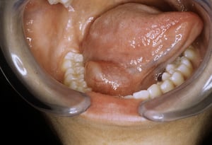
This photo shows a wart appearing as a pedunculated papilloma (exophytic growth, cauliflower-like) on the underside of the tongue. HPV can be a cause.
This photo shows a wart appearing as a pedunculated papilloma (exophytic growth, cauliflower-like) on the underside of
CLINICA CLAROS/SCIENCE PHOTO LIBRARY

Creamy white patches are seen inside the mouth and may bleed when scraped off. This finding is typical of thrush, which is caused by infection with Candida.
Creamy white patches are seen inside the mouth and may bleed when scraped off. This finding is typical of thrush, which
Image provided by Thomas Habif, MD.

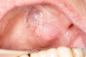
This photo shows a close-up of a ranula (center) in a person's mouth. Ranulas are mucoceles that occur in the floor of the mouth (generally larger than mucoceles that occur elsewhere in the oral cavity). The source of mucin content in ranulas is usually the sublingual gland (also occasionally from the submandibular gland duct).
This photo shows a close-up of a ranula (center) in a person's mouth. Ranulas are mucoceles that occur in the floor of
CLINICA CLAROS/SCIENCE PHOTO LIBRARY

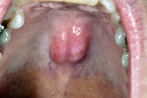
Torus palatinus is a common bony hard mass that occurs along the midline of the hard palate (often bilaterally, as is visible here).
Torus palatinus is a common bony hard mass that occurs along the midline of the hard palate (often bilaterally, as is v
DR P. MARAZZI/SCIENCE PHOTO LIBRARY

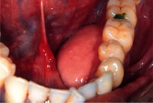
A benign growth of the lingual aspect of the mandible is visible here on the left side of the patient's mouth.
A benign growth of the lingual aspect of the mandible is visible here on the left side of the patient's mouth.
© Springer Science+Business Media
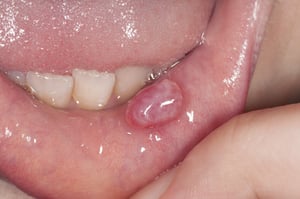
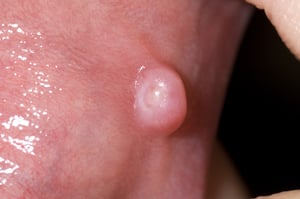
This photo shows a labial mucocele, a benign mass consisting of a swollen sac filled with mucus. Mucoceles are often fluctuant, although some can feel firmer to palpation.
This photo shows a labial mucocele, a benign mass consisting of a swollen sac filled with mucus. Mucoceles are often fl
DR P. MARAZZI/SCIENCE PHOTO LIBRARY

Pleomorphic adenoma is the most common intraoral benign neoplasm. The palatal mucosa is the most common site of occurrence.
Pleomorphic adenoma is the most common intraoral benign neoplasm. The palatal mucosa is the most common site of occurre
DR. P. MARAZZI/SCIENCE PHOTO LIBRARY

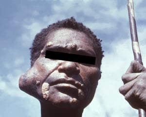
This patient has a large right-sided parotid salivary gland tumor. The whitish papules on the skin are an incidental finding due to yaws (a chronic cutaneous infection caused by Treponema pallidum subspecies pertenue).
This patient has a large right-sided parotid salivary gland tumor. The whitish papules on the skin are an incidental fi
Image courtesy of K. Mae Lennon and Clement Benjamin via the Public Health Image Library of the Centers for Disease Control and Prevention.
This photo shows an oral growth caused by chronic irritation. In this case, a fibroma on the inside of the cheek formed due to rubbing from orthodontic braces.
This photo shows an oral growth caused by chronic irritation. In this case, a fibroma on the inside of the cheek formed
DR P. MARAZZI/SCIENCE PHOTO LIBRARY
This example is a common oral wart (verruca vulgaris), appearing as a raised, rounded, flesh-colored lesion on the surface of the tongue. Common warts often arise in children but can develop later. It is often difficult to distinguish squamous papilloma from verruca vulgaris.
This example is a common oral wart (verruca vulgaris), appearing as a raised, rounded, flesh-colored lesion on the surf
DR P. MARAZZI/SCIENCE PHOTO LIBRARY
This photo shows a wart appearing as a pedunculated papilloma (exophytic growth, cauliflower-like) on the underside of the tongue. HPV can be a cause.
This photo shows a wart appearing as a pedunculated papilloma (exophytic growth, cauliflower-like) on the underside of
CLINICA CLAROS/SCIENCE PHOTO LIBRARY
Creamy white patches are seen inside the mouth and may bleed when scraped off. This finding is typical of thrush, which is caused by infection with Candida.
Creamy white patches are seen inside the mouth and may bleed when scraped off. This finding is typical of thrush, which
Image provided by Thomas Habif, MD.
This photo shows a close-up of a ranula (center) in a person's mouth. Ranulas are mucoceles that occur in the floor of the mouth (generally larger than mucoceles that occur elsewhere in the oral cavity). The source of mucin content in ranulas is usually the sublingual gland (also occasionally from the submandibular gland duct).
This photo shows a close-up of a ranula (center) in a person's mouth. Ranulas are mucoceles that occur in the floor of
CLINICA CLAROS/SCIENCE PHOTO LIBRARY
Torus palatinus is a common bony hard mass that occurs along the midline of the hard palate (often bilaterally, as is visible here).
Torus palatinus is a common bony hard mass that occurs along the midline of the hard palate (often bilaterally, as is v
DR P. MARAZZI/SCIENCE PHOTO LIBRARY
A benign growth of the lingual aspect of the mandible is visible here on the left side of the patient's mouth.
A benign growth of the lingual aspect of the mandible is visible here on the left side of the patient's mouth.
© Springer Science+Business Media
This photo shows a labial mucocele, a benign mass consisting of a swollen sac filled with mucus. Mucoceles are often fluctuant, although some can feel firmer to palpation.
This photo shows a labial mucocele, a benign mass consisting of a swollen sac filled with mucus. Mucoceles are often fl
DR P. MARAZZI/SCIENCE PHOTO LIBRARY
Pleomorphic adenoma is the most common intraoral benign neoplasm. The palatal mucosa is the most common site of occurrence.
Pleomorphic adenoma is the most common intraoral benign neoplasm. The palatal mucosa is the most common site of occurre
DR. P. MARAZZI/SCIENCE PHOTO LIBRARY
This patient has a large right-sided parotid salivary gland tumor. The whitish papules on the skin are an incidental finding due to yaws (a chronic cutaneous infection caused by Treponema pallidum subspecies pertenue).
This patient has a large right-sided parotid salivary gland tumor. The whitish papules on the skin are an incidental fi
Image courtesy of K. Mae Lennon and Clement Benjamin via the Public Health Image Library of the Centers for Disease Control and Prevention.
Chronic irritation can cause a persistent lump or raised area on the gingiva. Benign growths due to irritation are relatively common and, if necessary, can be removed by surgery. Benign growths on the gingiva can sometimes reappear because the irritant remains. Occasionally such irritation, particularly if it persists over a long period of time, can lead to premalignant or malignant changes.
Warts may occur in the mouth (oral squamous papillomas). Ordinary warts (verrucae vulgaris) can infect the mouth if a person sucks or chews one that is growing on a finger. Genital warts, caused by human papillomavirus infection (HPV), may also occur in the oral cavity when transmitted through oral sex. Oral warts may regress spontaneously but slowly (over months or years). Typical treatment is complete surgical excision when possible.
Oral candidiasis (thrush) often appears as white, cheesy plaques that stick tightly to the mucous membranes and leave red erosions when wiped off. Thrush is most common among patients with diabetes or immunocompromise and among those who are taking antibiotics.
Cysts of many kinds cause jaw pain and swelling. Often they are associated with an impacted wisdom tooth and can destroy considerable areas of the mandible as they expand (2). Certain types of cysts are more likely to recur after surgical removal. Various types of cysts may develop in the floor of the mouth. Often, these cysts are surgically removed because they make swallowing uncomfortable or because they are unattractive.
Mucoceles (mucus retention cysts and ranulas) are painless, benign, intraoral swellings due to cystic or pseudocystic accumulations of salivary gland mucus (3). They are often traumatic in origin. By far the most common lesion, mucoceles most often occur inside the lateral lower lip and often have a bluish translucent color due to presence of spilled mucin under the mucosa. They are usually the result of accidentally biting the (lower) lip and occur when salivary flow from a minor salivary gland is obstructed. Most mucoceles disappear in 1 or 2 weeks. Ranulas are large, usually bluish mucoceles on the floor of the mouth. Treatment is surgical excision.
A torus is a rounded projection of bone that forms in the midline of the hard palate (torus palatinus) or on the inner aspect of the mandible (torus mandibularis) (4). This osseous growth is common and benign. Even a large growth can be left alone unless it gets traumatized during eating or the person needs a denture that covers the area.
Oral osteomas are benign, slow-growing tumors of mature compact or cancellous bone occurring most commonly in the mandible and less commonly in the maxilla. They may resemble torus lesions. Multiple oral osteomas warrant evaluation for Gardner syndrome, which is a type of familial adenomatous polyposis (5).
Keratoacanthomas are growths that form on the lips and other sun-exposed areas, such as the face, forearms, and hands. A keratoacanthoma usually reaches its full size of about 1 to 3 cm or more in diameter within 1 or 2 months, then begins to shrink after another few months and may eventually disappear without treatment. Some pathologists consider keratoacanthoma an incipient variant of squamous cell carcinoma (6). As such, growths that do not resolve on their own should be excised and biopsied (6).
Odontomas are overgrowths of tooth-forming cells that look like small, misshapen extra teeth. In children, they may get in the way of normal tooth eruption. In adults, they may push teeth out of alignment. Large odontomas may also cause enlargement of the maxilla or mandible. They are usually removed surgically.
Salivary gland tumors are mostly benign, slow-growing, and painless (7). They usually occur as a single, soft, movable lump beneath normal-appearing skin or under the oral mucosa. Occasionally, when hollow and fluid-filled, they are firm. The most common type is a pleomorphic adenoma (mixed-type tumor) and it occurs mainly in women > age 40. Pleomorphic adenomas can become malignant and are removed surgically. Unless completely removed, this type of tumor is likely to recur. Other types of benign tumors are also removed surgically but are much less likely to become malignant or recur.
Oral potentially malignant disorders (premalignant [dysplastic] changes)
White, red, or mixed white-red areas that are not easily wiped away, persist for > 2 weeks, and are not definable as some other condition may be dysplastic and indicative of what are defined as oral potentially malignant disorders (OPMD) (8). The same risk factors are involved in dysplastic changes as well as in malignant growths, and dysplastic changes may become malignant if not removed.


This photo shows a thick plaque of leukoplakia extending widely over the dorsum of the tongue.
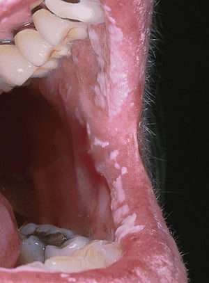
Leukoplakia is a flat white spot that may develop when the oral mucosa is irritated for a long period. The term leukoplakia is used when a white lesion on the oral mucosa cannot be characterized as any other definable lesion. The irritated spot appears white because it has a thickened layer of keratin, which normally is less abundant in the oral mucosa. Factors often associated with the development of idiopathic oral leukoplakia include tobacco use; alcohol consumption; deficiencies of vitamins C, B12, B6, B3; and endocrine disturbances. Leukoplakia that does not resolve spontaneously must be biopsied because it can progress to carcinoma (9).
Proliferative verrucous leukoplakia is an aggressive subtype of leukoplakia that typically involves the gingiva. It often becomes dysplastic and transforms into squamous cell carcinoma and has a higher transformation rate than conventional oral leukoplakia (10). It predominantly affects older females, while leukoplakia more commonly affects males.

Leukoplakia is a general term that can describe white hyperkeratotic plaques that develop in the mouth. The majority prove to be benign. However, in this image, squamous cell carcinoma is present in 1 of the leukoplakic lesions on the ventral surface of the tongue (arrow).
Erythroplakia is a red and flat or worn-away area that results when the oral mucosa thins. The area appears red because the underlying capillaries are more visible. Erythroplakia is a much more ominous predictor of oral cancer than leukoplakia.


Erythroplakia is a general term that can describe red, flat, or eroded velvety lesions that develop in the mouth. In this image, an exophytic squamous cell carcinoma on the tongue is surrounded by a margin of erythroplakia.
Mixed lesions show intermixed areas of leukoplakia and erythroplakia and also may be precursors of cancer.
Oral cancer
People who use tobacco, alcohol, or both are at much greater risk of oral cancer (11). For people who use chewing tobacco and snuff, the insides of the cheeks and lips are common sites. In other people, the most common sites for cancer include the lateral borders of the tongue, the floor of the mouth, and the oropharynx. Human papillomavirus (HPV) infection , especially with type 16, is a risk factor for oral cancer, primarily in the tonsils and at the base of the tongue (12). Rarely, cancers found in the oral region have metastasized from the lungs, breast, or prostate.
Oral cancer can have many different appearances but typically resembles dysplastic lesions (eg, white, red, or mixed white-red areas that are not easily wiped away).
Etiology references
1. Esmeili T, Lozada-Nur F, Epstein J. Common benign oral soft tissue masses. Dent Clin North Am. 2005;49(1):223-x. doi:10.1016/j.cden.2004.07.001
2. Rajendra Santosh AB. Odontogenic Cysts. Dent Clin North Am. 2020;64(1):105-119. doi:10.1016/j.cden.2019.08.002
3. Bowers EMR, Schaitkin B. Management of Mucoceles, Sialoceles, and Ranulas. Otolaryngol Clin North Am. 2021;54(3):543-551. doi:10.1016/j.otc.2021.03.002
4. Loukas M, Hulsberg P, Tubbs RS, et al. The tori of the mouth and ear: a review. Clin Anat. 2013;26(8):953-960. doi:10.1002/ca.22264
5. Gupta B, Chaudhari MA, Sohi HK, et al. Oral and Maxillofacial Manifestations of Gardner Syndrome: A Literature Analysis. J Craniofac Surg. Published online July 7, 2025. doi:10.1097/SCS.0000000000011639
6. Kwiek B, Schwartz RA. Keratoacanthoma (KA): An update and review. J Am Acad Dermatol. 74(6):1220-1233, 2016. doi: 10.1016/j.jaad.2015.11.033
7. Vamesu S, Ursica OA, Gurita AM, et al. A retrospective study of nonneoplastic and neoplastic disorders of the salivary glands. Medicine (Baltimore). 2023;102(42):e35751. doi:10.1097/MD.0000000000035751
8. Warnakulasuriya S, Kujan O, Aguirre-Urizar JM, et al. Oral potentially malignant disorders: A consensus report from an international seminar on nomenclature and classification, convened by the WHO Collaborating Centre for Oral Cancer. Oral Dis. 2021;27(8):1862-1880. doi:10.1111/odi.13704
9. Pimenta-Barros LA, Ramos-García P, González-Moles MÁ, Aguirre-Urizar JM, Warnakulasuriya S. Malignant transformation of oral leukoplakia: Systematic review and comprehensive meta-analysis. Oral Dis. 2025;31(1):69-80. doi:10.1111/odi.15140
10. Müller S. Oral epithelial dysplasia, atypical verrucous lesions and oral potentially malignant disorders: focus on histopathology. Oral Surg Oral Med Oral Pathol Oral Radiol. 2018;125(6):591-602. doi:10.1016/j.oooo.2018.02.012
11. Peres MA, Macpherson LMD, Weyant RJ, et al. Oral diseases: a global public health challenge. Lancet. 2019;394(10194):249-260. doi:10.1016/S0140-6736(19)31146-8
12. Pringle GA. The role of human papillomavirus in oral disease. Dent Clin North Am. 2014;58(2):385-399. doi:10.1016/j.cden.2013.12.008
Evaluation of Oral Growths
History
History of present illness includes questions about how long the growth has been present, whether it is painful, and whether there has been any injury to the area (eg, biting a cheek, scraping by a sharp tooth edge or dental restoration). Patients are asked about symptoms of systemic illness, particularly weight loss and malaise.
Past medical history should seek risk factors for candidiasis, including recent antibiotic use, diabetes, and HIV infection (or risk factors for HIV). The amount and duration of use of alcohol and tobacco are noted.
Physical examination
The physical examination focuses on the mouth and neck, inspecting and palpating all areas of the mouth and throat, including under the tongue. The neck is palpated for lymphadenopathy, which suggests possible cancer or chronic infection.
Red flags
The following findings are of particular concern:
Weight loss
Neck mass
Persistent sore throat
Difficulty swallowing
Interpretation of findings
The main concern is to not mistake an oral cancer or dysplastic lesion for a benign disorder. Clinicians should maintain a high degree of suspicion and refer the patient for biopsy if the lesion does not resolve in a few weeks. Additionally, any lesion that arouses suspicions of dysplasia (eg, nonhomogeneous in appearance, rapid growth, friability) should be referred immediately for biopsy.
Testing
Suspected candidiasis can be confirmed by finding yeast and pseudohyphae in 10% potassium hydroxide wet mounts of scrapings from a lesion. Other acute lesions, particularly those that appear related to local trauma or irritation, may be observed. However, most lesions that have been present for more than a few weeks, and those of unknown duration, should be biopsied because cancer is difficult to exclude clinically.
Treatment of Oral Growths
Treatment depends on the cause, aesthetic and functional impairments, pain, and malignant potential of the diagnosed growth.
Key Points
Most oral growths are benign.
Warts, candidal infections, and repeated trauma are common causes of benign growths.
Use of alcohol and tobacco and oral HPV infection are risk factors for cancer.
Because cancer is difficult to diagnose by inspection, biopsy is often necessary.


