Hypertrophic cardiomyopathy is a group of heart disorders, often genetically inherited, in which the walls of the left ventricle (the lower chamber of the heart) thicken (hypertrophy) and become stiff.
Most cases of hypertrophic cardiomyopathy is caused by an inherited genetic defect.
People experience fainting, chest pain, shortness of breath, and palpitations (awareness of irregular heartbeats).
Doctors suspect the diagnosis based on physical examination findings, but they use echocardiography or magnetic resonance imaging to confirm the diagnosis.
Medications that reduce the force of the heart’s forceful contractions are given.

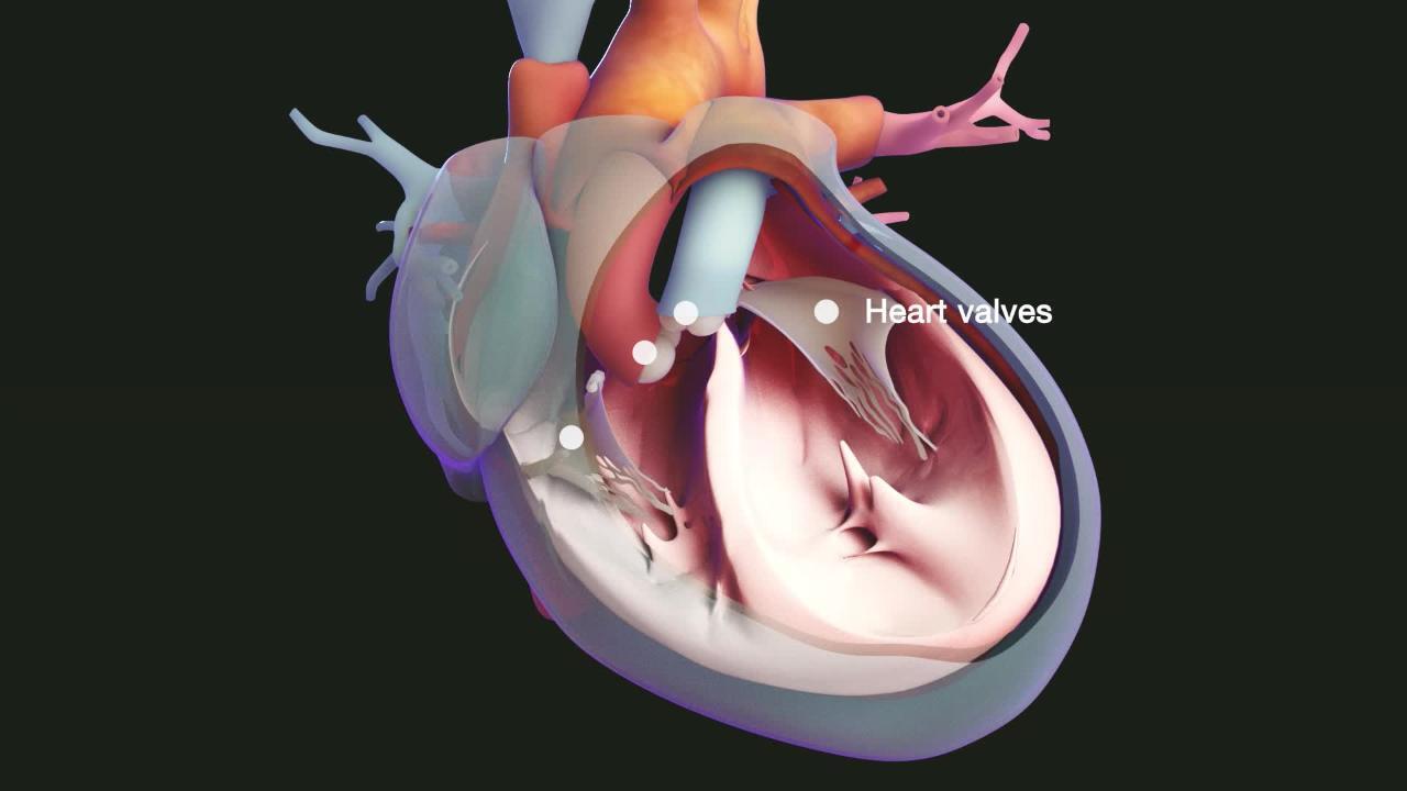
Cardiomyopathy refers to progressive impairment of the structure and function of the muscular walls of the heart chambers. There are 3 main types of cardiomyopathy. In addition to hypertrophic cardiomyopathy, there are dilated cardiomyopathy and restrictive cardiomyopathy (see also Overview of Cardiomyopathy).
The term cardiomyopathy is most often used when a disorder directly affects the heart muscle. Other disorders, such as high blood pressure and abnormal heart valves (such as aortic stenosis), also can eventually cause thickened heart muscle and heart failure.
Hypertrophic cardiomyopathy is a common cause of sudden death in young athletes. About 1 in 200 to 1 in 500 people is affected.


The illustration on the left shows a cutaway view of a normal heart.
The illustration on the right shows hypertrophic cardiomyopathy. Note how the walls of the ventricles are thickened.
© Nucleus Medical Media. All Rights Reserved.
Causes of Hypertrophic Cardiomyopathy
Although other conditions can cause thickening of the heart muscle, hypertrophic cardiomyopathy itself is usually caused by a genetic defect that affects the proteins in the heart muscle. The defect may be a:
Spontaneous genetic mutation
Inherited genetic defect
In some people with hypertrophic cardiomyopathy no genetic defect is identified.
Some other disorders that can cause thickening of the heart muscle are amyloidosis, hypertension, and aortic stenosis. Doctors use other tests to correctly diagnose and treat heart disease caused by those disorders.
Complications
The thick, stiff walls of the ventricles do not relax appropriately to allow the heart chambers to fill with blood. This difficulty becomes more severe when the heart beats quickly (as during exercise) because there is then even less time for the heart to fill. Because the heart does not fill properly, it pumps less blood with each beat and pressures inside the heart rise quickly during exercise.
Sometimes the thickened heart walls also interfere with blood flow out of the heart. This variation is called hypertrophic obstructive cardiomyopathy.
In addition, the mitral valve (the valve that opens between the left atrium and the left ventricle) may be unable to close normally, resulting in leakage of blood back into the left atrium (mitral regurgitation). This valve leakage and the enlarged ventricular walls typically cause abnormal heart sounds (heart murmurs).
Abnormal heart rhythms can occur, which can cause sudden death.
Symptoms of Hypertrophic Cardiomyopathy
Symptoms are highly variable, but when they occur, they usually develop when people are between ages 30 and 50 years. Symptoms first occur during exercise and can include:
Fainting (syncope)
Chest pain
Shortness of breath
Sensation of irregular heartbeats (palpitations)
Fainting usually occurs suddenly without any warning symptoms. Fainting or even sudden death may be the first sign that a person has this condition.
Diagnosis of Hypertrophic Cardiomyopathy
A doctor's examination
Echocardiography and/or magnetic resonance imaging (MRI) of the heart
Genetic testing
Doctors usually suspect the diagnosis of hypertrophic cardiomyopathy based on the person's symptoms and the results of a physical examination, electrocardiography (ECG), and a chest x-ray. The heart sounds and any murmurs heard through a stethoscope may be helpful.
Athletes should be evaluated for risk factors for hypertrophic cardiomyopathy (among other conditions) that indicate the need for further testing. In some areas, all athletes are screened with electrocardiography as well.
Echocardiography is the best way to confirm the diagnosis of hypertrophic cardiomyopathy, but MRI of the heart is often used because it may provide more detailed information, or may be able to see areas of the heart that are not clearly visible with an echocardiogram. MRI may also be helpful in identifying abnormalities in the heart muscle caused by other disorders.
Cardiac catheterization, an invasive procedure in which a catheter is threaded from a blood vessel in the arm, neck, or leg into the heart, is sometimes done to measure pressures in the heart chambers and the degree to which blood flow from the left ventricle is blocked due to the thickened walls.
Doctors may also monitor the person with ambulatory electrocardiography for 24 to 48 hours to detect abnormal heart rhythms. This testing is often repeated regularly to ensure abnormal heart rhythms are detected promptly.
Because hypertrophic cardiomyopathy is frequently caused by a gene mutation, genetic testing is often done to try and identify the genetic mutation causing the disease.
Family members of a person with hypertrophic cardiomyopathy should be tested for it as well. This may involve an echocardiogram every several years, or genetic testing, or a combination of both.
Treatment of Hypertrophic Cardiomyopathy
Medications such as a beta-blocker and/or a calcium channel blocker or a cardiac myosin inhibitor
Sometimes a procedure to improve blood flow
Sometimes an implantable cardioverter-defibrillator
There is no current treatment to correct the genetic cause of hypertrophic cardiomyopathy, although some gene therapy treatments are being studied. Treatment of heart muscle thickening caused by other disorders is directed toward those disorders.
Treatment of hypertrophic cardiomyopathy is aimed primarily at reducing the heart’s resistance to filling with blood between heartbeats, and, in some cases, at preventing sudden death.
Staying well hydrated, so that the amount of blood circulating is adequate to fill the ventricles, is one treatment strategy for hypertrophic cardiomyopathy. Sometimes, people are encouraged to modify or restrict their exercise habits as well.
Medications for hypertrophic cardiomyopathy
Beta-blockers and calcium channel blockers—taken separately or together—are the main treatment. Both reduce the force of heart muscle contraction. As a result, the heart can fill better and, if the thickened muscle was blocking blood flow, blood can flow out of the heart more easily. Also, beta-blockers and calcium channel blockers slow the heart rate, so that the heart has more time to fill. Sometimes, disopyramide, a medication that decreases the strength of heart contractions, is also used.
Other medicines, such as the cardiac myosin inhibitors mavacamten or aficamten, also decrease the contraction of the heart and relieve symptoms and increase exercise tolerance.
Amiodarone or other heart rhythm medicines are sometimes used to treat abnormal heart rhythms.
Myectomy
Surgery to remove some of the thickened heart muscle (myectomy) can improve the flow of blood from the heart, but surgery is done only when symptoms are incapacitating despite medications. Myectomy can relieve symptoms, but it does not reduce the risk of death. When myectomy is done in hospitals that have extensive experience doing the procedure, long-term results are excellent.
Alcohol septal ablation
Alcohol septal ablation (controlled destruction of a small area of heart muscle by injecting it with alcohol) is used in certain people to improve blood flow from the heart because it can be done during cardiac catheterization. Although cardiac catheterization is an invasive procedure in which a catheter is threaded into the heart, it has fewer risks in people who are at high risk of complications if they have heart surgery.
Implantable cardioverter-defibrillator
Some people with hypertrophic cardiomyopathy have especially high risk of developing deadly abnormal heart rhythms which can cause sudden death. Doctors may recommend an implantable cardioverter-defibrillator for these people.
Prognosis for Hypertrophic Cardiomyopathy
About 0.5% of adults with hypertrophic cardiomyopathy die each year. Children with hypertrophic cardiomyopathy are more likely to die than adults.
Death is usually sudden, presumably due to an abnormal heart rhythm. Death due to chronic heart failure is less common.
People who learn that they have inherited this disorder should have genetic counseling when they plan a family because they have a 50% chance of passing this disorder on to their offspring.
More Information
The following English-language resource may be useful. Please note that The Manual is not responsible for the content of this resource.
Drug Information for the Topic