Anal atresia (also called imperforate anus) is a type of birth defect in which the opening of the anus is missing or covered with a thin membrane of skin. This birth defect may be associated with other abnormalities of the rectum, urethra, or vagina.
The cause of anal atresia is not known.
Infants may develop intestinal obstruction.
The diagnosis is based on a physical examination and x-rays.
Surgery is needed to correct the defect.
The anus is the opening at the far end of the digestive tract through which stool leaves the body. The rectum is the pouch of large intestine that holds stool prior to defecation. In anorectal malformations, skin may be covering the area where the anus should be, and the skin may be several centimeters thick or just a thin membrane. The opening to the anus may be narrow or may be missing completely.
Anal atresia is part of a spectrum of anorectal malformations, which can also include narrowing of the anus or rectum, and abnormal connections called fistulas between the anus and the urethra, the area between the urethra and anus (the perineum), the vagina, or rarely the bladder. (See also Overview of Digestive Tract Birth Defects.)
Anal atresia occurs when the intestines do not develop properly while the fetus is growing. It is not known why the intestines do not develop properly.
Anal atresia commonly occurs along with other birth defects, such as defects of the spine, heart, kidneys, and limbs. Affected infants may also have tracheoesophageal fistula and esophageal atresia.
Infants with anal atresia do not defecate normally after birth. Eventually, if the defect is not treated, intestinal obstruction develops. Intestinal obstruction is a blockage that completely stops or seriously impairs the movement of material in the intestines. Symptoms of intestinal obstruction in infants include pain, irritability, vomiting, and a swollen abdomen.
Diagnosis of Anal Atresia
Physical examination
Imaging studies (x-ray and ultrasound)
Doctors often detect the malformation by looking at the anus when they first examine the baby after birth, before symptoms develop.
Using x-rays, a radiologist can see the location of and determine other details about a fistula. Doctors may also do an ultrasound to determine the type of malformation and to look for other defects.
Treatment of Anal Atresia
Surgery
Anal atresia usually requires immediate surgery to create a passage for stool and to close the fistula. Doctors determine whether the infant should undergo a permanent procedure or a temporary one. They base their decision on several factors, including the infant's gender and the presence and location of a fistula.
The temporary procedure is called a colostomy (see figure ). In this procedure, the surgeon makes a hole in the infant's abdominal wall and connects it to the colon to allow stool to flow into a plastic bag on the abdominal wall. The colostomy stays in place until the infant is older and the structures that need to be repaired have grown larger. The colostomy is closed when surgery to fully repair the defect is done.
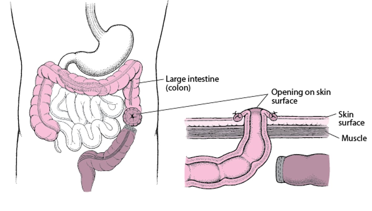
Understanding Colostomy
In a colostomy, the large intestine (colon) is cut. The healthy end of the large intestine, which is before the blockage, is brought to the skin surface through a surgically created opening in the abdominal wall. It is then stitched to the skin of the opening. Stool passes through the opening and into a disposable bag. The colostomy allows the remaining part of the large intestine to rest while the person recovers. After the person recovers from the surgery and the colon heals, the 2 ends can be reattached so that stool can pass normally. |