


Imaging tests of the liver, gallbladder, and biliary tract include ultrasonography, computed tomography (CT), magnetic resonance imaging (MRI), endoscopic retrograde cholangiopancreatography (ERCP), percutaneous transhepatic cholangiography, operative cholangiography, radionuclide scanning, and simple x-rays.
(See also Overview of the Liver and Gallbladder.)
Ultrasound
Ultrasound studies use sound waves to provide images of the liver, gallbladder, and bile ducts. Transabdominal ultrasound is better for detecting structural abnormalities affecting just certain parts of the liver, such as tumors, than for abnormalities that affect the entire liver uniformly, such as cirrhosis (severe scarring of the liver) or steatotic liver disease (excess fat in the liver). It is the least expensive and safest technique for creating images of the gallbladder and bile ducts.
Using ultrasound, a doctor can readily detect gallstones in the gallbladder. Ultrasound of the abdomen can distinguish whether jaundice (a yellowish discoloration of the skin and the whites of the eyes) is caused by obstructed bile ducts or by malfunctioning liver cells. If ultrasound shows bile ducts that are dilated (widened), the cause is typically obstruction. Ultrasound also provides guidance when doctors insert a needle to obtain a tissue sample for biopsy of the liver.
A type of ultrasound called Doppler ultrasound can show blood flowing through the blood vessels of the liver. Doppler ultrasound can detect blockages in the liver's arteries and veins, particularly the portal vein, which brings blood from the intestines to the liver. Doppler ultrasound can also detect the effects of high blood pressure within the portal vein (a condition called portal hypertension). Endoscopic ultrasound uses a tiny probe on the tip of an endoscope that is passed through the mouth into the stomach and the first segment of the small intestine (duodenum), bringing the probe closer to the liver and its surrounding organs.
Computed Tomography
Computed tomography (CT) provides excellent images of the liver and its blood vessels. It is particularly useful for detecting tumors and other masses in the liver. It can also detect collections of pus (abscesses) and some disorders that affect the entire liver uniformly, such as a steatotic liver disease (excess fat in the liver).
Magnetic Resonance Imaging
Magnetic resonance imaging (MRI) can detect certain liver disorders, such as hemochromatosis and steatotic liver disease, that affect all areas of the liver uniformly. MRI shows blood flow, providing information about blood vessel disorders. MRI is also useful for the detection of tumors.
MRI technology can also provide images of the bile ducts and nearby structures, using a technique called magnetic resonance cholangiopancreatography (MRCP). The images produced are as good as those produced by more invasive tests, in which a contrast agent is directly injected into the biliary and pancreatic ducts. Unlike CT, MRI tests do not involve exposure to x-rays, though they are more expensive than CT and take longer.
Endoscopic Retrograde Cholangiopancreatography
Endoscopic retrograde cholangiopancreatography (ERCP) involves passing an endoscope (a flexible viewing tube) through the mouth, esophagus, and stomach into the duodenum. A thin tube is then inserted through the endoscope into the biliary tract. Doctors inject a radiopaque contrast agent through the tube into the biliary tract, and, at the same time, x-rays are taken of the biliary tract and pancreatic duct.
ERCP is occasionally used simply to see the biliary tract structures, although doctors usually prefer MRCP when available because it is just as good and is safer. However, unlike other diagnostic tests, ERCP allows doctors to do biopsies and certain treatments because an endoscope is used during the procedure. For example, with the endoscope, a stone in a bile duct can be removed, or a tube (stent) can be inserted to bypass a bile duct blockage caused by inflammation or cancer. With ERCP, complications (such as inflammation of the pancreas [pancreatitis] or bleeding) can occur. Pancreatitis is the most common complication, occurring about 10% of the time. If a treatment is done during ERCP, complications can occur more often.
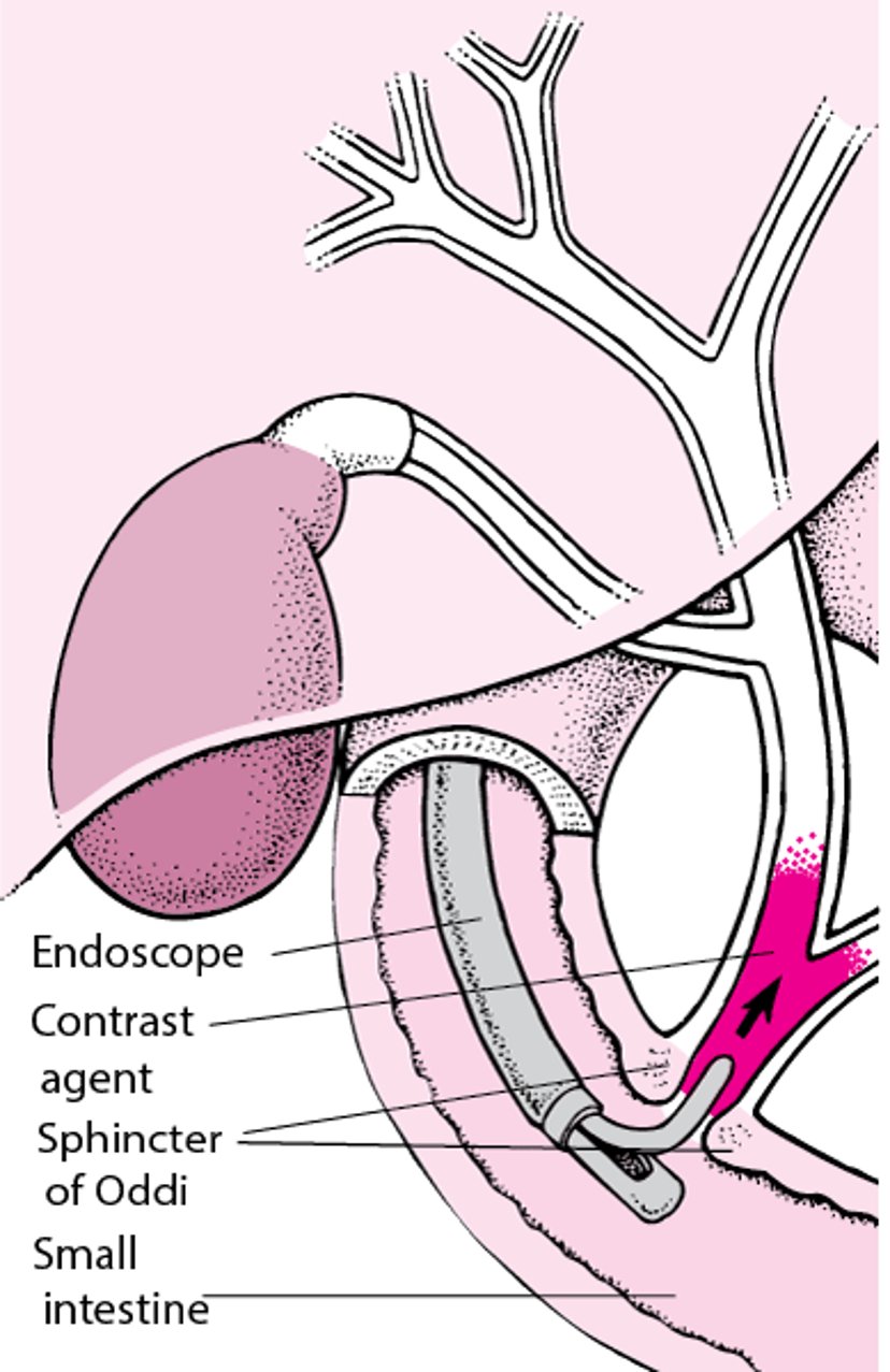
Understanding Endoscopic Retrograde Cholangiopancreatography (ERCP)
In endoscopic retrograde cholangiopancreatography (ERCP), a radiopaque contrast agent is introduced through an endoscope (a flexible viewing tube), which is inserted into the mouth and through the stomach into the duodenum (the first segment of the small intestine). The contrast agent is injected into the biliary tract just past the sphincter of Oddi. The contrast agent then flows back up the biliary tract and often shows the pancreatic ducts. Surgical instruments can also be used with the endoscope, allowing a doctor to remove a stone in a bile duct or insert a tube (stent) to bypass a bile duct blocked by scarring or cancer. |

Percutaneous Transhepatic Cholangiography
Percutaneous transhepatic cholangiography involves inserting a long needle through the skin into the liver and then injecting a radiopaque contrast agent into a bile duct in the liver, using ultrasound for guidance. The x-rays clearly reveal the biliary tract, particularly any blockage within the bile ducts. Like endoscopic retrograde cholangiopancreatography (ERCP), percutaneous transhepatic cholangiography is used more often for treatment or biopsy than to obtain images of the biliary tract. Complications of percutaneous transhepatic cholangiography, such as bleeding and internal damage, make it a less desirable method than ERCP, except in special circumstances.
Operative Cholangiography
Operative cholangiography involves the injection of a radiopaque contrast agent directly into the ducts of the biliary tract during gallbladder surgery. X-rays then reveal clear images of the biliary tract. This test is used only occasionally, when other, less invasive tests do not provide enough information.
Radionuclide (Radioisotope) Scanning
Radionuclide (radioisotope) scanning uses a substance containing a radioactive tracer that, when injected intravenously, collects in a particular organ. The radioactivity is detected by a gamma-ray camera, which is positioned over the upper abdomen and is attached to a computer that generates an image. A liver scan uses a radioactive substance that collects in liver cells.
Cholescintigraphy (hepatobiliary scintigraphy or scan), a type of radionuclide imaging, follows the movement of a radioactive substance as it is secreted from the liver and passes into the gallbladder and through the bile ducts into the duodenum (the first segment of the small intestine). This technique, which is done after the person fasts, can detect a blocked cystic duct (the tube that joins the gallbladder to the major bile duct—see figure View of the Liver and Gallbladder). Such a blockage indicates acute inflammation of the gallbladder (cholecystitis). However, cholescintigraphy is not used as much as it was in the past, now that MRI and MRCP are easily available.
X-rays of the Liver and Biliary Tract
Simple x-rays of the abdomen usually cannot detect disorders of the liver, gallbladder, or biliary tract.


