

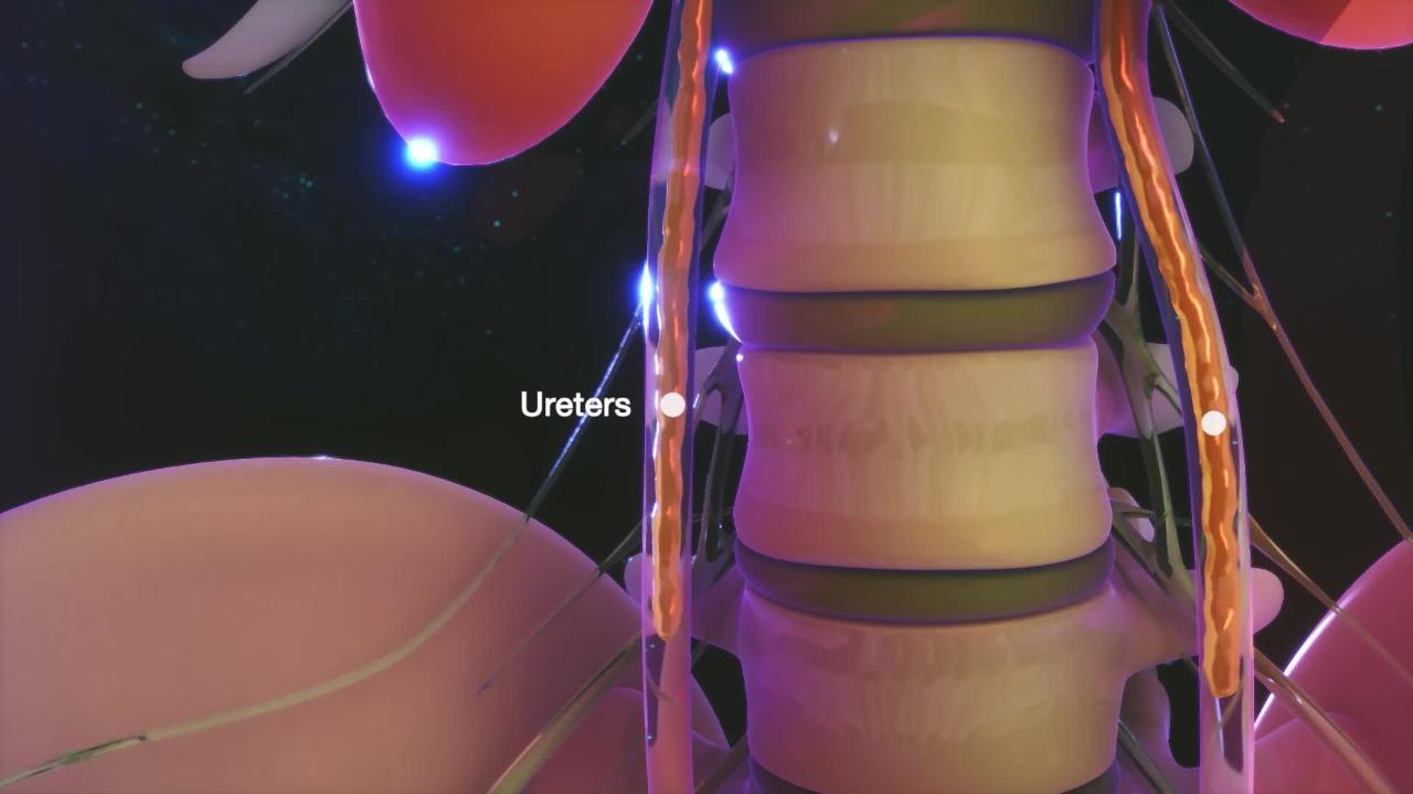
Ureters are the tubes that transport urine from the kidneys (the 2 organs that filter waste from the blood to make urine) to the bladder (the expandable, muscular sac that holds urine). People normally have 2 ureters. One ureter connects the left kidney to the bladder, and the other connects the right kidney to the bladder.
(See also Overview of Kidney and Urinary Tract Birth Defects.)
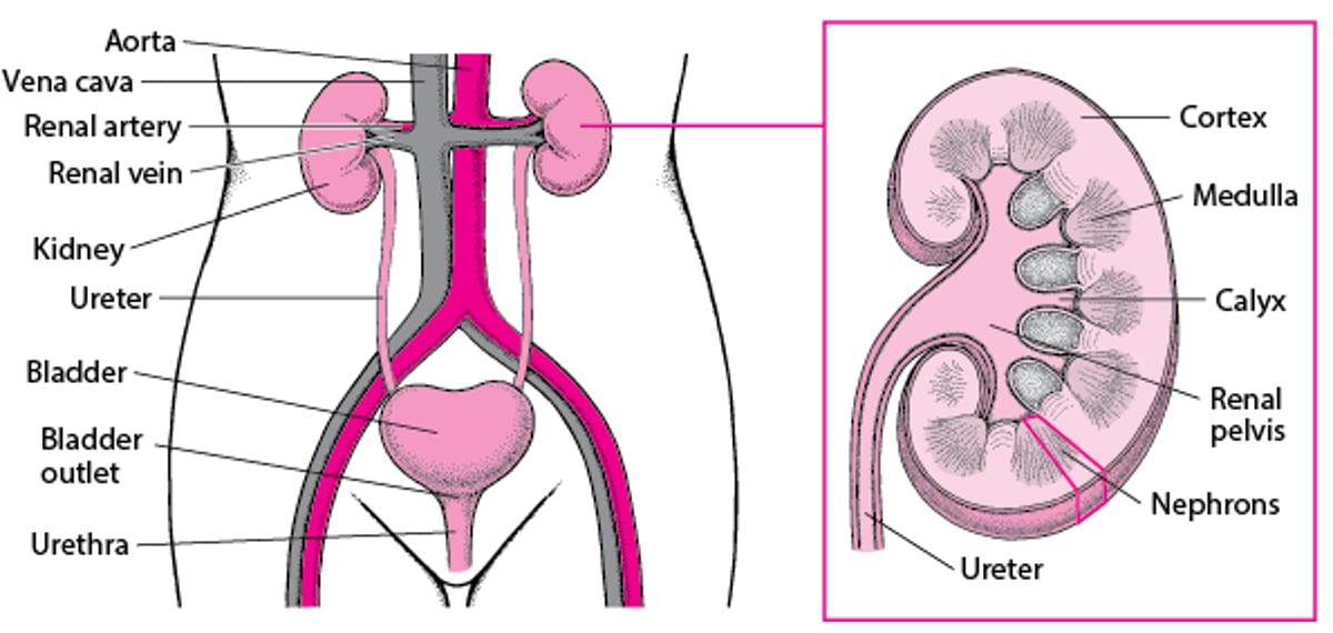
A Look Inside the Urinary Tract

Complications of birth defects of the ureters
There are many types of birth defects of the ureters. Many of these defects
Block or slow the flow of urine
Allow urine to flow backwards from the bladder to the kidneys (urinary reflux)
Any ureter defect that blocks or slows the flow of urine can cause urine to become stagnant, which can result in urinary tract infections (UTIs) or the formation of kidney stones. Blockage of urine flow also can raise the pressure inside the kidneys and damage them over time.
Urinary reflux usually happens when defects involve the junction where a ureter connects to the bladder. Normally the junction allows urine to flow only one way, from the kidneys to the bladder. Defects of the junction can allow urine to flow backward from the bladder into the kidney (urinary reflux). Reflux can affect one side or both sides.
Urinary reflux and/or frequent infections can damage the kidneys and ureters over time. Kidney damage can cause high blood pressure and rarely kidney failure.

Types of Birth Defects of the Ureters
Abnormalities of the ureters include
Extra ureters (duplication abnormalities)
Narrowed or widened ureters
Misplaced ureters
Bulging of the lower end of the ureter into the bladder (ureterocele)
Many children also have birth defects of the kidneys.
Extra ureters
Sometimes while a fetus is forming, the ureters split or duplicate, resulting in 2 ureters coming from a single kidney. Usually the 2 ureters enter the bladder (complete duplication), but sometimes the 2 ureters join together before they enter the bladder (partial duplication).
Many children who have duplicated ureters do not have symptoms. However, sometimes the connections between the duplicated ureters and the bladder are abnormal. Some abnormal connections block urine flow. Other abnormal connections allow urine to flow backward from the bladder into the kidneys (urinary reflux). Both types of abnormal connection increase risk of infection and kidney damage and may require surgery.
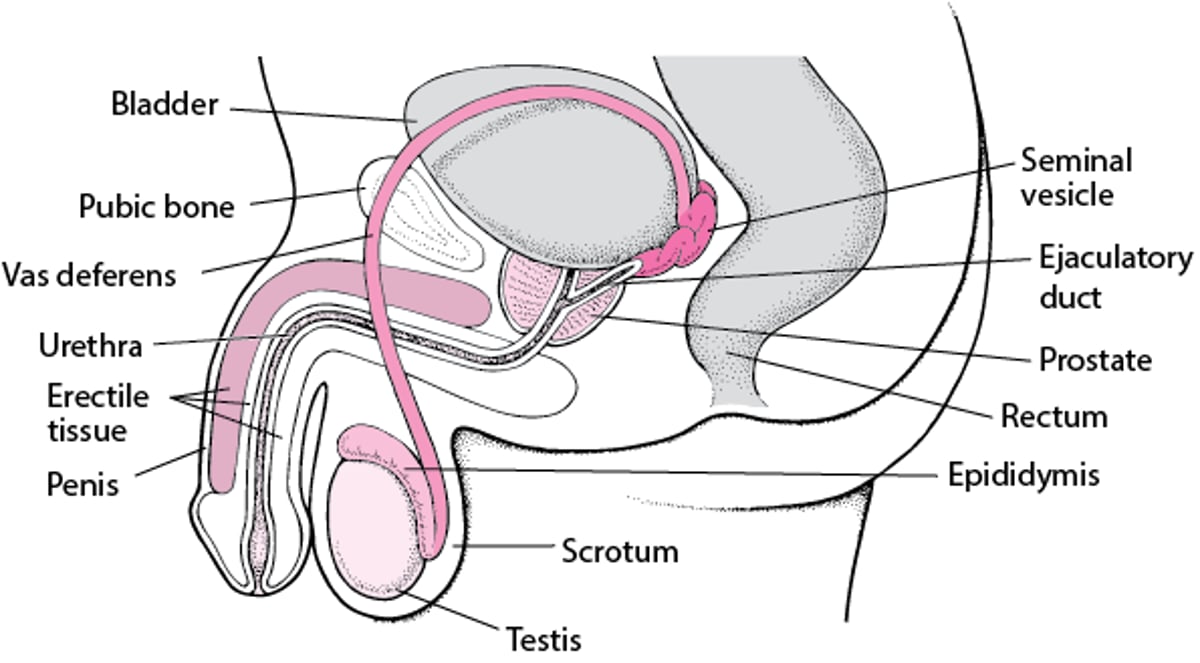
Less often, the duplicated ureter is attached to an area outside the bladder. In girls, the ureter may enter the vagina instead of the bladder, leading to constant dripping of urine from the vagina. In boys, the ureter may enter parts of the male reproductive system such as the vas deferens, seminal vesicles, or the ejaculatory ducts. Boys do not leak urine but may have recurring infections of the affected organ.
Male Reproductive Organs

Narrowed or widened ureters
A narrowed ureter prevents urine from passing normally from the kidney to the bladder. Narrowing usually occurs where the ureter joins the kidney or where the ureter joins the bladder. Narrowed ureters block urine flow, which increases risk of infection, kidney stones, and kidney damage. Narrowings usually lessen as children grow.
A widened ureter can result from an abnormality of the ureter itself or from the bladder being blocked. Widened ureters can allow urine to flow backward from the bladder into the kidneys (urinary reflux), which increases risk of infection and kidney damage.
Misplaced ureters
A misplaced ureter does not properly enter the bladder, which can allow urine to flow backward from the bladder into the kidneys (urinary reflux), which increases risk of infection and kidney damage.
Ureteroceles
A ureterocele is a bulging of the lower end of the ureter into the bladder. They may affect how well the ureter drains. If ureteroceles block urine flow, they increase the risk of infection, kidney stones, and kidney damage.
Diagnosis of Ureter Birth Defects
Prenatal ultrasound
Voiding cystourethrography
Before birth, defects of the ureters are often discovered by doctors during routine prenatal ultrasound.
After birth, if doctors suspect defects of the ureters, they do ultrasound of the kidneys, ureters, and bladder before and after the child urinates. Then they do a test called voiding cystourethrography (VCUG). For voiding cystourethrography, a catheter is passed through the urethra into the bladder, a liquid that shows up on x-rays (contrast agent) is put through the catheter, and x-rays are taken before, during, and after the child urinates. Magnetic resonance imaging (MRI) may also be done.
Treatment of Ureter Birth Defects
Sometimes preventive (prophylactic) antibiotics
Sometimes surgical procedures
Treatment depends on the specific birth defect and also on the severity of the complications.
Children who have few symptoms and no complications usually do not require treatment.
Children who have frequent urinary tract infections and/or signs of kidney damage typically need treatment.
If symptoms are not too severe, doctors sometimes give children daily preventive antibiotics to prevent infection and allow time to see if the defect corrects itself as the child grows.
Children with more severe symptoms usually need surgery to correct the problem and ensure urine drains properly.


