

Positron emission tomography (PET) is a type of medical imaging called radionuclide scanning. By detecting radiation after a radioactive material is administered, PET creates images that can provide information about a tissue’s function and can help to identify abnormal tissues.
In PET, a substance that the body uses (metabolizes), such as glucose or oxygen, is labeled with a radionuclide. The combination of this substance and the radionuclide is called a radioactive tracer. The tracer collects in specific tissues of the body. Generally, the more active the tissue (for example, the more glucose or oxygen it uses), the more tracer it collects and the more radiation it gives off.
The PET scanner contains several rings of detectors that record the radiation released. Data are recorded from many different angles. From these data, computers produce a series of 2-dimensional color images that look like slices of the body (called tomographs). The data can also be used to construct 3-dimensional images.
The images show different levels of activity in different intensities of color. Thus, PET can provide information about a tissue’s function and can identify abnormal tissues, which may be more or less active than normal tissues. However, PET does not show anatomic and structural detail of tissues and organs as well as computed tomography (CT) or magnetic resonance imaging (MRI).
(See also Overview of Imaging Tests.)
Procedure for PET
Before the procedure, people may be asked not to consume alcohol, caffeine, tobacco products, or any substances that might affect mental function (such as sedatives).
For PET, the radionuclide-labeled substance is injected into the person’s vein. The substances take about 30 to 60 minutes to reach the area being evaluated.
The person lies on a narrow, padded table that slides into the PET scanner, and the table is positioned so that area being evaluated is within the large circular opening of the PET scanner.
The person is asked to lie flat during most of the test, which may take 45 to 60 minutes. Depending on the area of the body being evaluated, the person may be asked to do certain activities, such as mental tasks to stimulate activity in the brain.
Uses of PET
PET is used to evaluate blood flow and activity in the heart and brain, as well as to detect cancer and other abnormalities.
Heart
PET of the heart can show how well the heart is functioning, which can help determine whether a person is a candidate for coronary artery bypass graft surgery or a heart transplant.
Brain
PET of the brain can show how well the brain is functioning and which areas of the brain are most active during certain activities—for example, during mathematical calculations.
PET is occasionally used to help doctors diagnose Alzheimer disease and Parkinson disease and help them evaluate seizure disorders.
Cancer
PET can show where a cancer is, where it has spread, and how it is responding to treatment.
Most PET scans are done to help doctors evaluate cancer. These cancers include lung cancer, colorectal cancer, esophageal cancer, head and neck cancer, lymphoma, and melanoma.
PET also helps doctors determine whether enlarged lymph nodes in people with cancer are due to the spread (metastasis) of the cancer or to another abnormality.
Variations of PET
PET computed tomography (PET-CT)
PET is usually combined with computed tomography (CT). PET-CT provides detailed 2-dimensional images showing anatomy (via CT) and function (via PET). The 2 images (CT and PET images) can be viewed separately, or one may be overlaid on top of the other. Thus, this technique provides useful information about both anatomy and function and can help doctors identify abnormalities that affect anatomy and/or function.
This technique is particularly useful for cancers in body parts that have many different tissues close together, such as the neck and pelvis. It helps precisely locate the cancer and can detect early recurrences.
This test usually takes less than 1 hour.

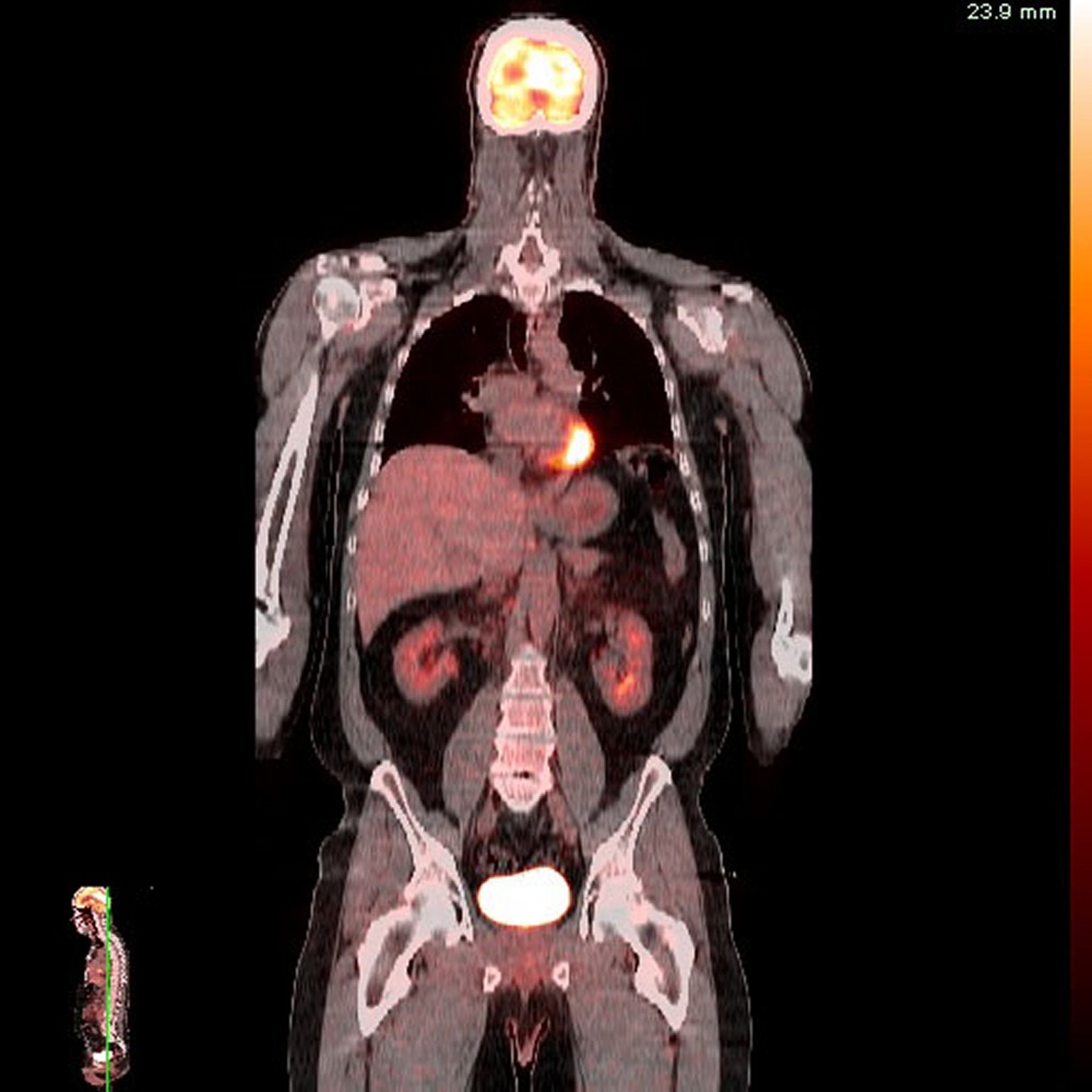
Image provided by Jon A. Jacobson, MD.
Disadvantages of PET
The amount of radiation exposure from PET is similar to that from CT. When PET and CT are done during a single examination, the radiation dose is significantly increased.
Because radionuclides used in PET give off radiation for only a short time, PET can be done only if the radionuclide is produced at a nearby location and can be obtained quickly.
PET is not widely available.


