


Noninvasive maternal screening, unlike invasive testing, has no risk of test-related complications. By more precisely assessing the risk of fetal abnormalities, noninvasive maternal screening can help women decide whether to have invasive testing. Noninvasive maternal screening for fetal chromosomal abnormalities should be offered to all pregnant women who have not already decided to have amniocentesis or chorionic villus sampling (CVS). However, even if CVS is to be done, maternal serum screening should still be offered to check for fetal neural tube defects.
Normal values vary with gestational age. Corrections for maternal weight, diabetes mellitus, race, and other factors may be necessary. Screening can be done during the
1st trimester
2nd trimester
Both trimesters (called sequential or integrated screening)
Any of the three approaches is acceptable. Maternal levels of alpha-fetoprotein should be measured during the 2nd trimester to check for neural tube defects.
Цінні поради та підводні камені
|
Скринінг при багатоплідних вагітностях
All forms of screening in singleton pregnancies (described above) are available to patients with a twin pregnancy. For twin pregnancies, screening performance using traditional methods (triple, quad) has lower sensitivity and specificity than in singleton pregnancies. Cell-free DNA (cfDNA) screening performance appears to be comparable for singleton and twin pregnancies. Because most dichorionic twin gestations are discordant for chromosome abnormalities, diagnostic testing is required to distinguish which twin is affected. However, screening for sex chromosome abnormalities in twin pregnancies is usually not available.
No serum screening or cfDNA screening protocols are validated for triplet or higher-order pregnancies.
Скринінг у І триместрі

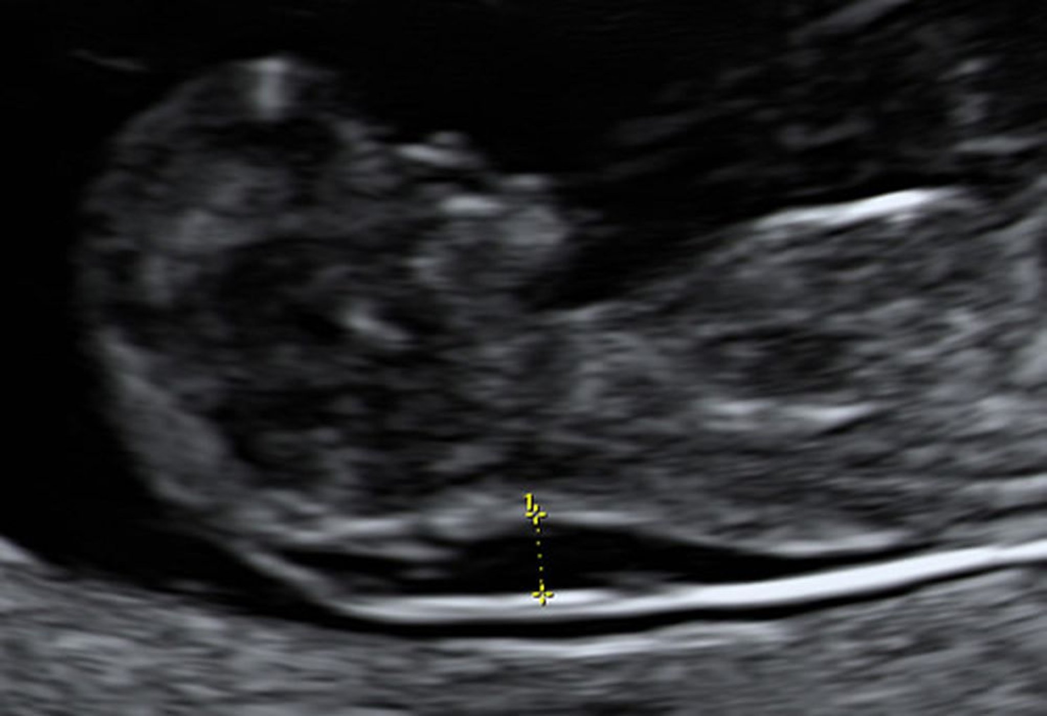
Photo from Jeffrey S. Dungan, MD.
Traditionally, 1st-trimester combined screening includes measurement of
Maternal serum beta-hCG (total or free)
Pregnancy-associated plasma protein A (PAPP-A)
Fetal nuchal translucency (by ultrasonography)
Fetal Down syndrome is typically associated with high levels of beta-hCG, low levels of PAPP-A, and enlarged fetal nuchal translucency. Although enlarged nuchal translucency is associated with increased risk of fetal Down syndrome, no threshold value for nuchal translucency is considered diagnostic.
In large prospective US trials involving women of various ages, overall sensitivity for detection of Down syndrome was about 85%, with a false-positive rate of 5%. Specialized ultrasound training and adherence to rigorous quality-assurance monitoring of nuchal translucency measurements are necessary to achieve this level of screening accuracy.
First-trimester screening should be offered to all pregnant women. It provides information early so that a definitive diagnosis can be made with CVS. An important advantage of 1st-trimester screening is that termination of pregnancy is safer during the 1st rather than the 2nd trimester.
Безклітинний тест на нуклеїнову кислоту плода
An increasingly used approach, called noninvasive prenatal screening or cell-free DNA (cfDNA) screening, can identify fetal chromosomal abnormalities in singleton pregnancies by analyzing circulating cell-free fetal nucleic acids in a maternal blood sample. This test can be done as early as 10 gestational weeks and is replacing traditional 1st- and 2nd-trimester noninvasive screening in many centers.
Cell-free fetal nucleic acids, most commonly DNA fragments, are shed into the maternal circulation during normal breakdown of placental trophoblast cells. Variation in amounts of fragments from particular chromosomes predicts fetal chromosomal abnormalities with higher accuracy than traditional 1st- and 2nd-trimester combined screening using serum analytes and ultrasonography. Also, sex chromosomal abnormalities (X, XXX, XYY, and XXY) can be identified in singleton pregnancies, although with somewhat lower accuracy. Early validation trials reported > 99% sensitivity and specificity for the identification of Down syndrome (trisomy 21) and trisomy 18 in high-risk pregnancies. Trisomy 13 can also be detected, although the sensitivity and specificity are somewhat lower (1).
Cell-free DNA (cfDNA) screening was historically recommended for women with preexisting risk factors for fetal trisomy. However, in a recent large multicenter trial that studied the effectiveness of cfDNA screening in a low-risk population, sensitivity for detection of fetal Down syndrome was equivalent to that in a high-risk population. Given the lower incidence of fetal Down syndrome in younger pregnant women, the specificity and positive predictive value were lower than if only high-risk women were screened. However, cfDNA screening was superior to traditional analyte screening in low-risk women in overall performance. Cell-free DNA screening has largely replaced serum analyte screening in high-risk women, and in low-risk women it is rapidly surpassing the use of traditional 1st- and 2nd-trimester combined screening with serum analytes and ultrasonography (1). The American College of Obstetricians and Gynecologists recommends offering cell-free DNA screening to all pregnant women (2).
Abnormal results from cfDNA screening should be confirmed with diagnostic karyotyping using fetal specimens obtained through invasive techniques. Negative results from cfDNA screening has reduced the use of routine invasive testing.
Довідкові матеріали щодо скринінгу у 1-му триместрі
Badeau M, Lindsay C, Blais J, Nshimyumukiza L, et al. Genomics-based non-invasive prenatal testing for detection of fetal chromosomal aneuploidy in pregnant women. Cochrane Database Syst Rev 11:CD011767. doi: 10.1002/14651858.CD011767.pub2, 2017
American College of Obstetricians and Gynecologists’ (ACOG) Committee on Practice Bulletins—Obstetrics; Committee on Genetics; Society for Maternal-Fetal Medicine: Screening for fetal chromosomal abnormalities: ACOG Practice Bulletin, Number 226. Practice Guideline. Obstet Gynecol 136 (4):e48-e69, 2020. doi: 10.1097/AOG.0000000000004084
Скринінг у 2 триместрі
Second-trimester screening may include cfDNA or the multiple marker screening approach, which includes
Screening for neural tube defects: Maternal levels of serum alpha-fetoprotein (MSAFP): MSAFP may be used independently as a screen for neural tube defects only, not for risk of Down syndrome. An elevated MSAFP level suggests open spina bifida, anencephaly, or abdominal wall defects. Unexplained elevations in MSAFP may be associated with increased risk of later pregnancy complications, such as stillbirth or intrauterine growth retardation.
Quadruple screening (aimed mainly at trisomy 21): Maternal levels of beta-hCG, unconjugated estriol, alpha-fetoprotein, and sometimes inhibin A: This screening may be used as an alternative or adjunct to 1st-trimester screening.
Second-trimester multiple marker screening is used to help assess the risk of Down syndrome, trisomy 18, and a few rarer single-gene syndromes (eg, Smith-Lemli-Opitz syndrome). Maternal serum tests are widely available, but detection rates for Down syndrome are not as high as those obtained with 1st-trimester screening or with cfDNA. Also, termination of pregnancy is riskier in the 2nd trimester than in the 1st trimester.
Second-trimester screening may also include
Targeted ultrasonography
Скринінг материнської сироватки на дефекти нервової трубки
An elevated level of MSAFP may indicate a fetal malformation such as open spina bifida. Results are most accurate when the initial sample is obtained between 16 and 18 weeks gestation, although screening can be done from about 15 to 20 weeks. Designating a cutoff value to determine whether further testing is warranted involves weighing the risk of missed abnormalities against the risk of complications from unnecessary testing. Usually, a cutoff value in the 95th to 98th percentile, or 2.0 to 2.5 times the normal pregnancy median (multiples of the median, or MOM), is used. This value is about 80% sensitive for open spina bifida and 90% sensitive for anencephaly. Closed spina bifida is usually not detected. Amniocentesis is eventually required in 1 to 2% of women originally screened. Lower cutoff values of MSAFP increase sensitivity but decrease specificity, resulting in more amniocenteses. Women who have been screened for fetal chromosome disorders by cfDNA should have serum screening with MSAFP alone, not with multiple marker screening.
Ultrasonography is the next step if further testing is warranted. Targeted ultrasonography with or without amniocentesis is done if no explanation can be determined with basic ultrasonography. Ultrasonography can
Confirm gestational age (which may be underestimated)
Detect multifetal pregnancy, fetal death, or congenital malformations
In some women, ultrasonography cannot identify a cause for elevated alpha-fetoprotein levels. Some experts believe that if high-resolution ultrasonography done by an experienced operator is normal, further testing is unnecessary. However, because this test occasionally misses neural tube defects, many experts recommend further testing by amniocentesis regardless of ultrasonography results.
Amniocentesis with measurement of alpha-fetoprotein and acetylcholinesterase levels in amniotic fluid is done if further testing is needed. Elevated alpha-fetoprotein in amniotic fluid suggests
A neural tube defect
Another malformation (eg, omphalocele, congenital nephrosis, cystic hygroma, gastroschisis, upper gastrointestinal atresia)
Contamination of the sample with fetal blood
Presence of acetylcholinesterase in amniotic fluid suggests
A neural tube defect
Another malformation
Elevated alpha-fetoprotein plus presence of acetylcholinesterase in amniotic fluid is virtually 100% sensitive for anencephaly and 90 to 95% sensitive for open spina bifida. Abnormal amniotic fluid markers indicate that a malformation is likely even if high-resolution ultrasonography (which can detect most of these malformations) does not detect a malformation, and parents should be informed.
Скринінг материнської сироватки на хромосомні порушення
During the 2nd trimester, the most common approach to screening is with cfDNA or multiple serum markers. These markers, adjusted for gestational age, are used mainly to refine estimates of Down syndrome risk beyond that associated with maternal age. With triple screening (ie, alpha-fetoprotein, hCG, and unconjugated estriol), sensitivity for Down syndrome is about 65 to 70%, with a false-positive rate of about 5%.
Quad screening is triple screening plus measurement of inhibin A. Quad screening increases sensitivity to about 80%, with a 5% false-positive rate.
If maternal serum screening suggests Down syndrome, ultrasonography is done to confirm gestational age, and risk is recalculated if the presumed gestational age is incorrect. If the original sample was drawn too early, another one must be drawn at the appropriate time. Amniocentesis is offered particularly if risk exceeds a specific prespecified threshold (usually 1 in 270, which is about the same as risk when maternal age is > 35).
Triple screening can also assess risk of trisomy 18, indicated by low levels of all 3 serum markers. Sensitivity for trisomy 18 is 60 to 70%; the false-positive rate is about 0.5%. Combining ultrasonography and serum screening increases sensitivity to about 80%.
Analysis of cfDNA does not depend on gestational age and thus is not prone to dating errors.
Прицільна УЗД
Targeted ultrasonography is offered at some perinatal centers and is used to assess risk of chromosomal abnormalities by searching for structural features associated with fetal aneuploidy (so-called soft markers). However, no structural finding is diagnostic for a given chromosomal abnormality, and all soft markers may also be seen in fetuses that are chromosomally normal. If results from prior trisomy screening were negative (risk-reducing), many of these soft markers have no clinical relevance and may be ignored (1). Nonetheless, the discovery of such a marker may lead to offering the woman amniocentesis to confirm or exclude a chromosomal abnormality. If a major structural malformation is present, a fetal chromosomal abnormality is more likely.
Disadvantages include unnecessary anxiety if a soft marker is detected and unnecessary amniocentesis. Several experienced centers report high sensitivity, but whether a normal ultrasound indicates a substantially reduced risk of fetal chromosomal abnormalities is unclear.
Довідкові матеріали щодо скринінгу у 2-му триместрі
1. American College of Obstetricians and Gynecologists/Committee on Genetics, and the Society for Maternal-Fetal Medicine: Practice bulletin no. 163: Screening for fetal aneuploidy. Committee on Practice Bulletins—Obstetrics, Obstet Gynecol 127 (5):e123–e137, 2016. doi: 10.1097/AOG.0000000000001406
Послідовний скринінг у 1-му та 2-му триместрі
Noninvasive 1st-trimester and 2nd-trimester quad screening can be combined sequentially, with invasive fetal genetic testing withheld until results of 2nd-trimester screening are available—whether 1st-trimester test results are abnormal or not. Sequential screening followed by amniocentesis for high-risk patterns increases sensitivity for Down syndrome to 95%, with a false-positive rate of only 5%.
A variation of sequential screening, called contingent sequential screening, is based on the level of risk indicated by 1st-trimester screening:
High risk: Invasive testing is offered without doing 2nd-trimester screening.
Intermediate risk: 2nd-trimester screening is offered.
Low risk (eg, < 1 in 1500): 2nd-trimester screening for Down syndrome is not offered because the 1st-trimester risk is so low.
Patients with abnormal 1st-trimester, 2nd-trimester, or sequential screening should be offered diagnostic testing (eg, amniocentesis). However, some patients may choose to pursue further testing for fetal trisomy with cfDNA (cell free DNA) analysis (1). Results of cfDNA testing may indicate low risk and be reassuring but are not definitive. Also, cfDNA testing may be inordinately expensive, and awaiting results of cfDNA testing delays definitive testing such as chorionic villus sampling or amniocentesis (2).
Довідкові матеріали щодо послідовного скринінгу у 1-му та 2-му триместрі
1. American College of Obstetricians and Gynecologists/Committee on Genetics, and the Society for Maternal-Fetal Medicine: Practice bulletin no. 163: Screening for fetal aneuploidy. Committee on Practice Bulletins—Obstetrics, Obstet Gynecol 127 (5):e123–e137, 2016. doi: 10.1097/AOG.0000000000001406
2. Norton ME, Jacobsson B, Swamy GK, et al: Cell-free DNA analysis for noninvasive examination of trisomy. N Engl J Med 372 (17):1589-1597, 2015.