


Bursitis is acute or chronic inflammation of a bursa (a fluid-filled sac lined by a synovial membrane that functions to reduce friction between tendons and either bone or skin). The cause of bursitis is usually unknown, but trauma, repetitive or acute, may contribute, as may infection, osteoarthritis, crystal-induced arthropathy, and certain inflammatory arthropathies. Symptoms include pain (particularly with motion or pressure), swelling, and tenderness. Diagnosis is usually clinical; however, ultrasound may be needed to evaluate deep bursae. Diagnosis of infection and crystal-induced disease requires analysis of bursal fluid. Treatment includes splinting, nonsteroidal anti-inflammatory drugs (NSAIDs), sometimes glucocorticoid injections, and treatment of the underlying cause.
Bursae are fluid-filled saclike cavities or potential cavities lined by a synovial membrane that are located where friction occurs (eg, where tendons or muscles pass over bony prominences). Bursae minimize friction between moving parts and facilitate movement. Some communicate with joints.

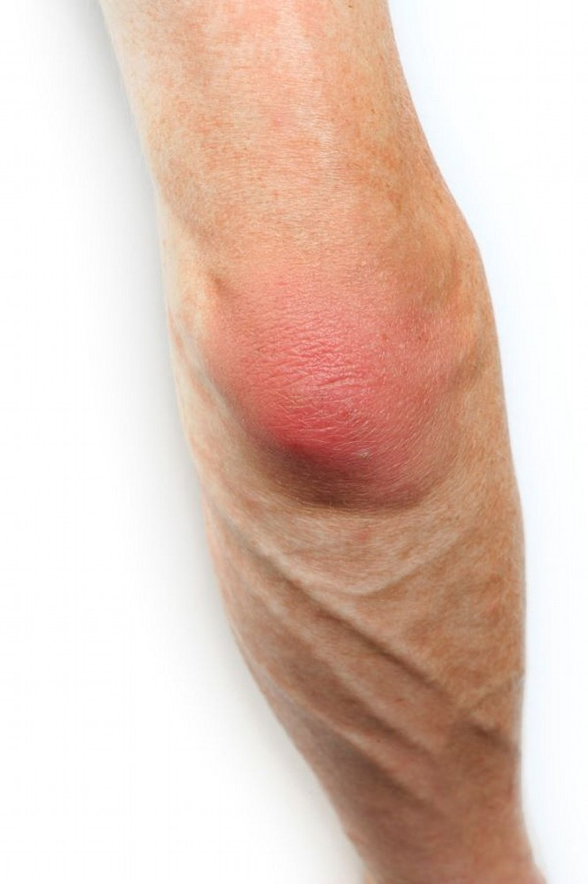
This photo shows a side view of erythema and swelling over the patella in a patient with prepatellar bursitis (housemaid's knee).
SCIENCE PHOTO LIBRARY
Bursitis may occur in the shoulder (subacromial or subdeltoid bursitis), particularly in patients with rotator cuff tendinopathy, which is usually the primary lesion in the shoulder. Other commonly affected bursae include olecranon (miner's or barfly’s elbow), prepatellar (housemaid’s knee), suprapatellar, retrocalcaneal, iliopectineal (iliopsoas), ischial (weaver’s bottom), greater trochanteric, pes anserine, and first metatarsal head (bunion) bursae. Occasionally, a bursa ruptures or develops a chronic communication with an adjacent joint.
Etiology of Bursitis
Bursitis may be caused by the following:
Injury
Osteoarthritis (eg, iliopsoas bursitis in hip osteoarthritis or pes anserine bursitis in knee osteoarthritis)
Chronic overuse and/or pressure
Inflammatory arthritis (eg, gout, pseudogout, rheumatoid arthritis, psoriatic arthritis, spondylitis)
Acute or chronic infection (eg, Staphylococcus aureus in acute infections and mycobacteria in chronic infections)
Idiopathic and traumatic causes are by far the most common. Acute bursitis may follow unusual exercise or strain and usually causes bursal effusion. The olecranon and prepatellar bursae are the bursae most susceptible to infection because of their superficial locations.
Chronic bursitis may develop after recurrent episodes of bursitis or from repeated trauma or gout. The bursal wall is thickened, with proliferation of its synovial lining; bursal adhesions, villus formation, tags, and milky urate deposits may develop.
Symptoms and Signs of Bursitis
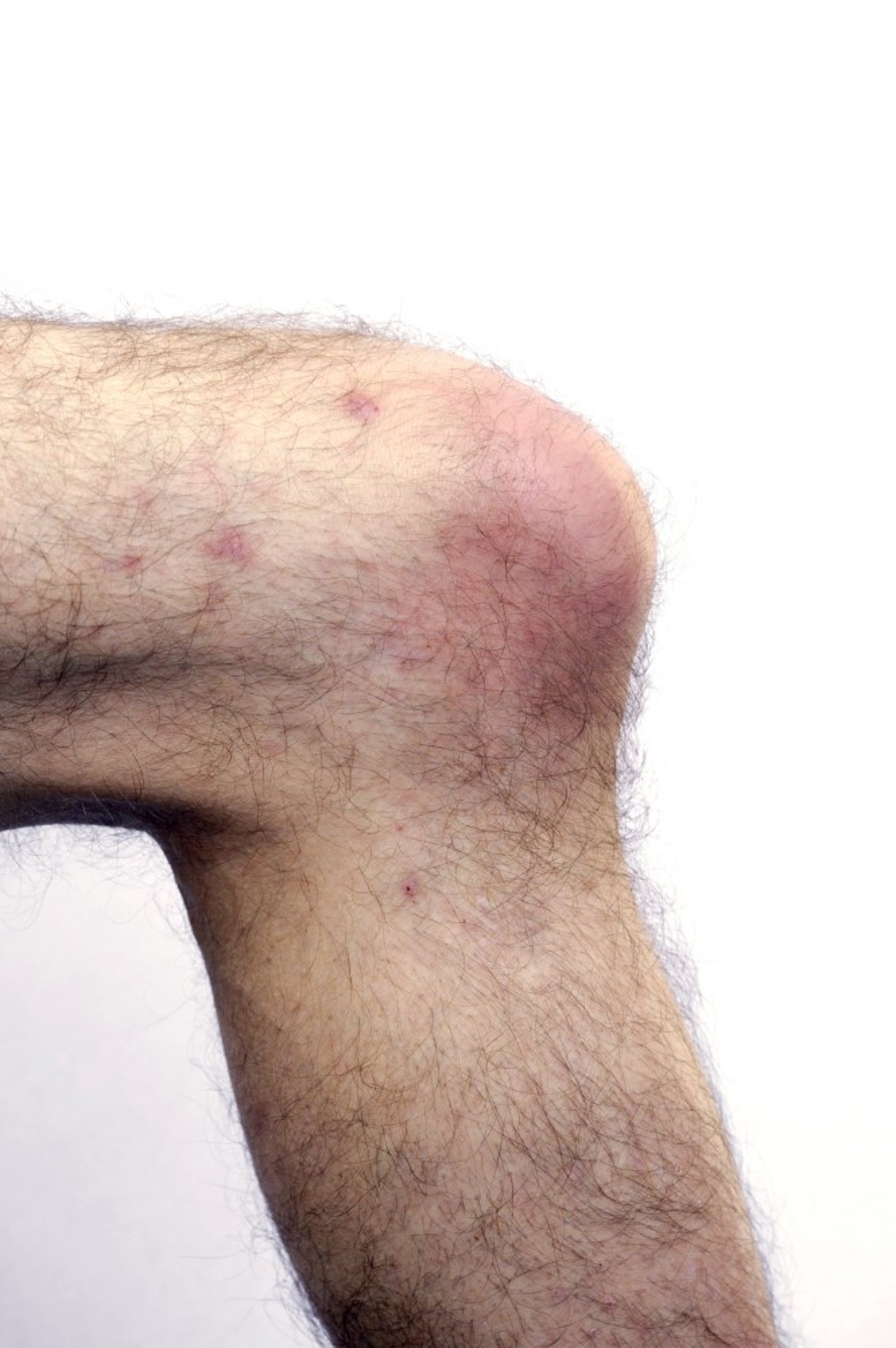
Acute bursitis causes pain, particularly when the bursa is compressed or stretched during motion, and often limits range of motion. Passive range of motion may still be normal (eg, patients with isolated olecranon bursitis have normal flexion-extension of the elbow). Swelling, sometimes with other signs of inflammation (eg, erythema), is common if the bursa is superficial (eg, prepatellar, olecranon). Swelling may be more prominent than pain in olecranon bursitis. Acute crystal- or bacterial-induced bursitis is usually accompanied by erythema, pitting edema, pain, and warmth in the area around the bursa.
This photo of a patient with olecranon bursitis shows signs of inflammation with erythema and swelling over the elbow bursa. The clinical concern is cellulitis with associated septic bursitis.
SCIENCE PHOTO LIBRARY
Chronic bursitis may last for several months and may recur frequently. Bouts may last a few days to several weeks. If inflammation persists near a joint, the joint’s range of motion may be limited. Prolonged limitation of motion may lead to muscle atrophy.
Diagnosis of Bursitis
Primarily history and physical examination
Sometimes ultrasound or MRI for deep bursitis (eg, subacromial bursa in the shoulder)
Aspiration for suspected infection, hemorrhage (due to trauma or anticoagulants), or crystal-induced bursitis
Superficial bursitis should be suspected in patients with swelling or signs of inflammation over bursae. Deep bursitis is suspected in patients with unexplained pain worsened by motion or pressure of palpation in a location compatible with bursitis. Usually, bursitis can be diagnosed clinically. Ultrasound or MRI can help confirm the diagnosis when deep bursae are not readily accessible for inspection, palpation, or aspiration. These tests are done to confirm a suspected diagnosis or exclude other possibilities. These imaging techniques increase the accuracy of identifying the involved structures.
If bursal swelling is particularly painful, erythematous, or warm, where the suspicion of infection or other inflammatory conditions like crystal-induced arthritis is very high, a bursal aspiration should be attempted. This can be done using surface anatomy or with radiographic or sonographic guidance. Aspiration is particularly important in patients who are immunosuppressed, because signs and symptoms of chronic infection may be minimal. After a local anesthetic is injected, fluid is withdrawn from the bursa using sterile techniques; analysis includes cell count, Gram stain and culture, and microscopic search for crystals. Gram stain, although helpful if positive, may not be specific, and white blood cell counts in infected bursae are usually lower than those in septic joints. Urate crystals are easily seen with polarized light microscopy, but the apatite crystals typical of calcific tendinitis appear only as shiny chunks that are not birefringent. Cholesterol plate crystals can be seen in chronic rheumatoid bursitis.


Signs of acute inflammation can be seen in and around the left knee. In some patients with septic bursitis, an overlying skin infection is present, which may be an infected laceration, abrasion, or, as in this image, cellulitis.
By permission of the publisher. From Gilliland B, Wener M: Atlas of Infectious Diseases: Skin, Soft Tissue, Bone and Joint Infections. Edited by G Mandell (series editor) and TP Bleck. Philadelphia, Current Medicine, 1995.
Acute bursitis should be distinguished from hemorrhage into a bursa, which should be considered, particularly when a patient taking anticoagulants develops acute bursal swelling. Hemorrhagic bursitis can cause similar manifestations because blood is inflammatory. Fluid in traumatic bursitis is usually serosanguinous. Cellulitis can cause signs of inflammation but does not normally cause bursal effusion; cellulitis overlying the bursa is a relative contraindication to bursal puncture through the cellulitis, but if septic bursitis is strongly suspected, aspiration must be performed and will likely provide culture identification of the infecting organism.
Treatment of Bursitis
Rest followed by physical therapy
Nonsteroidal anti-inflammatory drugs (NSAIDs)
Treatment of crystal-induced arthritis or infection
Sometimes glucocorticoid injection
For crystal-induced disease, see treatment of gout and pseudogout.
For suspected infection, empiric antibiotics effective against S. aureus should be given initially, after the bursa is aspirated and cultures are obtained (see treatment of staphylococcal infection). Subsequent choice of antibiotic is determined by results of Gram stain and culture. Infectious bursitis requires drainage or occasionally excision in addition to antibiotics.
Acute nonseptic bursitis is treated with activity modification and NSAIDs, sometimes with other analgesics. Physical therapy and voluntary movement should be increased as tolerated. This may accelerate restoration of range of motion. Pendulum exercises are helpful for the shoulder joint.
Glucocorticoid injection may be considered if bursitis (eg, subacromial bursitis) is persistent, infection has been excluded, and oral medications and rest are inadequate. Glucocorticoid injections have been shown to provide short-term pain relief (< 6 weeks) in patients with shoulder bursitis or related conditions (eg, rotator cuff tendinopathy) (1, 2).
Intrabursal injection of depot glucocorticoids 0.5 to 1 mL (eg, triamcinolone acetonide 40 mg/mL) is typically reserved for deep bursae (trochanteric, subacromial, or pes anserine). It is less commonly performed on superficial bursae (eg, olecranon, prepatellar). About 1 mL of local anesthetic (eg, 2% lidocaine) can be injected before or with the glucocorticoid injection. Dose and volume of the glucocorticoid may vary according to the size of the bursa. Infrequently, a flare of symptoms occurs within several hours of injection of a depot glucocorticoid (postinjection flare); the painful local reaction is thought to be due to a chemical synovitis in response to the crystals in the glucocorticoid suspension. It usually occurs in ≤ 24 hours of the injection and usually lasts ≤ 48 hours. Patients may use cold compresses plus analgesics for symptomatic relief.
Chronic bursitis is treated the same as acute bursitis, except that splinting and rest are less likely to help, and range-of-motion exercises are especially important. Rarely, the bursa needs to be excised.
Treatment references
1. Steuri R, Sattelmayer M, Elsig S, et al. Effectiveness of conservative interventions including exercise, manual therapy and medical management in adults with shoulder impingement: a systematic review and meta-analysis of RCTs. Br J Sports Med. 51(18):1340-1347, 2017. doi:10.1136/bjsports-2016-096515
2. Sun Y, Chen J, Li H, Jiang J, Chen S. Steroid Injection and Nonsteroidal Anti-inflammatory Agents for Shoulder Pain: A PRISMA Systematic Review and Meta-Analysis of Randomized Controlled Trials. Medicine (Baltimore). 94(50):e2216, 2015. doi:10.1097/MD.0000000000002216
Key Points
The usual causes of bursitis are injury and overuse, but infection and crystal-induced arthritis are possible.
Aspirate bursal fluid to diagnose bacterial or crystal-induced bursitis when the olecranon or prepatellar bursa is affected or when there is warmth, erythema, tenderness, and pitting edema.
If no infection is present, treat most cases with rest, high-dose NSAIDs, and sometimes intrabursal glucocorticoid injection.
Drug Information for the Topic


