

Granuloma inguinale (donovanosis) is a progressive infection of genital and perineal skin caused by Klebsiella granulomatis. The infection is characterized by slowly progressive skin lesions that are beefy red, raised, painless, and often ulcerated; regional lymphadenopathy is uncommon. Diagnosis is by physical examination and microscopic of evaluation of fluid from lesions. Treatment is with antibiotics, usually tetracyclines, macrolides, or trimethoprim/sulfamethoxazole.granulomatis. The infection is characterized by slowly progressive skin lesions that are beefy red, raised, painless, and often ulcerated; regional lymphadenopathy is uncommon. Diagnosis is by physical examination and microscopic of evaluation of fluid from lesions. Treatment is with antibiotics, usually tetracyclines, macrolides, or trimethoprim/sulfamethoxazole.
(See also Overview of Sexually Transmitted Infections.)
Infections with Klebsiella granulomatis are extremely rare in high-resource countries but are seen in India, southern Africa, and parts of South America (1).
General reference
1. Workowski KA, Bachmann LH, Chan PA, et al. Sexually Transmitted Infections Treatment Guidelines, 2021. MMWR Recomm Rep. 2021;70(4):1-187. Published 2021 Jul 23. doi:10.15585/mmwr.rr7004a1. Erratum: Vol. 70, No. RR-4. MMWR Morb Mortal Wkly Rep. 2023;72(4):107-108. Published 2023 Jan 27. doi:10.15585/mmwr.mm7204a5
Symptoms and Signs of Granuloma Inguinale
Sites of infection include the following:
Penis, scrotum, groin, and thighs in men
Vulva, vagina, and perineum in women
Anus and buttocks in patients who engage in anal-receptive intercourse
Face in both sexes
After an incubation period of about 1 to 12 weeks, a painless, red skin nodule slowly enlarges, becoming a raised, beefy red, moist, smooth, foul-smelling lesion. The lesion slowly enlarges, often ulcerates, and may spread to other skin areas. Lesions heal slowly, with scarring.
Secondary infections with other bacteria are common and can cause extensive tissue destruction.
Lymphadenopathy is uncommon.
Occasionally, granuloma inguinale spreads through the bloodstream to the bones, joints, or liver; without treatment, anemia, wasting, and, uncommonly, death may occur.


This photo shows the raised and ulcerated lesion of genital granuloma inguinale.
This photo shows the raised and ulcerated lesion of genital granuloma inguinale.
Image courtesy of Joe Miller and Dr. Cornelio Arevalo via the Public Health Image Library of the Centers for Disease Control and Prevention.


This photo shows the raised and ulcerated lesion of genital granuloma inguinale.
This photo shows the raised and ulcerated lesion of genital granuloma inguinale.
Image courtesy of Dr. Susan Lindsley via the Public Health Image Library of the Centers for Disease Control and Prevention.

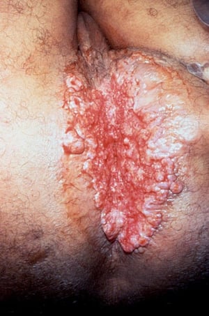
This photo shows the raised, red lesion of perianal granuloma inguinale.
This photo shows the raised, red lesion of perianal granuloma inguinale.
Image courtesy of Dr. Tabua via the Public Health Image Library of the Centers for Disease Control and Prevention.


This photo shows granuloma inguinale. In this person, the initial nodule slowly enlarged, eroded the surrounding tissues, and developed into a beefy, bulky, granulomatous mass.
This photo shows granuloma inguinale. In this person, the initial nodule slowly enlarged, eroded the surrounding tissue
© Springer Science+Business Media
This photo shows the raised and ulcerated lesion of genital granuloma inguinale.
This photo shows the raised and ulcerated lesion of genital granuloma inguinale.
Image courtesy of Joe Miller and Dr. Cornelio Arevalo via the Public Health Image Library of the Centers for Disease Control and Prevention.
This photo shows the raised and ulcerated lesion of genital granuloma inguinale.
This photo shows the raised and ulcerated lesion of genital granuloma inguinale.
Image courtesy of Dr. Susan Lindsley via the Public Health Image Library of the Centers for Disease Control and Prevention.
This photo shows the raised, red lesion of perianal granuloma inguinale.
This photo shows the raised, red lesion of perianal granuloma inguinale.
Image courtesy of Dr. Tabua via the Public Health Image Library of the Centers for Disease Control and Prevention.
This photo shows granuloma inguinale. In this person, the initial nodule slowly enlarged, eroded the surrounding tissues, and developed into a beefy, bulky, granulomatous mass.
This photo shows granuloma inguinale. In this person, the initial nodule slowly enlarged, eroded the surrounding tissue
© Springer Science+Business Media
Diagnosis of Granuloma Inguinale
Physical examination
Microscopic examination showing Donovan bodies in fluid from a lesion
Granuloma inguinale is suspected in patients from endemic areas with characteristic lesions.
Diagnosis of granuloma inguinale is confirmed microscopically by the presence of Donovan bodies (numerous bacilli in the cytoplasm of macrophages shown by Giemsa or Wright stain) in smears of fluid from scrapings from the edge of lesions. These smears contain many plasma cells.
Biopsy specimens are taken if the diagnosis is unclear or if adequate tissue fluid cannot be obtained because lesions are dry, sclerotic, or necrotic. The bacteria do not grow on ordinary culture media.
Treatment of Granuloma Inguinale
Antibiotics
Many oral antibiotics kill the bacteria, but tetracyclines, macrolides, and trimethoprim/sulfamethoxazole (TMP/SMX) are most effective, followed by ceftriaxone, aminoglycosides, fluoroquinolones, and chloramphenicol.Many oral antibiotics kill the bacteria, but tetracyclines, macrolides, and trimethoprim/sulfamethoxazole (TMP/SMX) are most effective, followed by ceftriaxone, aminoglycosides, fluoroquinolones, and chloramphenicol.
The recommended oral regimen is azithromycin 1 g once/week or 500 mg/day for > 3 weeks and continued until all lesions have completely healed (The recommended oral regimen is azithromycin 1 g once/week or 500 mg/day for > 3 weeks and continued until all lesions have completely healed (1).
Alternate oral regimens should be continued until all lesions have completely healed and include the following (1):
Doxycycline 100 mg orally 2 times a day for at least 3 weeksDoxycycline 100 mg orally 2 times a day for at least 3 weeks
Trimethoprim/sulfamethoxazole 1 double-strength (160 mg/800 mg) tablet orally 2 times a day for ≥ 3 weeksTrimethoprim/sulfamethoxazole 1 double-strength (160 mg/800 mg) tablet orally 2 times a day for ≥ 3 weeks
Erythromycin base 500 mg orally 4 times a day for ≥ 3 weeksErythromycin base 500 mg orally 4 times a day for ≥ 3 weeks
IV or IM antibiotics (eg, ceftriaxone) are an alternative.IV or IM antibiotics (eg, ceftriaxone) are an alternative.
Response to treatment should begin within 7 days, but healing of extensive disease may be slow and lesions may recur, requiring longer treatment. Patients with HIV infection may also require prolonged or intensive treatment. After apparently successful treatment, follow-up should continue for 6 months.
Sex partners within 60 days before onset of the patient's symptoms should be examined and, if infected, treated.
Treatment reference
1. Workowski KA, Bachmann LH, Chan PA, et al. Sexually Transmitted Infections Treatment Guidelines, 2021. MMWR Recomm Rep. 2021;70(4):1-187. Published 2021 Jul 23. doi:10.15585/mmwr.rr7004a1. Erratum: Vol. 70, No. RR-4. MMWR Morb Mortal Wkly Rep. 2023;72(4):107-108. Published 2023 Jan 27. doi:10.15585/mmwr.mm7204a5
Drug Information for the Topic


