


Langerhans cell histiocytosis (LCH) is a proliferation of dendritic mononuclear cells with infiltration into organs locally or diffusely. Most cases occur in children. Manifestations may include bone lesions; rashes; lung infiltrates; and hepatic, hematopoietic, and endocrine dysfunction. Diagnosis is based on biopsy. Factors predicting a poor prognosis include age < 2 years and dissemination, particularly involving the hematopoietic system, liver, spleen, or a combination of these. Treatments include supportive measures and chemotherapy or local treatment with surgery as indicated by the extent of disease.
(See also Pulmonary Langerhans Cell Histiocytosis.)
Langerhans cell histiocytosis (LCH) is a myeloid-derived dendritic cell (antigen-presenting cell) disorder. It can cause distinct clinical syndromes. Historically, these syndromes have been described as eosinophilic granuloma, Hand-Schüller-Christian disease, and Letterer-Siwe disease. Because these syndromes may be varied manifestations of the same underlying disorder and because most patients with LCH have manifestations of more than one of these syndromes, the designations of the separate syndromes (except for eosinophilic granuloma) are now mostly of historical significance. Estimates of the prevalence of LCH vary widely (eg, from about 1:50,000 to 1:200,000). Incidence is approximately 5 cases/million children (1).
All patients with LCH have evidence of activation of the mitogen-activated protein kinase (MAPK) pathway with abnormal RAS-RAF-MEK-ERK signaling (1, 2). This pathway is involved in regulation of normal myeloid cell differentiation and maturation. A mutation within the pathway results in constitutive activation of the pathway and negatively impacts cell maturation and apoptosis. Aberrant cell differentiation, uninhibited growth and a loss of apoptosis result in oncogenesis. BRAFV600E mutations are the most common mutation identified in approximately two-thirds of patients with LCH. This mutation is monoallelic and acts like a dominant driving oncogene. About 10 to 15% of patients have mutations in the gene that encodes MAP2K1, an enzyme in the pathway. Because of these mutations, LCH is considered an oncogene-driven cancer of myeloid lineage.
In LCH, abnormally proliferating myeloid-derived dendritic cells infiltrate one or more organs. Bones, skin, teeth, gingival tissue, ears, endocrine organs, lungs, liver, spleen, lymph nodes, and bone marrow may be involved. Organs may be affected by infiltration, causing dysfunction, or by compression from adjacent enlarged structures. In about half of patients, more than one organ is involved.
General references
1. Allen CE, Merad M, McClain KL: Langerhans-cell histiocytosis. N Engl J Med 379(9):856–868, 2018. doi: 10.1056/NEJMra1607548
2. Sconocchia T, Foßelteder J, Sconocchia G, Reinisch A: Langerhans cell histiocytosis: current advances in molecular pathogenesis. Front Immunol 2023 Oct 26;14:1275085. doi: 10.3389/fimmu.2023.1275085. PMID: 37965340; PMCID: PMC10642229.
Symptoms and Signs of Langerhans Cell Histiocytosis
Symptoms and signs of Langerhans cell histiocytosis vary considerably depending on which organs are infiltrated (1).
Patients are divided into 2 groups based on organ involvement (2):
Single system
Multisystem
Single system disease is unifocal or multifocal involvement of one of the following organs: bone, skin, lymph nodes, lungs, central nervous system, or other, rare locations (eg, thyroid, thymus). An example of single system disease of the bone is eosinophilic granuloma.
Multisystem disease is disease in 2 or more organ systems. Risk-organs are those in which involvement portends a worse prognosis and include the liver, spleen, and bone marrow. Multisystem disease without risk-organ involvement is considered low-risk disease and used to be called Hand-Schüller-Christian disease. Multisystem disease with risk-organ involvement is high-risk disease and used to be known as Letterer-Siwe disease.
Here, the syndromes are described by their historical designations, but few patients present with classic manifestations, and other than eosinophilic granuloma, these designations are no longer used.

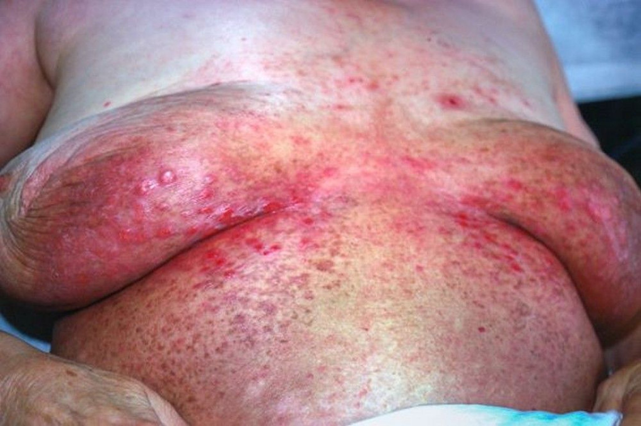
This image shows an erythematous papular rash resulting from skin manifestations of histiocytosis.
Image courtesy of Karen McKoy, MD.
Eosinophilic granuloma (single-system disease)
Unifocal or multifocal single-system lesions occurs predominantly in older children and young adults, usually by age 30; incidence peaks between ages 5 and 10 years. Lesions most frequently involve bones, often with pain, the inability to bear weight, or both and with overlying tender (sometimes warm) swelling.
Congenital self-healing reticulohistiocytosis (single-system disease)
Congenital self-healing reticulohistiocytosis (previously called Hashimoto-Pritzker disease) is a single-system disease with isolated skin lesions that occurs in neonates. Lesions generally resolve on their own or respond to topical steroid treatment. Patients should be evaluated to exclude multisystem disease.
Multisystem disease without risk-organ involvement (Hand-Schüller-Christian disease)
This syndrome occurs in children aged 2 to 5 years and in some older children and adults. Classic findings in this systemic disorder include involvement of the bones of the skull, ribs, pelvis, scapula, or a combination. Long bones and lumbosacral vertebrae are less frequently involved; the wrists, hands, knees, feet, and cervical vertebrae are rarely involved. In patients with classic findings, patients may have proptosis caused by orbital tumor mass. Rarely, vision loss or strabismus is caused by optic nerve or orbital muscle involvement. Tooth loss caused by apical and gingival infiltration is common in adult patients.
Chronic otitis media and otitis externa due to involvement of the mastoid and petrous portions of the temporal bone with partial obstruction of the auditory canal are fairly common. Antidiuretic hormone (ADH) deficiency (central diabetes insipidus), a component of the classic triad that also includes flat bone involvement and proptosis, affects 5 to 50% of patients, with higher percentages in children who have systemic disease and involvement of the orbit and skull. Up to 40% of children with systemic disease have short stature. Hyperprolactinemia and hypogonadism can result from hypothalamic infiltration.
Multisystem disease with risk-organ involvement (Letterer-Siwe disease)
This syndrome, a systemic disorder, is the most severe form of Langerhans cell histiocytosis. Typically, a child < 2 years presents with a scaly seborrheic, eczematoid, sometimes purpuric rash involving the scalp, ear canals, abdomen, and intertriginous areas of the neck and face. Denuded skin may facilitate microbial invasion, leading to sepsis. Frequently, there is ear drainage, lymphadenopathy, hepatosplenomegaly, and, in severe cases, hepatic dysfunction with hypoproteinemia and diminished synthesis of clotting factors. Anorexia, irritability, failure to thrive, and pulmonary manifestations (eg, cough, tachypnea, pneumothorax) may also occur. Significant anemia and sometimes neutropenia occur; thrombocytopenia is of grave prognostic significance. Parents frequently report precocious eruption of teeth, when in fact the gums are receding to expose immature dentition. Patients may appear abused or neglected.
Symptoms and signs references
1. Rodriguez-Galindo C: Clinical features and treatment of Langerhans cell histiocytosis. Acta Paediatr 110(11):2892–2902, 2021. doi:10.1111/apa.16014
2. Sconocchia T, Foßelteder J, Sconocchia G, Reinisch A: Langerhans cell histiocytosis: current advances in molecular pathogenesis. Front Immunol 14:1275085, 2023. doi: 10.3389/fimmu.2023.1275085
Diagnosis of Langerhans Cell Histiocytosis
Biopsy
Langerhans cell histiocytosis is suspected in patients (particularly young patients) with unexplained pulmonary infiltrates, bone lesions, or ocular or craniofacial abnormalities; and in children < 2 years with typical rashes or severe, unexplained multiorgan disease.

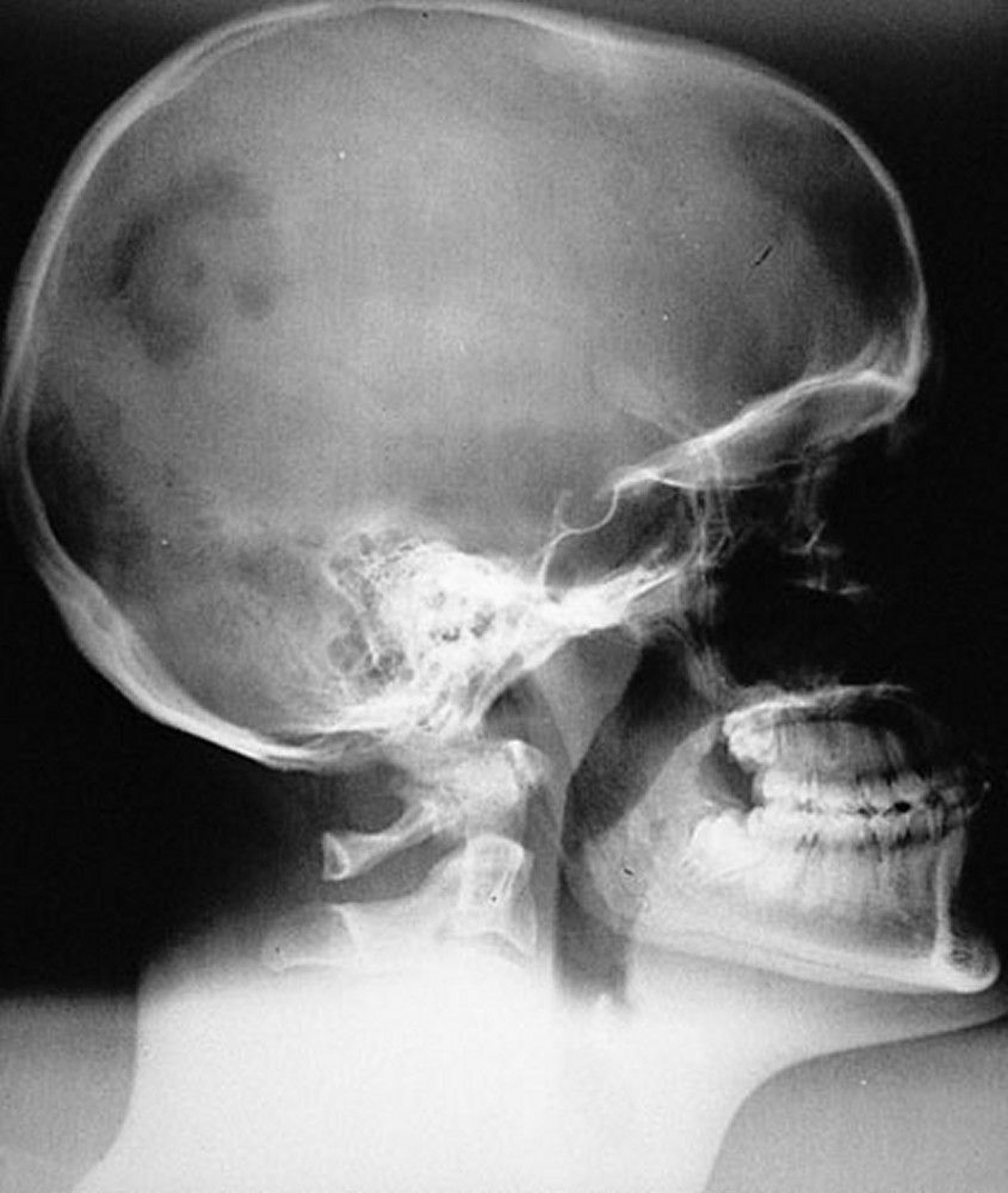
Radiographic appearance of typical monostotic unifocal LCH lesion (eosinophilic granuloma) of the skull. The lesion is sharply marginated but not beveled.
By permission of the publisher. From Swearingen B, Schaefer P, Primavera J, Klibanski A. In Atlas of Clinical Endocrinology: Neuroendocrinology and Pituitary Disease. Edited by S Korenman (series editor) and ME Molitch. Philadelphia, Current Medicine, 2000.

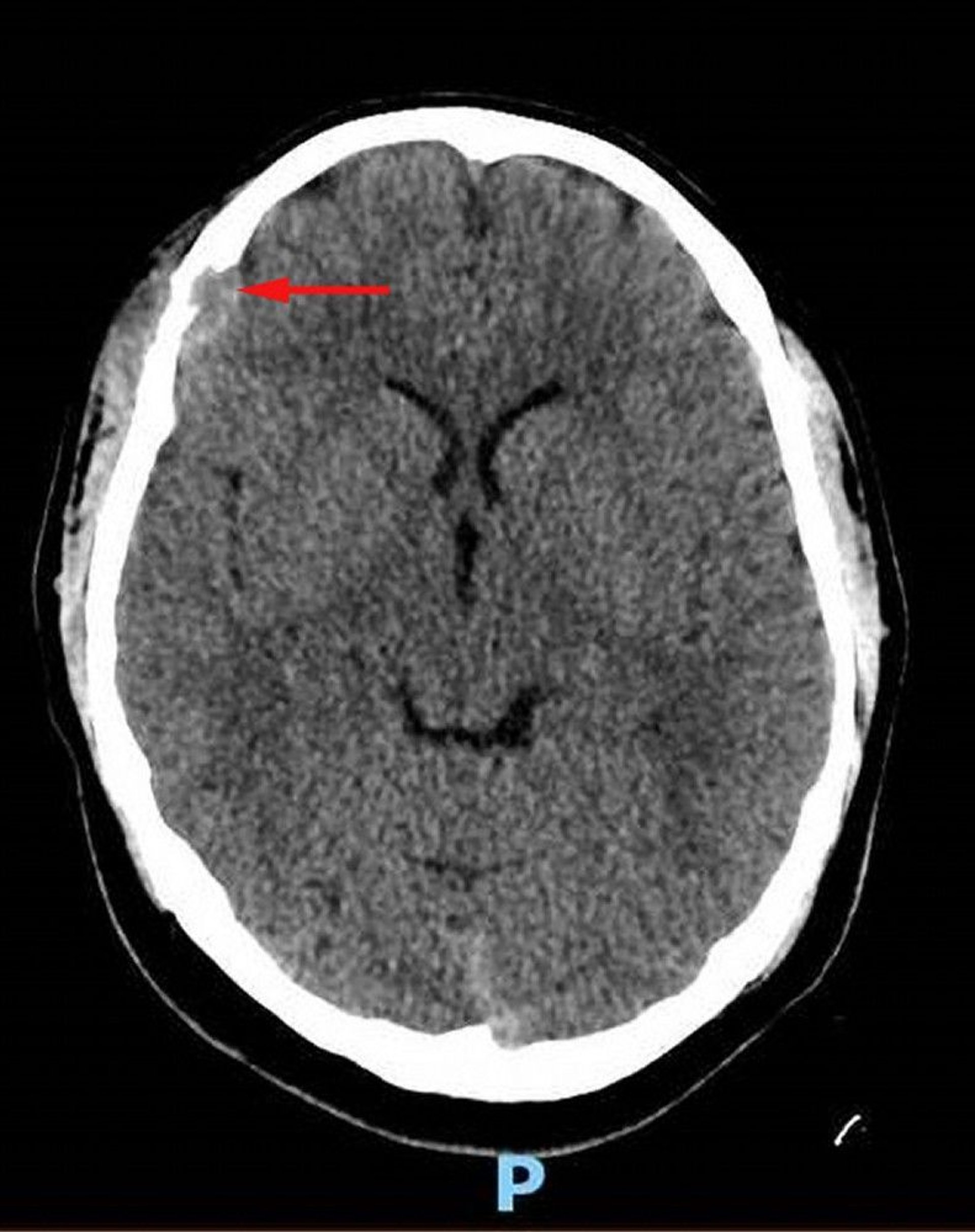
This image shows a soft-tissue lesion in the right frontal bone with associated osseous destruction and intracranial extension of soft tissue into the dural space (red arrow). Asymmetric overlying scalp soft tissue is present. Biopsy was diagnostic for Langerhans cell histiocytosis.
Image courtesy of Carolyn Fein Levy, MD, and Jeffrey M. Lipton, MD, PhD.

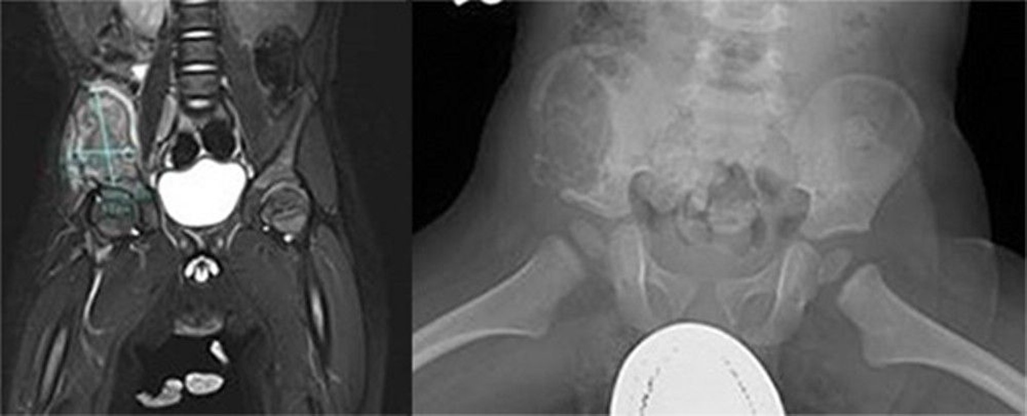
This image shows a large aggressive lesion within the right iliac wing. An MRI revealed the mass was heterogenous on T2 and low T1 signal with heterogenous enhancement of the mass and surrounding musculature with deviation of the right psoas muscle medially, suggesting an aggressive bone lesion (left). A large lytic lesion within the anterior and lateral ala of the ileum extending anterior and inferior to the ischium was visible on x-ray (right). There are expanded cortex and cortical irregularities. Biopsy was diagnostic for Langerhans cell histiocytosis.
Images courtesy of Carolyn Fein Levy, MD, and Jeffrey M. Lipton, MD, PhD.
Radiographs are often obtained because of presenting symptoms. Bone lesions are usually sharply marginated, and round or oval, with a beveled edge giving the appearance of depth. However, some lesions are radiographically indistinguishable from Ewing sarcoma, osteosarcoma, other benign and malignant conditions, or osteomyelitis.
Diagnosis is based on biopsy. Langerhans cells are usually prominent, except in older lesions. These cells are identified by a pathologist experienced in the diagnosis of LCH according to their immunohistochemical characteristics, which include cell surface CD1a, CD207 (langerin), and S-100 (although not specific). Tumor tissue should be tested for BRAFV600E mutation and other MAPK pathway mutations via next generation sequencing (NGS). Once the diagnosis is established, the extent of disease must be determined by appropriate laboratory studies and imaging.
Laboratory studies used to define the extent of disease include the following:
Complete blood count with differential (bone marrow aspiration in patients with unexplained anemia or thrombocytopenia)
Comprehensive metabolic panel
Coagulation studies
Early morning urinalysis and urine osmolality
Imaging studies include the following:
Skeletal survey, including chest radiography
Ultrasound of the abdomen
CT of the chest (if the chest radiograph is abnormal or pulmonary symptoms are present)
CT or MRI of the abdomen (if examination reveals hepatosplenomegaly or if liver test results are abnormal)
PET/CT if available (because it can identify bone lesions not seen on skeletal survey)
MRI of the spine (if clinically indicated or lesions identified on skeletal survey or PET/CT)
MRI of the head (to evaluate the pituitary gland, temporal bones, and orbit)
Treatment of Langerhans Cell Histiocytosis
Supportive care
Sometimes hormone replacement therapy for hypopituitarism, most commonly antidiuretic hormone deficiency
Chemotherapy for multisystem involvement, single system multifocal involvement, and involvement in certain CNS risk sites such as skull-based lesions
Sometimes surgery with curettage, corticosteroid injection, or very rarely, radiation therapy (usually for unifocal bone involvement)
General supportive care is essential and may include scrupulous hygiene to limit ear, cutaneous, and dental lesions. Debridement or resection of severely affected gingival tissue limits oral involvement. Seborrhea-like dermatitis of the scalp may diminish with use of a selenium-based shampoo twice a week. If shampooing is ineffective, topical corticosteroids are used in small amounts and briefly in small areas.
Patients with systemic disease are monitored for potential chronic disabilities, such as cosmetic or functional orthopedic and cutaneous disorders and neurologic lesions as well as for psychologic problems that may require psychosocial support.
Many patients require hormone replacement for diabetes insipidus or other manifestations of hypopituitarism.
Chemotherapy is indicated for patients with multisystem involvement, single system multifocal involvement, and disease in certain CNS risk sites, such as the bones of the skull (including zygomatic, orbital, sphenoid, temporal, and ethmoid bones—1, 2). CNS risk lesions imparts a higher risk of neurodegenerative disease. Therefore even when these present as single bone lesions treatment is recommended (2, 3). Protocols sponsored by the Histiocyte Society are used; treatment protocols vary according to the risk category.
Imaging studies are repeated at 6 and 12 weeks to assess response to therapy. Patients with a good response will continue on therapy (4). Patients with a poor response or progression during therapy should have more intensive therapy. Protocols for poor responders with goals of early aggressive salvage are under study (5).
Local surgery, corticosteroid injection, curettage, or very rarely, radiation therapy is used for disease involving a single bone. These treatments should be done by specialists experienced in treating Langerhans cell histiocytosis. Easily accessible lesions in noncritical locations undergo surgical curettage. Surgery should be avoided when it may result in significant cosmetic deformities, orthopedic deformities, or loss of function.
Radiation therapy was sometimes given to patients at risk of vision loss secondary to proptosis, skeletal deformity from vertebral collapse, or spinal cord injury or to patients with severe pain. However, with the use of chemotherapy and targeted medications, it is rarely necessary.
Patients with Langerhans cell histiocytosis that progresses despite standard therapy usually respond to more aggressive chemotherapy. Patients who do not respond to salvage chemotherapy may undergo reduced-intensity hematopoietic stem cell transplantation, experimental chemotherapy, or immunosuppressive or other immunomodulatory therapy. Patients with BRAFV600E mutations in whom multiple lines of therapy fail may be candidates for BRAF inhibitors (eg, vemurafenib, dabrafenib) alone or in combination with a MEK inhibitor (eg, trametinib) (6), and those with other mutations or without mutations can be considered for MAPK inhibitor therapy (eg, trametinib, cobimetinib—7, 8). The successful use of targeted therapy with dabrafenib and trametinib as first-line therapy has been reported (9), but further studies on larger patient populations are needed.
Treatment references
1. Minkov M, Grois N, McClain K, et al: Langerhans cell histiocytosis: Histocyte Society evaluation and treatment guidelines. April 2009.
2. Haupt R, Minkov M, Astigarraga I, et al: Langerhans cell histiocytosis (LCH): guidelines for diagnosis, clinical work-up, and treatment for patients till the age of 18 years. Pediatr Blood Cancer 60(2):175–184, 2013. doi: 10.1002/pbc.24367
3. Allen CE, Ladisch S, McClain KL: How I treat Langerhans cell histiocytosis. Blood 126(1):26–35, 2015. doi:10.1182/blood-2014-12-569301
4. Gadner H, Minkov M, Grois N, et al: Therapy prolongation improves outcome in multisystem Langerhans cell histiocytosis. Blood 121(25):5006–5014, 2013. doi: 10.1182/blood-2012-09-455774
5. Gulati N, Allen CE: Langerhans cell histiocytosis: Version 2021. Hematol Oncol 39 Suppl 1(Suppl 1):15–23, 2021. doi:10.1002/hon.2857
6. Whitlock JA, Geoerger B, Dunkel IJ, et al: Dabrafenib, alone or in combination with trametinib, in BRAF V600-mutated pediatric Langerhans cell histiocytosis. Blood Adv 7(15):3806–3815, 2023. doi:10.1182/bloodadvances.2022008414
7. Suh JK, Kang S, Kim H, et al: Recent advances in the understanding of the molecular pathogenesis and targeted therapy options in Langerhans cell histiocytosis. Blood Res 56(S1):S65–S69, 2021. doi: 10.5045/br.2021.2021013
8. Diamond EL, Durham BH, Ulaner GA, et al: Efficacy of MEK inhibition in patients with histiocytic neoplasms. Nature 567(7749):521–524, 2019. doi:10.1038/s41586-019-1012-y
9. Cournoyer E, Ferrell J, Sharp S, et al: Dabrafenib and trametinib in Langerhans cell histiocytosis and other histiocytic disorders. Haematologica 109(4):1137–1148, 2024. doi:10.3324/haematol.2023.283295
Prognosis for Langerhans Cell Histiocytosis
Generally, patients with single system disease (unifocal, multifocal, and central nervous system [CNS] risk lesions) and multisystem disease without risk-organ involvement are considered low risk. Patients with multisystem disease and risk-organ involvement are considered high risk. Response to therapy has been shown to be a more important risk factor than age.
With treatment, the overall survival rate for patients with multisystem disease without risk organ involvement is 100%, but event-free survival is about 70% (1). Death is rare among patients with organ involvement who do not respond to initial therapy. However, disease recurrence is more common in multisystem disease patients with risk organ involvement. A chronic remitting and exacerbating course may occur, particularly among adults.
Some evidence suggests that BRAFV600E mutations are more likely to be present in patients with high-risk multisystem disease with risk-organ involvement and have an increased risk of treatment failure and relapse (2). Circulating BRAFV600E levels have been shown to accurately reflect disease activity and affect response to therapy (3, 4). Further investigations are ongoing to assess circulating cell-free BRAFV600E as a biomarker of disease and incorporation of these findings into therapeutic decision making.
Prognosis references
1. Haupt R, Minkov M, Astigarraga I, et al: Langerhans cell histiocytosis (LCH): guidelines for diagnosis, clinical work-up, and treatment for patients till the age of 18 years. Pediatr Blood Cancer 60(2):175–184, 2013. doi: 10.1002/pbc.24367
2. Héritier S, Emile JF, Barkaoui MA, et al: BRAF Mutation Correlates With High-Risk Langerhans Cell Histiocytosis and Increased Resistance to First-Line Therapy. J Clin Oncol 34(25):3023–3030, 2016. doi:10.1200/JCO.2015.65.9508
3. Héritier S, Hélias-Rodzewicz Z, Lapillonne H, et al: Circulating cell-free BRAFV600E as a biomarker in children with Langerhans cell histiocytosis. Br J Haematol 178(3):457–467, 2017. doi:10.1111/bjh.14695
4. Wang CJ, Zhu T, Zhao CZ, et al: BRAF-V600E mutations in plasma and peripheral blood mononuclear cells correlate with prognosis of pediatric Langerhans cell histiocytosis treated with first-line therapy. Pediatr Blood Cancer 71(9):e31099, 2024. doi:10.1002/pbc.31099
Key Points
Langerhans cell histiocytosis (LCH) involves a proliferation of myeloid-derived dendritic cells that infiltrate one or more organs.
Manifestations vary significantly depending on the organ(s) affected.
Bone lesions cause pain; lesions at the skull base may affect vision, hearing, and pituitary function (particularly causing diabetes insipidus).
Liver, spleen, lymph nodes, and bone marrow may be affected, resulting in a worse prognosis.
Use surgery or curettage with or without corticosteroid injection for single bone lesions.
Use chemotherapy in patients with multisystem, multifocal and skull-based site involvement.
Mutations that can be identified using next-generation sequencing is helpful in diagnosis, prognosis, and treatment with targeted therapy.
More Information
The following English-language resources may be useful. Please note that The Manual is not responsible for the content of these resources.
Histiocyte Society: International society for research into treatment of histiocytic diseases
North American Consortium for Histiocytosis: Conducts clinical and translational studies on histiocytosis and supports researchers and clinicians working in the field
PDQ® Pediatric Treatment Editorial Board: PDQ Langerhans Cell Histiocytosis Treatment. Bethesda, MD: National Cancer Institute. Updated June 14, 2024. Available at: https://www.cancer.gov/types/langerhans/hp/langerhans-treatment-pdq. [PMID: 26389240].
Drug Information for the Topic


