


Hyperkalemia is a serum potassium concentration > 5.5 mEq/L (> 5.5 mmol/L), usually resulting from decreased renal potassium excretion or abnormal movement of potassium out of cells. There are usually several simultaneous contributing factors, including increased potassium intake, medications that impair renal potassium excretion, and acute kidney injury or chronic kidney disease. Hyperkalemia can also occur in metabolic acidosis (eg, as in diabetic ketoacidosis). Clinical manifestations are generally neuromuscular, resulting in muscle weakness and cardiac toxicity that, when severe, can degenerate to ventricular fibrillation or asystole. Diagnosis is by measuring serum potassium. Treatment may involve decreasing potassium intake, adjusting medications, giving a cation exchange resin and, in emergencies, giving calcium gluconate, insulin, and dialysis.
(See also Overview of Disorders of Potassium Concentration.)
Etiology of Hyperkalemia
A common cause of increased serum potassium concentration is:
Pseudohyperkalemia
Pseudohyperkalemia is most often caused by hemolysis of red blood cells in a blood sample. Pseudohyperkalemia can also occur as a result of prolonged application of a tourniquet or excessive fist clenching when venous blood is drawn. Thrombocytosis can cause pseudohyperkalemia in serum (platelet potassium is released during clotting), as can extreme leukocytosis.
Normal kidneys eventually excrete potassium loads, so sustained, nonartifactual hyperkalemia usually implies diminished renal potassium excretion. However, other factors usually contribute. They can include increased potassium intake, increased potassium release from cells, or both (see table ). When sufficient potassium chloride is rapidly ingested or given parenterally, severe hyperkalemia may result even when renal function is normal, but hyperkalemia is usually temporary.
Factors Contributing to Hyperkalemia
Description | Cause | Examples |
|---|---|---|
Decreased potassium excretion | Medications | Angiotensin-converting enzyme inhibitors Angiotensin II receptor blockers Cyclosporine Direct renin inhibitor (aliskiren) Heparin Lithium Potassium-sparing diuretics Nonsteroidal anti-inflammatory drugs Tacrolimus Trimethoprim |
Hypoaldosteronism | ||
Kidney disorders | ||
Other disorders | Decreased effective circulating volume | |
Increased potassium intake (usually iatrogenic) | Oral intake | Dietary Oral potassium supplements |
IV intake | Blood transfusions IV fluids with supplemental potassium Parenteral nutrition Potassium citrate solutions Potassium-containing medications (eg, penicillin G) | |
Increased potassium movement out of cells | Medications | Beta-blockers Digoxin toxicity |
Increased tissue catabolism | Acute intravascular hemolysis Bleeding into soft tissues or gastrointestinal tract | |
Inherited disorders | ||
Insulin deficiency | Fasting | |
Other disorders | Exercise |
Hyperkalemia due to total body potassium excess is particularly common in oliguric states (especially acute kidney injury) and with rhabdomyolysis, burns, bleeding into soft tissue or the gastrointestinal tract, and adrenal insufficiency. In chronic kidney disease, hyperkalemia is uncommon until the glomerular filtration rate falls to < 10 to 15 mL/minute unless dietary or IV potassium intake is excessive.
Symptoms and Signs of Hyperkalemia
Although flaccid paralysis occasionally occurs, hyperkalemia is usually asymptomatic until cardiac arrhythmias develop.
In the rare disorder hyperkalemic familial periodic paralysis, weakness frequently develops during attacks and can progress to frank paralysis.
Diagnosis of Hyperkalemia
Serum potassium measurement
ECG
Review of medications
Assessment of renal function
Hyperkalemia (serum potassium > 5.5 mEq/L [> 5.5 mmol/L]) may be found on routine serum electrolyte measurement. It should be suspected in patients with typical changes on an ECG or in patients at high risk, such as those with chronic kidney disease, acute kidney injury, advanced heart failure, or urinary obstruction, or treated with angiotensin-converting enzyme (ACE) inhibitors and potassium-sparing diuretics.
Pseudohyperkalemia should be considered in patients without risk factors or ECG abnormalities. Hemolysis may be reported by the laboratory. When pseudohyperkalemia is suspected, potassium concentration should be repeated, taking measures to avoid hemolysis of the sample (such as avoiding small-gauge needles or tourniquet use and limiting fist clenching), and blood should be promptly processed by the laboratory.
ECG
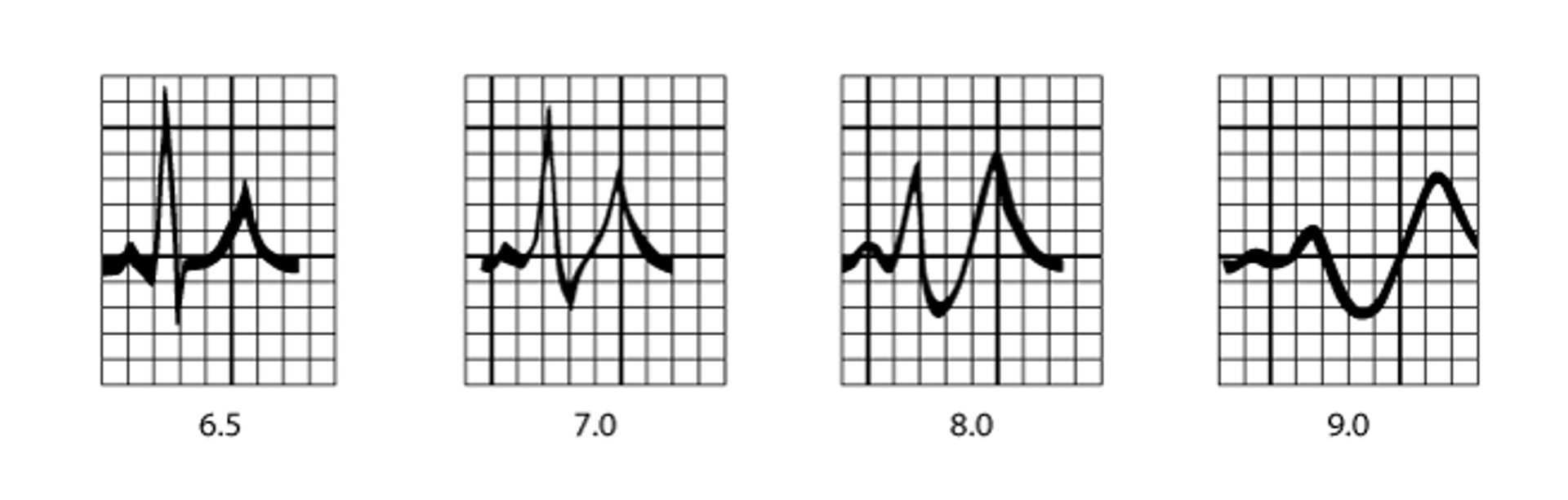
ECG should be done on patients with hyperkalemia. ECG changes (see figure ) are frequently visible when serum potassium is > 5.5 mEq/L (> 5.5 mmol/L). Slowing of conduction is characterized by an increased PR interval and shortening of the QT interval. Tall, symmetric, peaked T waves are visible initially. Potassium > 6.5 mEq/L (> 6.5 mmol/L) causes further slowing of conduction with widening of the QRS interval, disappearance of the P wave, and nodal and escape ventricular arrhythmias. Finally, the QRS complex degenerates into a sine wave pattern, and ventricular fibrillation or asystole ensues.
ECG Patterns in Hyperkalemia
Typical progression of ECG findings in hyperkalemia. Serum potassium concentrations (in mEq/L and mmol/L) vary widely among patients with similar ECG changes. |

Diagnosis of cause
Diagnosis of the cause of hyperkalemia requires a detailed history, including a review of medications, a physical examination with emphasis on volume status, and measurement of electrolytes, blood urea nitrogen (BUN), and creatinine. In cases in which kidney failure is present, additional tests, including renal ultrasound to exclude obstruction, are needed.
Treatment of Hyperkalemia
Treatment of the cause
For mild hyperkalemia, sodium polystyrene sulfonate, patiromer, or sodium zirconium cyclosilicate
For moderate or severe hyperkalemia, IV insulin and glucose, an IV calcium solution, possibly an inhaled beta 2-agonist, and usually hemodialysis
Mild hyperkalemia
Patients with serum potassium < 6 mEq/L (< 6 mmol/L) and no ECG abnormalities may respond to diminished potassium intake or stopping potassium-elevating medications. The addition of a loop diuretic enhances renal potassium excretion as long as volume depletion is not present.
Sodium polystyrene sulfonate in sorbitol can be given (15 to 30 g in 30 to 70 mL of 70% sorbitol orally every 4 to 6 hours). It acts as a cation exchange resin and removes potassium through the gastrointestinal mucosa. Sorbitol is administered with the resin to ensure passage through the gastrointestinal tract. Patients unable to take medications orally because of nausea or other reasons may be given similar doses by enema. Enemas are not as effective at lowering potassium in patients with ileus. Enemas should not be used if acute abdomen is suspected. Approximately 1 mEq (1 mmol) of potassium is removed per gram of resin given. Resin therapy is slow and often fails to lower serum potassium significantly in hypercatabolic states. Because sodium is exchanged for potassium when sodium polystyrene sulfonate is used, sodium overload (see Hypernatremia) may occur, particularly in patients with oliguria and preexisting volume overload.
In patients with recurrent hyperkalemia, avoidance of medications that can induce hyperkalemia (see table ) is generally all that is needed. In patients who need ACE inhibitors and angiotensin II receptor blockers (eg, patients with chronic heart failure or diabetic nephropathy), the polymer resin patiromer can be taken daily to help decrease gut absorption of potassium and prevent hyperkalemia. Sodium zirconium cyclosilicate may also be used. It is a polymer matrix that binds to potassium in the gut. It decreases serum potassium over several hours and has few gastrointestinal adverse effects.
Moderate to severe hyperkalemia
Serum potassium between 6 and 6.5 mEq/L (6 and 6.5 mmol/L) needs prompt attention, but the actual treatment depends on the clinical situation.
If no ECG changes are present and renal function is intact, maneuvers as for mild hyperkalemia are usually effective. Follow-up serum potassium measurements are needed to ensure that the hyperkalemia has been successfully treated.
If serum potassium is > 6.5 mEq/L (> 6.5 mmol/L), more aggressive therapy is required. Administration of regular insulin 5 to 10 units IV is followed immediately by or administered simultaneously with rapid infusion of 50 mL 50% glucose. Infusion of 10% dextrose in water should follow at 50 mL/hour to prevent hypoglycemia. The effect on serum potassium peaks in 1 hour and lasts for several hours.
If ECG changes include the loss of P-wave or widening of the QRS complex, treatment with IV calcium as well as insulin and glucose is indicated; 10 to 20 mL of 10% calcium gluconate (or 5 to 10 mL of 22% calcium gluceptate) is given IV over 5 to 10 minutes. If the ECG shows a sine wave pattern or asystole, calcium gluconate may be given more rapidly (5 to 10 mL IV over 2 minutes). Calcium antagonizes the effect of hyperkalemia on cardiac muscle. Calcium should be given with caution to patients taking digoxin because of the risk of precipitating hypokalemia-related arrhythmias. Calcium chloride can also be used but can be irritating to peripheral veins and cause tissue necrosis if extravasated. Calcium chloride should be given only through a correctly positioned central venous catheter.
The benefits of calcium occur within minutes but last only 20 to 30 minutes. Calcium infusion is a temporizing measure while awaiting the effects of other treatments or initiation of hemodialysis and may need to be repeated.
A high-dose beta 2-agonist, such as albuterol 10 to 20 mg inhaled over 10 minutes (5 mg/mL concentration), can lower serum potassium by 0.5 to 1.5 mEq/L (0.5 to 1.5 mmol/L) and may be a helpful adjunct. The peak effect occurs in 90 minutes. However, beta 2-agonists are contraindicated in patients with unstable angina or acute myocardial infarction.
Administration of IV sodium bicarbonate (NaHCO3) is frequently used to treat hyperkalemia, but evidence supporting its use is limited. It may lower serum potassium over several hours. Reduction may result from alkalinization or from the hypertonicity due to the concentrated sodium in the preparation, some of which is reabsorbed, causing potassium to be excreted to preserve electrical neutrality. The amount of sodium contained in the infusion may be harmful for patients being treated with dialysis who also may have volume overload. Another possible complication of IV sodium bicarbonate is that it acts to acutely lower the ionized calcium concentration, which further exacerbates the cardiotoxicity of hyperkalemia. When sodium bicarbonate is given, the typical dose is 3 ampules of 7.5% sodium bicarbonate in one liter of 5% dextrose in water infused over 2 to 4 hours. Bicarbonate therapy has little effect when used by itself in patients with severe renal insufficiency unless acidemia is also present.
In addition to strategies for lowering potassium by shifting it into cells, maneuvers to remove potassium from the body should also be done early in the treatment of severe or symptomatic hyperkalemia. Potassium can be removed via the gastrointestinal tract by administration of sodium polystyrene sulfonate, but because the rate of potassium removal is somewhat unpredictable, close monitoring is needed.
Patiromer and sodium zirconium silicate are not recommended for use as an emergency treatment to acutely lower potassium because of these medications' delayed onset of action.
Hemodialysis should be instituted promptly when emergency medical treatment is ineffective especially in patients with severe chronic kidney disease and/or acute kidney injury. Dialysis should be considered early in patients with end-stage renal disease and hyperkalemia because they are at increased risk of progression to more severe hyperkalemia and serious cardiac arrhythmias. Peritoneal dialysis is relatively inefficient at removing potassium acutely.
Key Points
Common causes of hyperkalemia include potassium-retaining medications, renal insufficiency, adrenal insufficiency, and disorders involving cellular breakdown (eg, rhabdomyolysis, burns, bleeding into soft tissue or the gastrointestinal tract).
Hyperkalemia is usually asymptomatic until cardiac toxicity develops, although some patients have weakness.
ECG changes begin with an increased PR interval, shortening of the QT interval, and tall, symmetric, peaked T waves; with potassium > 6.5 mEq/L (> 6.5 mmol/L), QRS interval widens, and P wave disappears; ultimately, the QRS complex degenerates into a sine wave pattern, and ventricular fibrillation or asystole ensues.
Give sodium polystyrene sulfonate, patiromer, or sodium zirconium cyclosilicate for mild hyperkalemia.
Give IV insulin, glucose, and calcium, and possibly an inhaled beta 2-agonist for moderate to severe hyperkalemia.
Use hemodialysis for patients with chronic kidney disease and those with significant ECG changes.
Drug Information for the Topic


