


Echinococcosis is caused by the dog tapeworms Echinococcus granulosus and Echinococcus multilocularis. In people, the tapeworms can cause fluid-filled cysts or masses to form in the liver or other organs.
People are usually infected when they accidentally consume soil, water, or food that has been contaminated by Echinococcus eggs passed in dog stool.
Cysts form in the liver, lungs, or another organ and cause various symptoms, including pain.
Doctors do imaging tests (such as ultrasonography or computed tomography) to check for cysts, blood tests to check for antibodies to the tapeworm, and analysis of fluid from the cyst to confirm the diagnosis.
Treatment usually involves removing cysts or draining them, injecting a salt solution to kill the parasite and then removing it, and giving the medication albendazole.Treatment usually involves removing cysts or draining them, injecting a salt solution to kill the parasite and then removing it, and giving the medication albendazole.
(See also Overview of Parasitic Infections and Tapeworm Infection.)
Adult tapeworm species called Echinococcus granulosus and Echinococcus multilocularis live in the intestine of dogs or other canines (such as foxes or coyotes). These tapeworms sometimes infect people, causing cysts in the liver or other organs.
Echinococcus granulosus is common in the sheep-raising areas of the Mediterranean, Middle East, Australia, New Zealand, South Africa, and South America. It also occurs rarely in some parts of North America.
Echinococcus multilocularis occurs mainly in Central Europe, Alaska, Canada, and Siberia. It also occurs rarely in the continental United States in Wyoming, the Dakotas, and the upper Midwest.
Dog tapeworm life cycle
Dogs, particularly herd dogs, become infected when they consume cysts of the tapeworms in tissues of infected animals (such as sheep, goats, cattle, or pigs). The cysts (called hydatid cysts) develop into adult tapeworms in the dog's intestine. Infected dogs pass tapeworm eggs in their stool. Sheep, cattle, goats, or pigs consume tapeworm eggs in soil contaminated with dog stool. Inside these animals, the eggs hatch and develop into cysts in the animal's internal organs.
People (often shepherds) are infected when they accidentally consume soil, water, or food that has been contaminated by Echinococcus eggs passed in dog stool.
Echinococcus eggs remain alive in soil for up to a year. Eggs may also be present on the fur of infected animals. After people touch an infected animal, they may pick up eggs, transfer the eggs from their hands to their mouth or to food, and thus become infected.
The eggs hatch in the intestine and release spheres that contain tapeworm larvae (called oncospheres). The spheres penetrate the wall of the intestine and travel through the bloodstream to various organs, such as the liver and lungs. In these organs, the spheres develop into cysts, which enlarge gradually and which, in people, can cause symptoms. The resulting infection is called echinococcosis.

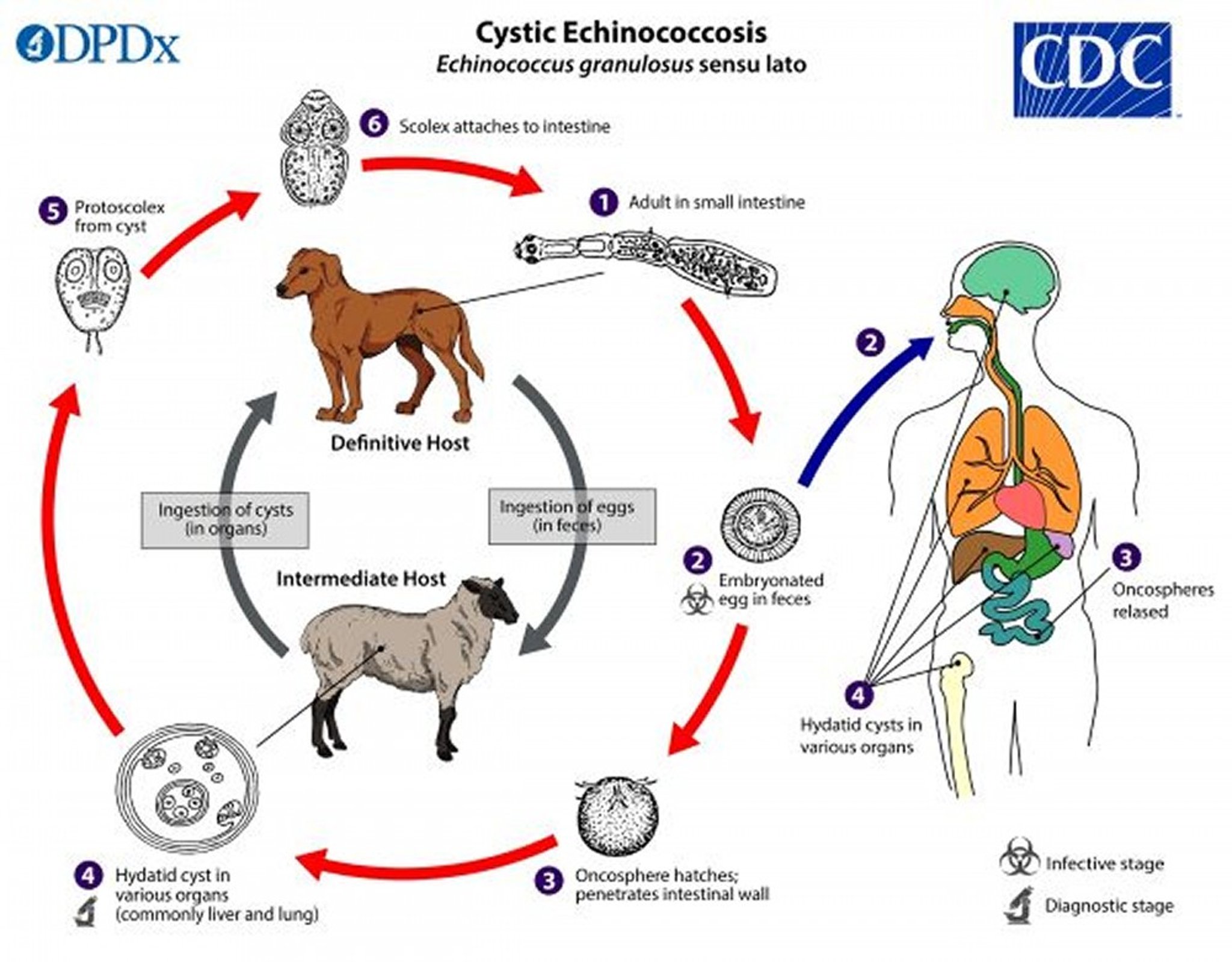
1. The adult dog tapeworm lives in the intestine of dogs and other canines (called the definitive hosts).
2. The adult tapeworms release eggs, which are passed in stool.
3. After the eggs are consumed by other animals (called intermediate hosts)—usually, sheep, goats, pigs, cattle, horses, camels, or people—the eggs hatch in the intestine and release spheres (called oncospheres) that contain tapeworm larvae. The spheres penetrate the wall of the intestine.
4. Then, the spheres travel through the bloodstream to various organs, such as the liver and lungs. In these organs, the spheres develop into cysts, which enlarge gradually and which, in people, can cause symptoms. Larvae (called protoscolices) and smaller cysts form within the cyst. Dogs and other canines (such as foxes or coyotes) become infected by consuming cysts in the organs of the infected intermediate host (such as a sheep, goat, or pig).
5–6. After a dog or other canine consumes the cysts, the cysts release the protoscolices, which attach to the wall of the intestine and develop into adults.
Image from the Centers for Disease Control and Prevention Image Library, Global Health, Division of Parasitic Diseases and Malaria.
Symptoms of Echinococcosis
Echinococcosis symptoms include the following:
Abdominal pain and yellowing of the skin and whites of the eyes (jaundice) if cysts form in the liver
Chest pain and coughing up blood or the contents of cysts if cysts form in the lungs
Hives or a severe life-threatening allergic reaction (anaphylaxis)
Diagnosis of Echinococcosis
X-rays, ultrasonography, computed tomography, or magnetic resonance imaging
Blood tests
Cysts in the liver or other tissues can be seen using ultrasonography, computed tomography (CT), or magnetic resonance imaging (MRI). Echinococcosis cysts in the lungs can be seen on chest x-rays and are sometimes discovered when a routine x-ray is taken.
Blood tests for antibodies to Echinococcus may also be helpful. Antibodies are proteins produced by the immune system to help defend the body against attack, including that by parasites.
Treatment of Echinococcosis
Surgical removal or drainage of the cyst
Injection of a salt solution to kill the parasite
AlbendazoleAlbendazole
Doctors can often surgically remove the Echinococcus granulosus cyst or drain the cyst with a needle. To drain the cyst, they use ultrasound to guide placement of the needle. They then remove cyst fluid, inject a salt solution into the cyst to kill the parasites, and drain the solution out. Masses due to Echinococcus multilocularis are removed surgically.
Albendazole is an oral prescription medication used to treat a variety of parasitic worm infections. Albendazole is an oral prescription medication used to treat a variety of parasitic worm infections.Albendazole is given before and during procedures, such as surgery or drainage of a cyst with a needle, to prevent the infection from spreading if the cyst's contents spill during the procedure. Albendazole is usually continued for 1 to 6 months after the procedure. It reduces the likelihood that a cyst will come back or spread. Albendazole alone can kill some cysts. It is also used to suppress the growth of cysts that cannot be removed surgically or drained.
Occasionally, when the infection is severe, liver transplantation is done.
Blood tests (complete blood count and liver enzymes) are monitored during albendazole therapy.Blood tests (complete blood count and liver enzymes) are monitored during albendazole therapy.
Prevention of Echinococcosis
Echinococcosis can be prevented by
Carefully washing hands
Not consuming food or water that may be contaminated with dog stool in areas where echinococcosis occurs
Educating sheep farmers and administering niclosamide to dogs that are in regular contact with sheep
Drug Information for the Topic


