

Lumbar spinal stenosis is narrowing of the spinal canal in the lower back. The narrowing squeezes (compresses) the nerves that travel through the lower back into the legs.
Osteoarthritis, injuries, spondylolisthesis, and spondylolysis can cause narrowing of the spinal canal.
Pain is felt in the low back and may travel down one or both legs.
The diagnosis is based on a doctor's evaluation and sometimes on the results of imaging or electrodiagnostic tests.
Treatment includes measures to relieve pain and sometimes surgery.
The spinal canal runs through the center of the spine and contains the spinal cord and the bundle of nerves that extends downward from the bottom of the spinal cord in the lower back. The word lumbar means lower, and stenosis means narrowing.

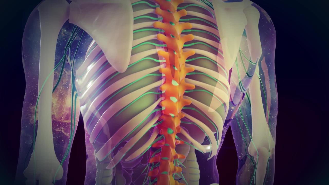
Along the length of the spinal cord are the spinal nerves. The spinal nerves emerge from the sides through spaces between the vertebrae to connect with nerves throughout the body. The part of the spinal nerve nearest the spinal cord is called the spinal nerve root. Because of their location, spinal nerve roots can be squeezed when the spinal canal is narrowed, resulting in pain.
Lumbar spinal stenosis is a common cause of low back pain in older people and may also cause sciatica. Spinal stenosis also develops in middle-aged people who were born with a narrow spinal canal.
The most common causes of lumbar spinal stenosis include osteoarthritis, spondylolisthesis, and spondylolysis. Paget disease of bone is a less common cause.
The Spine
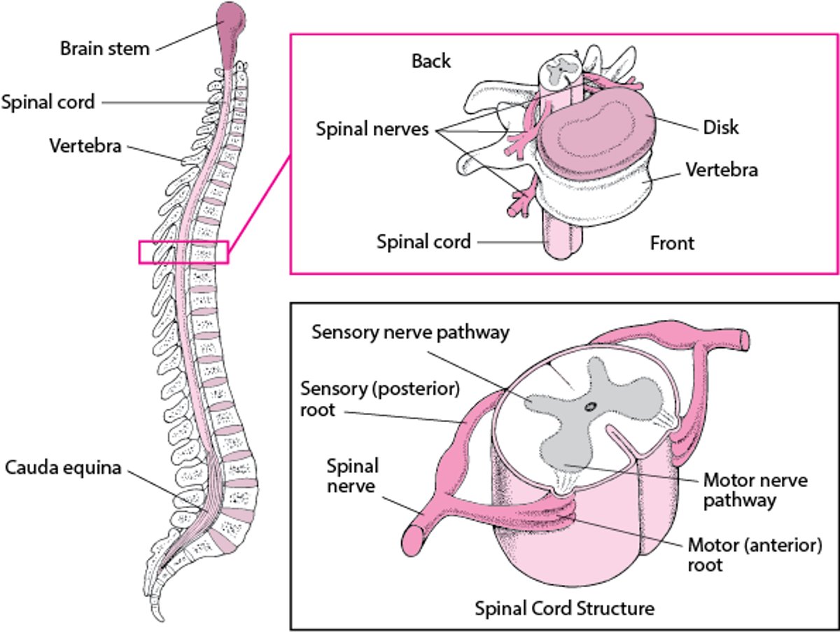
A column of bones called vertebrae make up the spine (spinal column). The vertebrae protect the spinal cord (a long, fragile structure contained in the spinal canal), which runs through the center of the spine. Between the vertebrae are discs composed of cartilage, which help cushion the spine and give it some flexibility. Spinal nerves: Emerging from the spinal cord between the vertebrae are 31 pairs of spinal nerves. Each nerve emerges in two short branches (roots)—motor and sensory—which join to form a spinal nerve. The motor roots carry commands from the brain and spinal cord to other parts of the body, particularly to skeletal muscles. The sensory roots carry information to the brain from other parts of the body. Cauda equina: The spinal cord ends about three fourths of the way down the spine, but a bundle of nerves extends beyond the cord. This bundle is called the cauda equina because it resembles a horse's tail. The cauda equina carries nerve impulses to and from the legs, lower intestine, and bladder. |

Symptoms of Lumbar Spinal Stenosis
Symptoms of lumbar spinal stenosis vary depending on which part of the spinal cord and nerves are affected. People may have pain, pins-and-needles sensations, weakness, and reduced reflexes in a foot or leg.
Pain may be felt over the lower back and be worsened by straightening the back (as when walking or leaning back), is relieved by leaning forward or sitting, and may travel down one leg or both legs. Similarly, walking up hills is less painful than walking down.
Pain also occurs in the buttocks, thighs, or calves during walking, running, climbing stairs, or even standing. The pain is not relieved by standing still but by flexing the back or by sitting (although people may continue to have pins-and-needles sensations). People often experience some relief when pushing a shopping cart or using a rolling walker because the back is slightly flexed. Similarly, walking up hills is less painful than walking down.
Rarely, sudden compression of a nerve rootlets can cause cauda equina syndrome. If the cauda equina (the bundle of nerves extending from the bottom of the cord in the lower back) is affected, control of bladder and bowels can be lost. A lower leg can become paralyzed, and feeling may be lost in and around the groin. If these serious symptoms develop, medical attention is required immediately.
Diagnosis of Lumbar Spinal Stenosis
A doctor's evaluation
Sometimes imaging tests, electrodiagnostic studies, or both
Doctors typically make the diagnosis of lumbar spinal stenosis based on the characteristic pain. During a physical examination, doctors check a person's strength and reflexes.
Doctors may do other tests if people have weakness or numbness or if their symptoms have lasted for more than 6 weeks. Magnetic resonance imaging (MRI) and computed tomography (CT) are imaging tests that can help doctors identify abnormalities of the spine that are causing lumbar spinal stenosis. Tests of the nerves and muscles (electrodiagnostic tests), such as nerve conduction studies and electromyography, can help doctors identify the affected area of stenosis and spinal nerve root compression, and the severity of the damage.
Treatment of Lumbar Spinal Stenosis
Measures to relieve pain
Sometimes surgery for severe pain
Measures to relieve pain
One to two days of bed rest may provide pain relief. Longer bed rest weakens the core muscles (abdominal muscles, which run from the bottom of the rib cage to the pelvis, muscles in the back of the spine, and muscles in the buttocks) and increases stiffness, thus worsening back pain and prolonging recovery. Sleeping in a comfortable position on a medium mattress is recommended. People who sleep on their back can place a pillow under their knees. People who sleep on their side should use a pillow to support their head in a neutral position (not tilted down toward the bed or up toward the ceiling). They should place another pillow between their knees with their hips and knees bent slightly if that relieves their back pain. People can continue to sleep on their stomach if they are comfortable doing so.
Applying cold (such as ice packs) or heat (such as a heating pad) or using over-the-counter analgesics (such as acetaminophen and nonsteroidal anti-inflammatory drugs [NSAIDs]) may help relieve the pain. Some people may be helped by medications that reduce nerve pain, such as gabapentin, antiseizure medications, or certain antidepressants. If pain is severe or persists, doctors may give corticosteroid injections into the epidural space (between the spine and the outer layer of tissue covering the spinal cord).(such as a heating pad) or using over-the-counter analgesics (such as acetaminophen and nonsteroidal anti-inflammatory drugs [NSAIDs]) may help relieve the pain. Some people may be helped by medications that reduce nerve pain, such as gabapentin, antiseizure medications, or certain antidepressants. If pain is severe or persists, doctors may give corticosteroid injections into the epidural space (between the spine and the outer layer of tissue covering the spinal cord).
Physical therapy and stretching the hamstring muscles gently after warming up may help relieve muscle spasms. (See also Low Back Pain: Prevention.)
Surgery
When measures to relieve pain are not effective in people with lumbar stenosis, surgery may be needed to relieve pressure on the spinal cord and spinal nerves. One surgical procedure is called lumbar laminectomy. In this procedure, a small incision is created in the skin along the lower bones in the back (the lumbar vertebrae). The muscles are separated, and the bone is exposed. A portion of the vertebrae called the lamina is removed, taking pressure off the spinal cord and spinal nerves. Sometimes, when removing the lamina in several vertebrae is necessary, the vertebrae can be fused together with another piece of bone. In some cases, pressure can be relieved by just making a hole in the lamina rather than removing it entirely. This procedure is called lumbar laminotomy. Both lumbar laminectomy and lumbar laminotomy can be done through very small incisions, which shortens recovery time.


