

Gout is a disorder caused by the precipitation of monosodium urate crystals in and around joints, most often causing recurrent acute or chronic arthritis. The initial attack (flare) of gout is usually monoarticular and often involves the first metatarsophalangeal joint. Symptoms of gout include acute, severe pain, tenderness, warmth, redness, and swelling. Hyperuricemia (serum urate > 6.8 mg/dL [> 0.4 mmol/L]) is almost always present. Definite diagnosis requires identification of crystals in synovial fluid. Treatment of acute flares is with anti-inflammatory drugs. Acute flares can be treated with nonsteroidal anti-inflammatory drugs (NSAIDs), colchicine, glucocorticoids, and/or occasionally IL-1 inhibitors (eg, anakinra). Long-term management to prevent recurrent flares involves persistent lowering of the serum urate level below its saturation level (< 6.8 mg/dL [< 0.4 mmol/L]) with allopurinol, febuxostat, or uricosuric drugs such as probenecid.Gout is a disorder caused by the precipitation of monosodium urate crystals in and around joints, most often causing recurrent acute or chronic arthritis. The initial attack (flare) of gout is usually monoarticular and often involves the first metatarsophalangeal joint. Symptoms of gout include acute, severe pain, tenderness, warmth, redness, and swelling. Hyperuricemia (serum urate > 6.8 mg/dL [> 0.4 mmol/L]) is almost always present. Definite diagnosis requires identification of crystals in synovial fluid. Treatment of acute flares is with anti-inflammatory drugs. Acute flares can be treated with nonsteroidal anti-inflammatory drugs (NSAIDs), colchicine, glucocorticoids, and/or occasionally IL-1 inhibitors (eg, anakinra). Long-term management to prevent recurrent flares involves persistent lowering of the serum urate level below its saturation level (allopurinol, febuxostat, or uricosuric drugs such as probenecid.
Gout is more common among men than women. Usually, gout develops during middle age in men and after menopause in women (estrogen has a partially protective effect). Gout is rare in younger people but is often more severe in people who develop the disorder before age 30. There is a strong hereditary component to developing hyperuricemia and gout. Patients with metabolic syndrome are also at increased risk of gout.
Pathophysiology of Gout
The greater the degree and duration of hyperuricemia, the greater the likelihood that gout will develop (1). Urate levels can be elevated because of
Decreased renal (most common) or gastrointestinal excretion (2)
Increased production (rare)
Increased purine intake (usually in combination with decreased excretion) is generally only a minor contributor
Why only some people with elevated serum uric acid (urate) levels develop gout flares is not known.
Decreased renal excretion is by far the most common cause of hyperuricemia (2). It may be hereditary (eg, due to variations in uric acid transporter efficiency) and also occurs in patients receiving thiazide and loop diuretics and in diseases that decrease the glomerular filtration rate (GFR). Ethanol increases purine catabolism in the liver and increases the formation of lactic acid, which blocks urate secretion by the renal tubules, and ethanol may also stimulate liver urate synthesis (). It may be hereditary (eg, due to variations in uric acid transporter efficiency) and also occurs in patients receiving thiazide and loop diuretics and in diseases that decrease the glomerular filtration rate (GFR). Ethanol increases purine catabolism in the liver and increases the formation of lactic acid, which blocks urate secretion by the renal tubules, and ethanol may also stimulate liver urate synthesis (3).
Increased production of urate may be caused by increased nucleoprotein turnover in hematologic conditions (eg, lymphoma, leukemia, hemolytic anemia) and in conditions with increased rates of cellular proliferation and cell death (eg, psoriasis, cytotoxic cancer therapy, tumor lysis syndrome [4], radiation therapy). Increased urate production may also occur as a primary hereditary abnormality and in obesity, because urate production correlates with body surface area (5). In most cases, the cause of urate overproduction is unknown, but rarely can be attributable to enzyme abnormalities; deficiency of hypoxanthine-guanine phosphoribosyltransferase (complete deficiency is Lesch-Nyhan syndrome) is a possible rare cause, as is overactivity of phosphoribosylpyrophosphate synthetase (6).
Increased intake of purine-rich foods (eg, shellfish, red meat, liver, kidney, anchovies, asparagus, consommé, herring, meat gravies and broths, mushrooms, mussels, sardines, sweetbreads) can contribute to hyperuricemia (7). Beer, including nonalcoholic beer, is particularly rich in guanosine, a purine nucleoside (8). However, a strict low-purine diet lowers serum urate by only about 1 mg/dL (0.1 mmol/L) and thus is rarely sufficient therapy for patients with gout. High-fructose corn syrup also stimulates liver synthesis of urate. While diet in most patients is a mild contributor to the serum urate level, overconsumption of fructose may alter the inflammatory milieu and increase the likelihood of the immune system reacting to uric acid deposits, causing a flare.
Urate precipitates as needle-shaped monosodium urate (MSU) crystals, which are deposited extracellularly in avascular tissues (eg, cartilage) or in relatively avascular tissues (eg, tendons, tendon sheaths, ligaments, walls of bursae) and skin around cooler distal joints and tissues (eg, ears, finger pads). In severe, long-standing hyperuricemia, MSU crystals may be deposited in larger central joints and in the parenchyma of organs such as the kidney. At the acid pH of urine, urate precipitates readily as small platelike or diamond-shaped uric acid crystals that may aggregate to form gravel or stones, which may obstruct urine outflow. Tophi are MSU crystal aggregates that most often develop in joint and cutaneous tissue. They are usually encased in a fibrous granulomatous matrix, which prevents them from causing acute inflammation.
Acute gouty arthritis may be triggered by trauma, medical stress (eg, infection), surgery, use of thiazide diuretics or medications with hypouricemic effects (eg, allopurinol, febuxostat, probenecid, nitroglycerin), or indulgence in purine-rich food or alcohol (Acute gouty arthritis may be triggered by trauma, medical stress (eg, infection), surgery, use of thiazide diuretics or medications with hypouricemic effects (eg, allopurinol, febuxostat, probenecid, nitroglycerin), or indulgence in purine-rich food or alcohol (9). Flares are often precipitated by a sudden increase or, more commonly, a sudden decrease in serum urate levels (10). Why acute flares follow some of these precipitating conditions is unknown. Tophi in and around joints can limit motion and cause deformities, called chronic tophaceous gouty arthritis. Gout increases the risk of developing secondary osteoarthritis (11).
Pathophysiology references
1. Choi HK, Mount DB, Reginato AM; American College of Physicians; American Physiological Society. Pathogenesis of gout. Ann Intern Med. 2005;143(7):499-516. doi:10.7326/0003-4819-143-7-200510040-00009
2. Perez-Ruiz F, Calabozo M, Erauskin GG, Ruibal A, Herrero-Beites AM. Renal underexcretion of uric acid is present in patients with apparent high urinary uric acid output. Arthritis Rheum. 2002;47(6):610-613. doi:10.1002/art.10792
3. Yamamoto T, Moriwaki Y, Takahashi S. Effect of ethanol on metabolism of purine bases (hypoxanthine, xanthine, and uric acid). . Effect of ethanol on metabolism of purine bases (hypoxanthine, xanthine, and uric acid).Clin Chim Acta. 2005;356(1-2):35-57. doi:10.1016/j.cccn.2005.01.024
4. Howard SC, Jones DP, Pui CH. The tumor lysis syndrome [published correction appears in N Engl J Med. 2018 Sep 13;379(11):1094.]. N Engl J Med. 2011;364(19):1844-1854. doi:10.1056/NEJMra0904569
5. Choi HK, Ford ES. Prevalence of the metabolic syndrome in individuals with hyperuricemia. Am J Med. 2007;120(5):442-447. doi:10.1016/j.amjmed.2006.06.040
6. Torres RJ, Puig JG. Hypoxanthine-guanine phosophoribosyltransferase (HPRT) deficiency: Lesch-Nyhan syndrome. Orphanet J Rare Dis. 2007;2:48. Published 2007 Dec 8. doi:10.1186/1750-1172-2-48
7. Choi HK, Atkinson K, Karlson EW, Willett W, Curhan G. Purine-rich foods, dairy and protein intake, and the risk of gout in men. N Engl J Med. 2004;350(11):1093-1103. doi:10.1056/NEJMoa035700
8. Choi HK, Atkinson K, Karlson EW, Willett W, Curhan G. Alcohol intake and risk of incident gout in men: a prospective study. Lancet. 2004;363(9417):1277-1281. doi:10.1016/S0140-6736(04)16000-5
9. Helget LN, Mikuls TR. Environmental Triggers of Hyperuricemia and Gout. Rheum Dis Clin North Am. 2022;48(4):891-906. doi:10.1016/j.rdc.2022.06.009
10. Urano W, Yamanaka H, Tsutani H, et al. The inflammatory process in the mechanism of decreased serum uric acid concentrations during acute gouty arthritis. J Rheumatol. 2002;29(9):1950-1953.
11. Yokose C, Chen M, Berhanu A, Pillinger MH, Krasnokutsky S. Gout and Osteoarthritis: Associations, Pathophysiology, and Therapeutic Implications. Curr Rheumatol Rep. 2016;18(10):65. doi:10.1007/s11926-016-0613-9
Symptoms and Signs of Gout

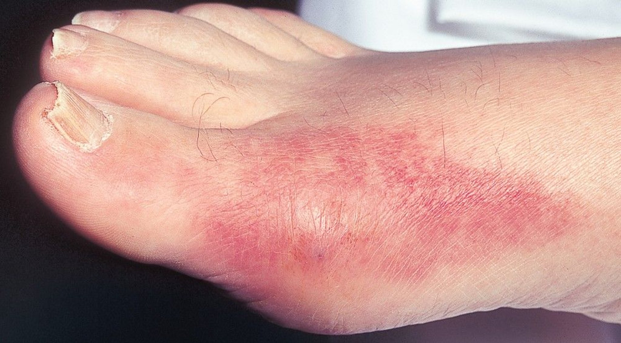
Podagra, or acute pain in the first metatarsophalangeal joint that is accompanied by redness, tenderness, and swelling, is a common manifestation of acute gout.
© Springer Science+Business Media
Acute gouty arthritis usually begins with sudden onset of pain (often nocturnal). The metatarsophalangeal joint of a great toe is most often involved (called podagra), but the instep, ankle, knee, wrist, and elbow are also common sites. Rarely, the hip, shoulder, sacroiliac, sternoclavicular, or cervical spine joints are involved. The pain becomes progressively more severe, usually over a few hours, and is often excruciating. Swelling, warmth, redness, and exquisite tenderness may suggest infection. The overlying skin may become tense, warm, shiny, and red or purplish. Fever, tachycardia, chills, and malaise sometimes occur.
Course
The first few flares usually affect only a single joint and typically resolve spontaneously over 7 to 10 days (1). Later flares may affect several joints simultaneously or sequentially and persist up to 3 weeks if untreated. Subsequent flares develop after progressively shorter symptom-free intervals. Eventually, multiple flares may occur each year. If ongoing urate-lowering therapy is not initiated, patients can develop chronic deforming arthritis from tophaceous gout due to ongoing urate deposition.
Tophi

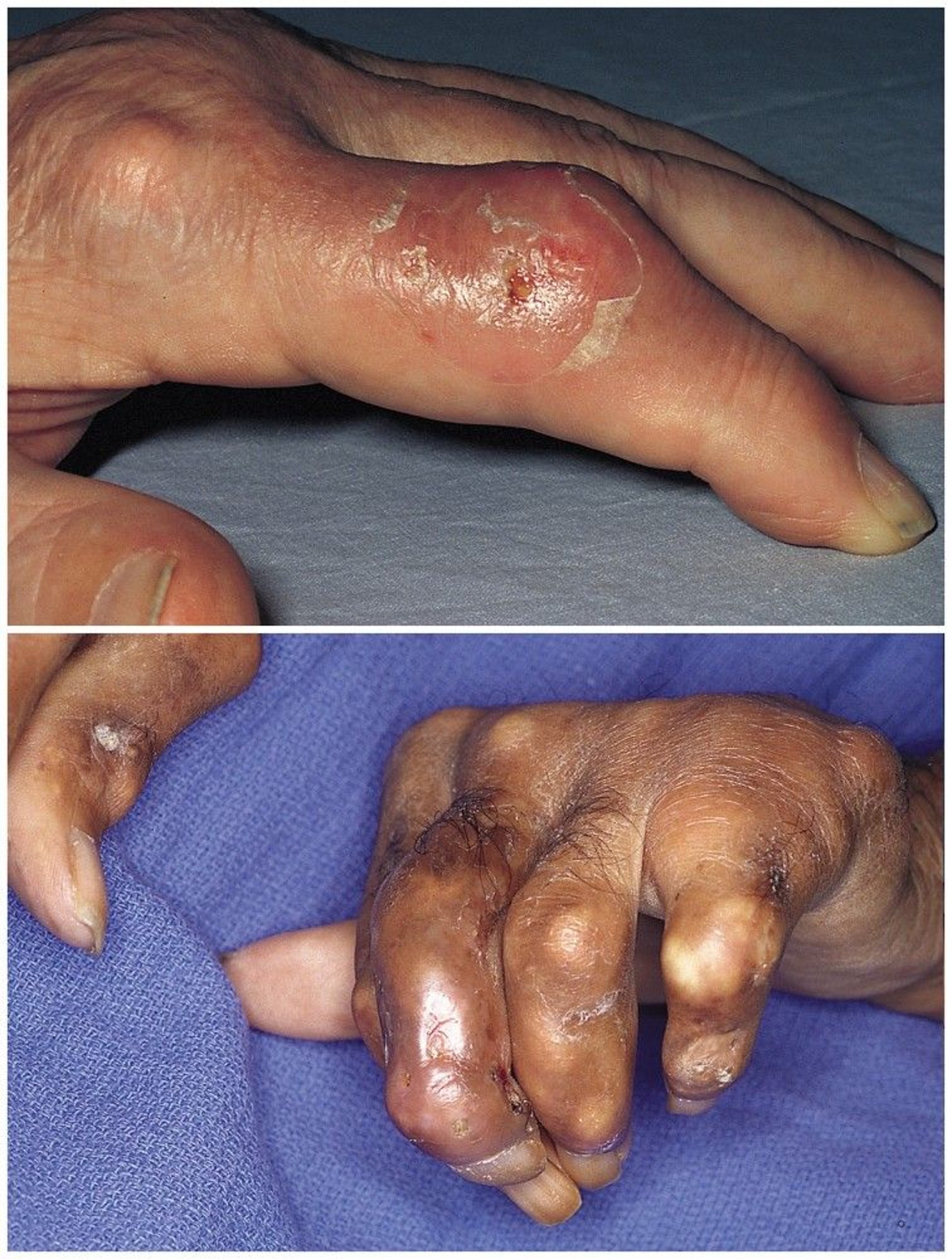
The top photo shows firm nodules, swelling, and serous drainage in the finger caused by tophaceous gout. The bottom photo shows tophaceous gout with flexion deformities in the hands, which can also be accompanied by destructive erosions of bone and cartilage in multiple joints.
© Springer Science+Business Media

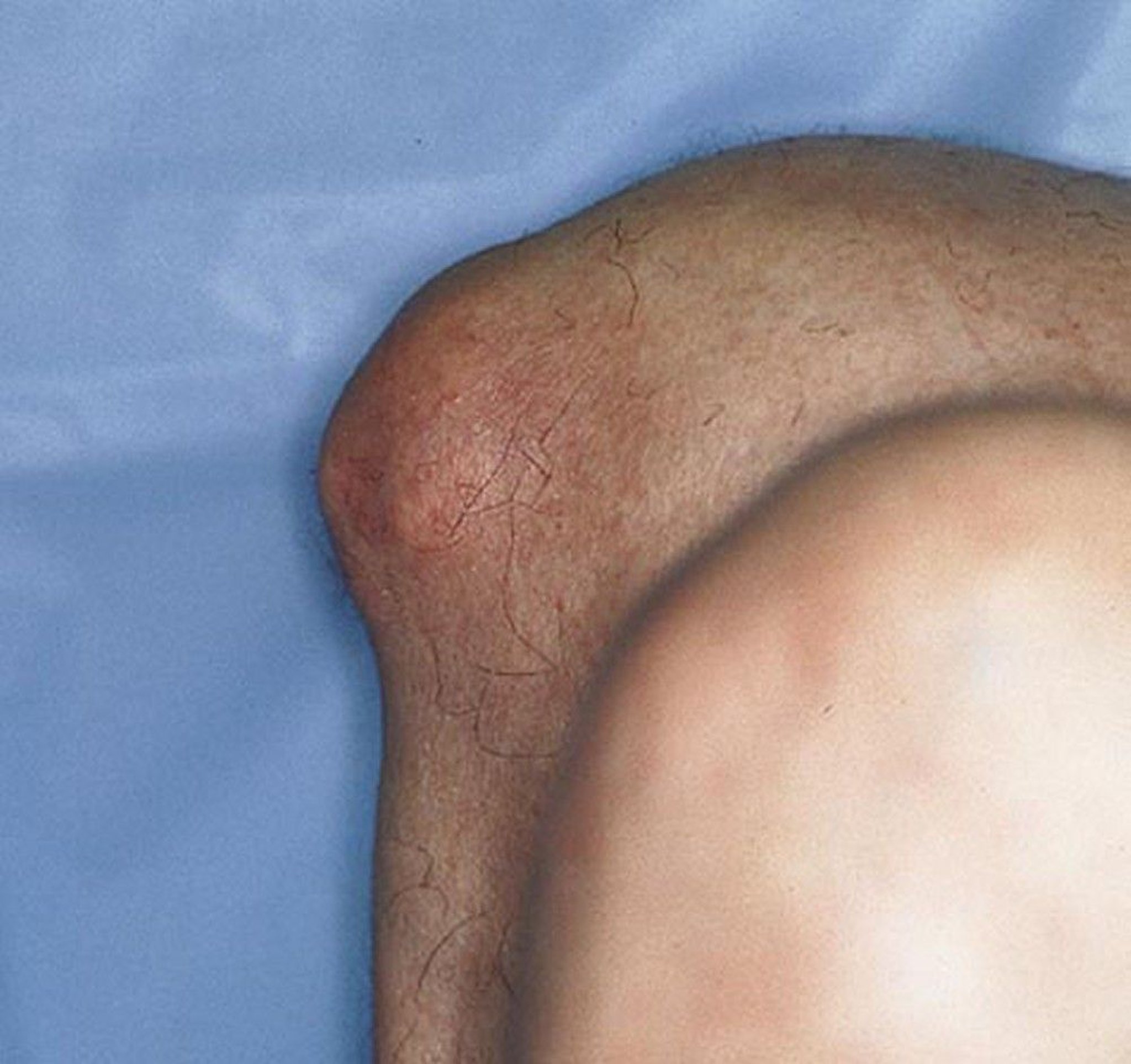
This image shows prominent tophaceous deposits in the prepatellar bursa of the right knee.
© Springer Science+Business Media
Palpable tophi develop in patients with gout and can rarely occur in patients who have never had acute gouty arthritis. They are usually firm yellow or white papules or nodules, single or multiple. They can develop in various locations, commonly the fingers, hands, feet, and around the olecranon or Achilles tendon. Tophi can also develop in the kidneys and other organs and under the skin on the ears. Patients with osteoarthritic Heberden nodes may develop tophi in the nodes. This development occurs most often in older women taking diuretics, and these can become dramatically inflamed and misdiagnosed as inflammatory osteoarthritis. Normally painless, tophi, especially in the olecranon bursae, can become acutely inflamed and painful, often after mild or inapparent injury. Tophi may erupt through the skin, discharging chalky masses of urate crystals. These sinus tracts can become infected. Tophi in and around joints may eventually cause deformities and secondary osteoarthritis.
Complications of gout
Gouty arthritis can cause pain, deformity, and limited joint motion. Inflammation can be flaring in some joints while subsiding in others. Patients with gout may develop urolithiasis with uric acid stones or calcium oxalate stones.
Complications of gout include renal obstruction and infection, with secondary tubulointerstitial disease. Untreated progressive renal dysfunction, most often related to coexisting hypertension or, less often, some other cause of nephropathy, further impairs excretion of urate, accelerating crystal deposition in tissues.
There is an increased incidence of myocardial infarction following acute flares (2). Cardiovascular disease, obstructive sleep apnea, metabolic dysfunction–associated liver disease (MASLD), and components of metabolic syndrome are common among patients with gout.
Symptoms and signs references
1. Neogi T. Clinical practice. Gout. N Engl J Med. 2011;364(5):443-452. doi:10.1056/NEJMcp1001124
2. Cipolletta E, Tata LJ, Nakafero G, Avery AJ, Mamas MA, Abhishek A. Association Between Gout Flare and Subsequent Cardiovascular Events Among Patients With Gout. JAMA. 2022;328(5):440-450. doi:10.1001/jama.2022.11390
Diagnosis of Gout
History and physical examination
Synovial fluid analysis
The diagnosis of gout should be suspected in patients with acute monoarticular arthritis or oligoarticular arthritis, particularly older adults or those with other risk factors. Podagra and recurrent instep inflammation are particularly suggestive. Previous flares that began explosively and resolved spontaneously within 7 to 10 days are also characteristic. Similar symptoms can result from the following:
Acute calcium pyrophosphate arthritis (calcium pyrophosphate dihydrate (CPPD) crystal deposition disease) (however, calcium pyrophosphate deposition generally occurs in larger joints, is not associated with tophi, and its clinical course is often milder but protracted)
Acute rheumatic fever with joint involvement and juvenile idiopathic arthritis (however, these disorders occur mostly in young people, who rarely get gout, and the former is often migratory with minimal joint effusions)
Rheumatoid arthritis (RA) (however, RA tends to be symmetrical and persistent, with more affected joints during a flare, flares persisting for longer periods of time, and flares in all joints subsiding together; whereas with gout, inflammation is usually flaring in some joints while subsiding in others)
Acute fracture in patients unable to provide a history of injury (particularly metatarsal stress fractures)
Acute infectious arthritis or chronic infectious arthritis (differentiation requires synovial fluid analysis)
Palindromic rheumatism
Acute calcific periarthritis caused by basic calcium phosphate
Palindromic rheumatism is characterized by acute, recurrent flares of inflammation in or near one or occasionally several joints or tendon sheaths with spontaneous resolution; pain can be as severe as in gout. Flares often subside spontaneously and completely but may last days to weeks. Such flares may herald the onset of RA, and the presence of rheumatoid factor and anticyclic citrullinated peptide (anti-CCP) antibodies can help in differentiation; they are positive in about 50% of patients (1).

This photo shows large erosions caused by tophi involving the distal metatarsal and proximal phalanx of the great toe.
Image courtesy of N. Lawrence Edwards, MD.

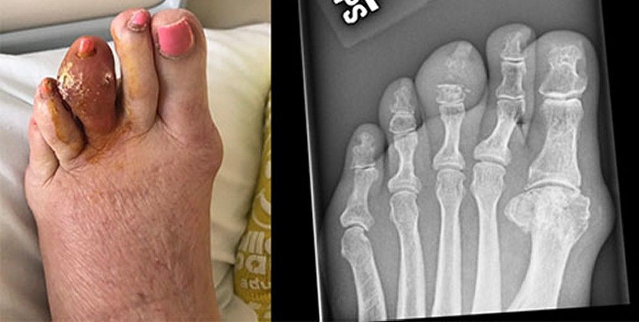
This photo shows a large tophus of the left third toe, which has broken down and released hardened uric acid. The radiograph on the right shows erosion of the middle phalanx and distal interphalangeal joint.
Images courtesy of Brian F. Mandell, MD.
Synovial fluid analysis
If acute gouty arthritis is suspected, arthrocentesis and synovial fluid analysis should be performed at the initial presentation (2). A typical recurrence in a patient with previously documented gout does not mandate arthrocentesis, but it should be performed if there is any question of the diagnosis or if the patient’s risk factors or any clinical characteristics suggest infectious arthritis (3, 4). In some cases, a diagnosis of gout may be reasonably presumed based on the patient’s history and clinical features or on imaging results in cases when joint fluid cannot be obtained; however, every attempt should be made to document the presence of MSU crystals in synovial fluid from an affected joint.

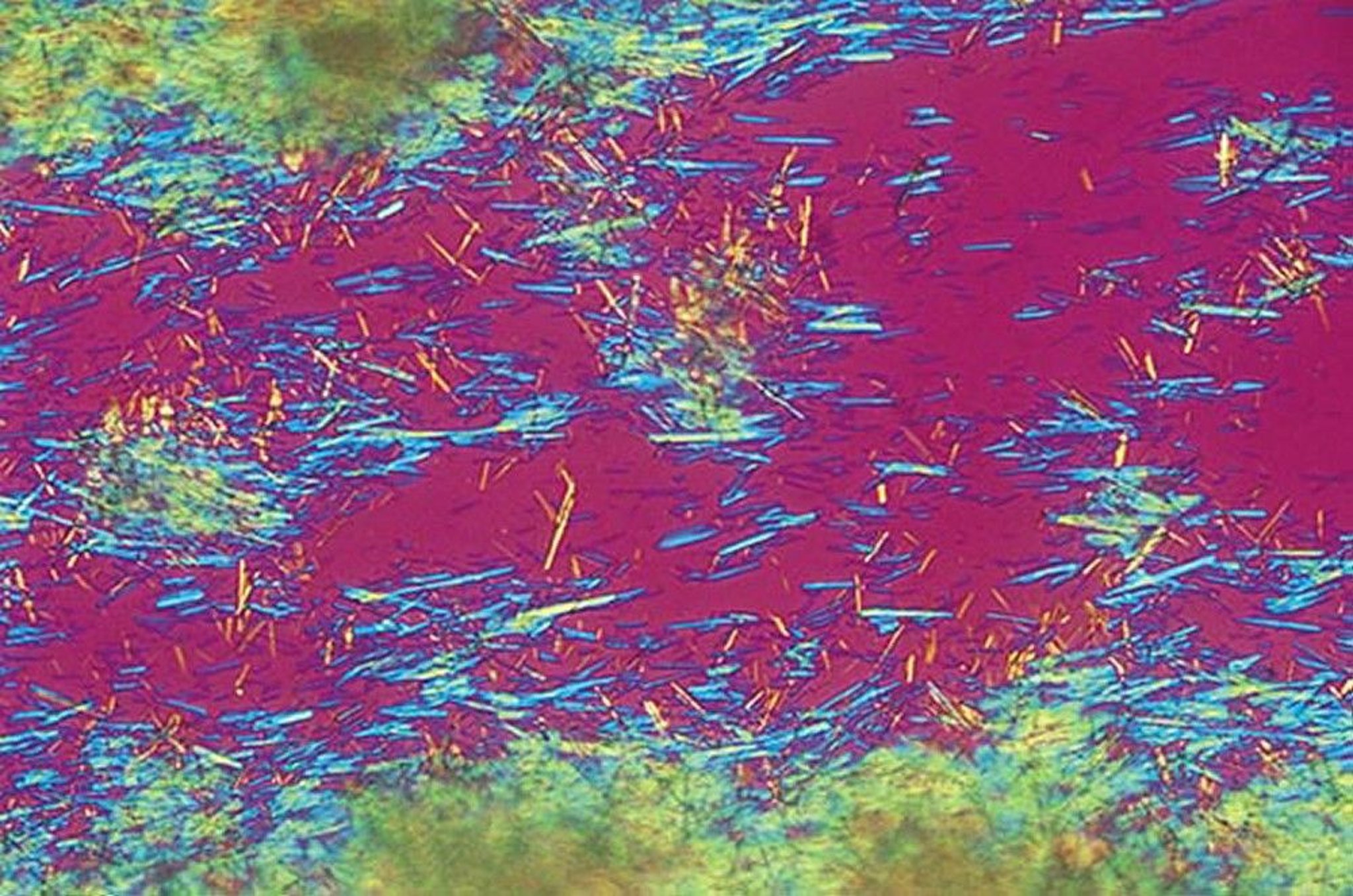
Brightly birefringent, needle-shaped, urate crystals aspirated from a gouty tophus can be seen in this image taken with a polarizing microscope and a red compensator filter. The negatively birefringent crystals appear yellow when they lie parallel to the optical axis of the compensator and blue when perpendicular to it.
By permission of the publisher. From Myers S: Atlas of Rheumatology. Edited by G Hunder. Philadelphia, Current Medicine, 2005.
Synovial fluid analysis can confirm the diagnosis by identifying needle-shaped, strongly negatively birefringent urate crystals that are free in the fluid or engulfed by phagocytes. Synovial fluid during flares has inflammatory characteristics (see table ), usually 2,000 to 100,000 white blood cells/mcL, with > 80% polymorphonuclear white blood cells ( 5). These findings overlap considerably with infectious arthritis, which must be excluded by Gram stain (which is insensitive) and culture ( 4).
Microscopic Examination of Crystals in Joints
Crystal Type | Birefringence | Elongation* | Shape | Length (micrometer) |
|---|---|---|---|---|
Monosodium urate | Strong | Negative | Needle- or rod-shaped | 2–15 |
Weak or often not birefringent | Positive | Rhomboid- or rod-shaped | 2–15 | |
Calcium oxalate (rare)† | Weak or strong | Positive or indeterminate | Bipyramidal | 5–30 |
Not birefringent with polarized light | — | Shiny, coinlike, or slightly irregular clumps (often too small to delineate) | 3–65 (aggregates) | |
* Elongation is determined by inserting an additional red plate or compensator between the 2 polarizing plates in the microscope and aligning a crystal with an orienting arrow denoting the axis of slow vibration. Crystals that have negative elongation are yellow parallel to the axis of slow vibration marked on the compensator; positive elongation appears blue in the same direction. In clinical practice, these have also been termed negative or positive birefringence. | ||||
† These crystals occur primarily in patients with kidney failure. | ||||
Serum urate level
An elevated serum urate level supports the diagnosis of gout but is neither specific nor sensitive; at least 30% of patients have a normal serum urate level during an acute flare in part due to the uricosuric properties of the proinflammatory cytokine interleukin-6 (IL-6) or because a sudden lowering of the serum urate precipitated the flare (6). However, the baseline serum urate level between flares reflects the size of the extracellular miscible urate pool. The level should be measured on 2 or 3 occasions in patients with newly proven gout to establish a baseline. The serum urate level can be low immediately after initiation of therapy, but flares can continue to occur as long as tissue deposits remain. The dissolution of the urate deposits may take many months after initiating therapy.
Diagnosis of chronic gouty arthritis
Synovial fluid findings from chronic effusions of affected joints are usually diagnostic. Radiographs of the affected joint are unnecessary if the diagnosis of acute gout has been established by synovial fluid analysis and rarely show erosions at the time of first flares.
Chronic gouty arthritis should be considered in patients with persistent unexplained joint disease or subcutaneous or bony tophi. Radiographs of the first metatarsophalangeal joint or other affected joint may be useful. These radiographs may show punched-out lesions of subchondral bone with overhanging bony margins, most commonly in the first metatarsophalangeal joint; lesions must be ≥ 5 mm in diameter to be visible on radiographs. Joint space is typically preserved until very late in the course of disease.
Diagnostic ultrasonography is increasingly used to detect a typical "double-contour sign" suggesting urate crystal deposition, but sensitivity is operator-dependent and differentiation from calcium pyrophosphate crystal deposits may be more difficult to do conclusively. Urate deposition over the articular cartilage (double-contour sign) and clinically inapparent tophi are characteristic changes. These findings may be evident even before the first gout flare. Dual-energy CT scans (DECTs) can also reveal uric acid deposits and can be useful if the diagnosis is unclear based on standard clinical evaluation and testing ( 7), particularly if synovial fluid aspiration and analysis cannot be done. No imaging modality is completely accurate, particularly when evaluating a first flare.
Diagnosis references
1. Salvador G, Gomez A, Vinas O, et al. Prevalence and clinical significance of anti-cyclic citrullinated peptide and antikeratin antibodies in palindromic rheumatism. An abortive form of rheumatoid arthritis?. Rheumatology (Oxford). 2003;42(8):972-975. doi:10.1093/rheumatology/keg268
2. Qaseem A, McLean RM, Starkey M, et al. Diagnosis of Acute Gout: A Clinical Practice Guideline From the American College of Physicians. Ann Intern Med. 2017;166(1):52-57. doi:10.7326/M16-0569
3. Horowitz DL, Katzap E, Horowitz S, Barilla-LaBarca ML. Approach to septic arthritis. Am Fam Physician. 2011;84(6):653-660.
4. Becker JA, Daily JP, Pohlgeers KM. Acute Monoarthritis: Diagnosis in Adults. Am Fam Physician. 2016;94(10):810-816.
5. Dore RK. The gout diagnosis. Cleve Clin J Med. 2008;75 Suppl 5:S17-S21. doi:10.3949/ccjm.75.suppl_5.s17
6. Zhang J, Sun W, Gao F, et al. Changes of serum uric acid level during acute gout flare and related factors.Front Endocrinol (Lausanne). 2023;14:1077059. Published 2023 Feb 21. doi:10.3389/fendo.2023.1077059
7. Singh JA, Budzik JF, Becce F, Pascart T. Dual-energy computed tomography vs ultrasound, alone or combined, for the diagnosis of gout: a prospective study of accuracy. Rheumatology (Oxford). 2021;60(10):4861-4867. doi:10.1093/rheumatology/keaa923
Treatment of Gout
Termination of an acute flare with nonsteroidal anti-inflammatory drugs (NSAIDs), colchicine, glucocorticoids, or an interleukin-1 (IL-1) inhibitorTermination of an acute flare with nonsteroidal anti-inflammatory drugs (NSAIDs), colchicine, glucocorticoids, or an interleukin-1 (IL-1) inhibitor
Prevention of further deposition of monosodium urate (MSU) crystals, reduction in flare incidence, and resolution of existing tophi by lowering the serum urate level (by decreasing urate production with allopurinol or febuxostat, dissolving deposits with uricase replacement therapy, or increasing urate excretion with probenecid)Prevention of further deposition of monosodium urate (MSU) crystals, reduction in flare incidence, and resolution of existing tophi by lowering the serum urate level (by decreasing urate production with allopurinol or febuxostat, dissolving deposits with uricase replacement therapy, or increasing urate excretion with probenecid)
Prevention of recurrent acute flares with daily colchicine or an NSAIDPrevention of recurrent acute flares with daily colchicine or an NSAID
Treatment of coexisting hypertension, hyperlipidemia, and obesity and avoidance of excess dietary purines
Treatment of acute flares
The approach to treatment of an acute gout flare is rapid initiation of anti-inflammatory therapy to resolve pain and restore function, with first-line options including colchicine, NSAIDs, and glucocorticoids (The approach to treatment of an acute gout flare is rapid initiation of anti-inflammatory therapy to resolve pain and restore function, with first-line options including colchicine, NSAIDs, and glucocorticoids (1, 2). The choice of these agents is individualized, largely based on comorbidities and contraindications. Interleukin-1 (IL-1) inhibitors (eg, anakinra) may be used for patients who do not tolerate or have contraindications to first-line therapies, or first-line therapies are ineffective.). The choice of these agents is individualized, largely based on comorbidities and contraindications. Interleukin-1 (IL-1) inhibitors (eg, anakinra) may be used for patients who do not tolerate or have contraindications to first-line therapies, or first-line therapies are ineffective.
Nonsteroidal anti-inflammatory drugs (NSAIDs) are effective in treating acute flares and are generally well-tolerated. However, they can have adverse effects, including gastrointestinal upset or bleeding, hyperkalemia, increases in creatinine, and fluid retention. Older and dehydrated patients are at particular risk, especially if there is a history of renal disease. Virtually any NSAID used in anti-inflammatory (high) doses is effective and is likely to exert an analgesic effect beginning within a few hours. Treatment should be continued for several days after the pain and signs of inflammation have resolved to prevent relapse.
Oral colchicine,Oral colchicine, a traditional therapy, produces a dramatic response in some patients if begun soon after the onset of symptoms; it is most effective if started within 12 to 24 hours of an acute flare. A dose of 1.2 mg can be followed with 0.6 mg 1 hour later; joint pain tends to decrease after 12 to 24 hours and sometimes ceases within 3 to 7 days, but continued dosing is generally needed to achieve resolution, which may take time. If colchicine is tolerated, 0.6 to 1.2 mg once a day can be continued as the flare subsides. Renal insufficiency and drug interactions, especially with CYP3A4 inhibitors (such as clarithromycin, antifungals, and grapefruit juice) and some statins may warrant reduction of dosage or use of other treatments. Gastrointestinal upset and diarrhea are common adverse effects.a traditional therapy, produces a dramatic response in some patients if begun soon after the onset of symptoms; it is most effective if started within 12 to 24 hours of an acute flare. A dose of 1.2 mg can be followed with 0.6 mg 1 hour later; joint pain tends to decrease after 12 to 24 hours and sometimes ceases within 3 to 7 days, but continued dosing is generally needed to achieve resolution, which may take time. If colchicine is tolerated, 0.6 to 1.2 mg once a day can be continued as the flare subsides. Renal insufficiency and drug interactions, especially with CYP3A4 inhibitors (such as clarithromycin, antifungals, and grapefruit juice) and some statins may warrant reduction of dosage or use of other treatments. Gastrointestinal upset and diarrhea are common adverse effects.
Glucocorticoids are used to treat acute flares. Aspiration of affected joints, followed by an intra-articular glucocorticoid injection (in patients who do not have suspected septic arthritis), is very effective, particularly for monoarticular symptoms; triamcinolone acetonide or methylprednisolone acetate can be used, with dose depending on the size of the affected joints. Oral prednisone (about 0.5 mg/kg once a day with taper) or methylprednisolone, IM or IV glucocorticoids can be effective, particularly if multiple joints are involved. As with NSAID therapy, glucocorticoids should be continued for a few days after the flare fully resolves to prevent relapse.are used to treat acute flares. Aspiration of affected joints, followed by an intra-articular glucocorticoid injection (in patients who do not have suspected septic arthritis), is very effective, particularly for monoarticular symptoms; triamcinolone acetonide or methylprednisolone acetate can be used, with dose depending on the size of the affected joints. Oral prednisone (about 0.5 mg/kg once a day with taper) or methylprednisolone, IM or IV glucocorticoids can be effective, particularly if multiple joints are involved. As with NSAID therapy, glucocorticoids should be continued for a few days after the flare fully resolves to prevent relapse.
If monotherapy is ineffective or doses (eg, of NSAIDs) are limited by toxicity, colchicine can be combined with NSAIDs or glucocorticoids.If monotherapy is ineffective or doses (eg, of NSAIDs) are limited by toxicity, colchicine can be combined with NSAIDs or glucocorticoids.
In addition to NSAIDs or glucocorticoids, supplementary analgesics, rest, ice application, and splinting of the inflamed joint may be helpful. If patients are taking urate-lowering drugs when an acute flare begins, the medications should be continued at the same dose; dose adjustments are deferred until the flare has subsided. There is no contraindication to initiating urate-lowering therapy during an acute flare if appropriate anti-inflammatory therapy is being provided.
If glucocorticoids, colchicine, and NSAIDs are contraindicated or ineffective, an If glucocorticoids, colchicine, and NSAIDs are contraindicated or ineffective, anIL-1 inhibitor, such as anakinra, can be used (, such as anakinra, can be used (3). Anakinra may hasten resolution of a flare and shorten the hospital stay of a patient with multiple comorbidities that limit the use of the other medications. Anakinra is typically given as 100 mg subcutaneously once a day until symptoms resolve. Anakinra has the advantage of not affecting glucose levels or kidney function or causing fluid retention and can be used in a patient with an active infection that is being appropriately treated. Due to cost considerations anakinra is not typically used to treat acute gout flares in the outpatient setting.). Anakinra may hasten resolution of a flare and shorten the hospital stay of a patient with multiple comorbidities that limit the use of the other medications. Anakinra is typically given as 100 mg subcutaneously once a day until symptoms resolve. Anakinra has the advantage of not affecting glucose levels or kidney function or causing fluid retention and can be used in a patient with an active infection that is being appropriately treated. Due to cost considerations anakinra is not typically used to treat acute gout flares in the outpatient setting.
Prevention of recurrent flares
The frequency of acute flares is reduced by taking 0.6 mg of colchicine once or twice a day (maximum 1.2 mg per day depending on tolerance and renal function). An extra two 0.6-mg tablets of colchicine taken at the first suggestion of a flare may abort flares. If the patient is taking prophylactic doses of colchicine and has taken higher doses of colchicine to treat an acute flare within the past 2 weeks, an NSAID or glucocorticoid should be used instead to try to abort the flare.The frequency of acute flares is reduced by taking 0.6 mg of colchicine once or twice a day (maximum 1.2 mg per day depending on tolerance and renal function). An extra two 0.6-mg tablets of colchicine taken at the first suggestion of a flare may abort flares. If the patient is taking prophylactic doses of colchicine and has taken higher doses of colchicine to treat an acute flare within the past 2 weeks, an NSAID or glucocorticoid should be used instead to try to abort the flare.
A (reversible) neuropathy and/or myopathy can develop during chronic colchicine ingestion. This condition is more likely to occur in patients with renal insufficiency, and in patients also receiving certain statins or macrolides, but rarely occurs in patients without any of these risk factors. Colchicine should be dose-reduced or avoided in patients with renal insufficiency or those who are on CYP3A4 inhibitors. A (reversible) neuropathy and/or myopathy can develop during chronic colchicine ingestion. This condition is more likely to occur in patients with renal insufficiency, and in patients also receiving certain statins or macrolides, but rarely occurs in patients without any of these risk factors. Colchicine should be dose-reduced or avoided in patients with renal insufficiency or those who are on CYP3A4 inhibitors.
Flare frequency can also be decreased with daily low-dose NSAIDs if kidney function allows. Chronic glucocorticoid use is not an ideal prophylactic therapy because of its adverse effect potential.
Lowering the serum urate level
Colchicine, NSAIDs, and glucocorticoids do not slow the progressive joint damage caused by tophi, because they do not lower the serum urate level or dissolve monosodium urate deposits. Joint damage can be prevented and, if present, may be reversed with urate-lowering medications. Tophaceous deposits are resorbed by lowering serum urate or dissolved with uricase replacement therapy. Maintaining a serum urate level below the saturation point (target is usually < 6 mg/dL [< 0.35 mmol/L]) will ultimately decrease the frequency of acute arthritic flares as the deposits are dissolved. This decrease is accomplished byColchicine, NSAIDs, and glucocorticoids do not slow the progressive joint damage caused by tophi, because they do not lower the serum urate level or dissolve monosodium urate deposits. Joint damage can be prevented and, if present, may be reversed with urate-lowering medications. Tophaceous deposits are resorbed by lowering serum urate or dissolved with uricase replacement therapy. Maintaining a serum urate level below the saturation point (target is usually < 6 mg/dL [< 0.35 mmol/L]) will ultimately decrease the frequency of acute arthritic flares as the deposits are dissolved. This decrease is accomplished by
Blocking urate production with xanthine oxidase inhibitors (XOI) (allopurinol or febuxostat)Blocking urate production with xanthine oxidase inhibitors (XOI) (allopurinol or febuxostat)
Increasing urate excretion with a uricosuric drug (probenecid or losartan)Increasing urate excretion with a uricosuric drug (probenecid or losartan)
Using both types of drugs together in severe tophaceous gout or in patients intolerant of higher doses of a XOI
Increasing urate excretion by converting urate to allantoin, which is more soluble and readily excreted, with uricase replacement therapy in patients such as those who have severe tophaceous gout or did not respond to other urate-lowering therapy
Urate-lowering therapy is indicated for patients with
Tophaceous deposits
Evidence of joint damage due to gout on imaging studies
Frequent or disabling flares (eg, ≥ 2 flares a year) of gouty arthritis
Urolithiasis
Patients with infrequent flares but whose serum uric acid level is > 9 mg/dL (> 0.5 mmol/L) or for whom having any flares poses a particular hardship
Multiple comorbidities (eg, peptic ulcer disease, chronic kidney disease) that are relative contraindications to the medications used to treat recurrent acute flares (NSAIDs or glucocorticoids)
Hyperuricemia is not usually treated in the absence of gout flares or uric acid nephrolithiasis.
The goal of urate-lowering therapy is to lower the serum urate level. If tophi are not present, a reasonable target level is < 6 mg/dL (< 0.35 mmol/L), which is below the level of saturation (> 6.8 mg/dL [0.4 mmol/L] at normal core body temperature and pH). Compelling data demonstrate decreased frequency of flares when the serum urate level decreases to < 6 mg/dL with this treat-to-target strategy. Two randomized trials have established that significantly fewer patients with a serum urate level < 6 mg/dL experienced gout flares than those with a higher serum urate level. The patients with the target serum urate (< 6 mg/dL) who did flare had fewer gout flares than patients who were above this serum urate threshold (4).
If palpable tophi are present or if there is marked disability from tophaceous deposits, a reasonable goal is to dissolve them more rapidly, and this requires an even lower target level. The lower the serum urate level, the faster tophi resolve. After presumed complete dissolution of deposits, the serum urate can be allowed to increase to a level < 6 mg/dL.
Medications are effective in lowering serum urate; dietary restriction of purines is less effective, but high intake of high-purine food, alcohol (beer in particular), and nonalcoholic beer should be avoided. Carbohydrate restriction (especially avoidance of high-fructose corn syrup) and weight loss can lower serum urate level, particularly in patients with insulin resistance because high insulin levels suppress urate excretion. Intake of low-fat dairy products should be encouraged. Because acute flares tend to develop during the first months of urate-lowering therapy, such therapy should be started in conjunction with once or twice a day colchicine or NSAIDs. levels suppress urate excretion. Intake of low-fat dairy products should be encouraged. Because acute flares tend to develop during the first months of urate-lowering therapy, such therapy should be started in conjunction with once or twice a day colchicine or NSAIDs.
Resolution of tophi may take many months even with maintenance of serum urate at low levels. Serum urate should be measured periodically, usually monthly while determining required medication dosage and then at least yearly to confirm the effectiveness of therapy or more often if there are medication changes or weight gain. Urate-lowering therapy should not be stopped if a patient has a flare.
Allopurinol,Allopurinol, a xanthine oxidase inhibitor of urate synthesis, is the most commonly prescribed and preferred initial urate-lowering therapy. Uric acid stones or gravel may dissolve during allopurinol treatment. Treatment usually begins with 50 to 100 mg orally once a day and can be slowly dose-escalated up to 800 mg orally once a day. The dose can be split if single daily dosing causes gastrointestinal distress. Some clinicians recommend decreasing the starting dose in patients with renal insufficiency (eg, 50 mg orally once a day if creatinine clearance is < 60 mL/min/1.73 m2) to potentially decrease the incidence of rare but severe systemic hypersensitivity reactions; however, data that confirm the effectiveness of this approach is limited. The final dose of allopurinol should be determined by the target serum urate level. The most commonly used daily dose is 300 mg, but this dose is effective in lowering serum uric acid levels to < 6 mg/dL (< 0.35 mmol/L) in fewer than 40% of patients with gout. Absorption of allopurinol may decrease at doses higher than 300 mg, so split dosing (eg, twice daily dosing) should be considered. should be determined by the target serum urate level. The most commonly used daily dose is 300 mg, but this dose is effective in lowering serum uric acid levels to allopurinol may decrease at doses higher than 300 mg, so split dosing (eg, twice daily dosing) should be considered.
Adverse effects of allopurinol include mild gastrointestinal distress and rash, which can be a harbinger of Stevens-Johnson syndrome, life-threatening hepatitis, vasculitis, or leukopenia. Adverse effects are more common among patients with renal dysfunction. HLA-B*5801 carriers are at higher risk of Adverse effects of allopurinol include mild gastrointestinal distress and rash, which can be a harbinger of Stevens-Johnson syndrome, life-threatening hepatitis, vasculitis, or leukopenia. Adverse effects are more common among patients with renal dysfunction. HLA-B*5801 carriers are at higher risk ofallopurinol hypersensitivity syndrome (which includes severe cutaneous adverse reactions), and HLA-B*5801 prevalence varies by race (5). Therefore, the American College of Rheumatology recommends testing for the for the HLA B*5801 allele prior to treatment in patients of Southeast Asian descent (eg, Han Chinese, Korean, Thai) and African American patients (6). The use of allopurinol is not recommended in HLA B*5801 positive patients unless the benefits clearly outweigh the risks. Alternative urate-lowering therapies (eg, febuxostat) may be used depending on patient-specific factors such as cardiovascular disease status, renal function, and disease severity. ). The use of allopurinol is not recommended in HLA B*5801 positive patients unless the benefits clearly outweigh the risks. Alternative urate-lowering therapies (eg, febuxostat) may be used depending on patient-specific factors such as cardiovascular disease status, renal function, and disease severity.Allopurinol is contraindicated in patients taking azathioprine or mercaptopurine because it can decrease metabolism of these medications and thus potentiate their immunosuppressive and cytolytic effects. Hepatic transaminase levels can become elevated and should be measured periodically. is contraindicated in patients taking azathioprine or mercaptopurine because it can decrease metabolism of these medications and thus potentiate their immunosuppressive and cytolytic effects. Hepatic transaminase levels can become elevated and should be measured periodically.
FebuxostatFebuxostat is costlier (in the United States) but more potent xanthine oxidase inhibitor of urate synthesis. It is especially useful in patients who do not tolerate allopurinol, who have contraindications to allopurinol, or in whom allopurinol does not sufficiently decrease urate levels. is costlier (in the United States) but more potent xanthine oxidase inhibitor of urate synthesis. It is especially useful in patients who do not tolerate allopurinol, who have contraindications to allopurinol, or in whom allopurinol does not sufficiently decrease urate levels.Febuxostat appears to prevent acute flares as efficaciously as allopurinol (7). Febuxostat is begun at 20–40 mg orally once a day and increased to 80 to 120 mg orally once a day if urate does not decrease to < 6 mg/dL (< 0.35 mmol/L). Febuxostat (like allopurinol) is contraindicated in patients taking azathioprine or mercaptopurine because it can decrease metabolism of these drugs. Compared with ) is contraindicated in patients taking azathioprine or mercaptopurine because it can decrease metabolism of these drugs. Compared withallopurinol, febuxostat increased the risk of mortality in one study of patients with known cardiovascular disease (8), but several additional studies have not confirmed this observation (9). Transaminase levels can become elevated and should be measured periodically.
PegloticasePegloticase is a pegylated form of recombinant uricase. Uricase is an enzyme, absent in humans, that converts urate to allantoin, which is more soluble. Pegloticase is expensive and is used primarily in patients with gout in whom other treatments have been unsuccessful in lowering the serum urate level. Pegloticase can also be used in patients who have a high burden of tophaceous deposits that would not likely be dissolved in a reasonable time period by other urate-lowering therapies. It is given IV every 2 weeks for many months (typically at least 6 to 9 months) to totally deplete the excess urate deposits; it often lowers the serum urate level to < 1 mg/dL (< 0.1 mmol/L). Pegloticase is contraindicated in patients with G6PD deficiency because it can cause hemolysis and methemoglobinemia. Pegloticase infusion can rarely be associated with symptoms consistent with anaphylaxis. The effectiveness of the currently available preparation is limited by a high rate of development of drug-neutralizing antibodies. Failure of urate levels to decrease to < 6 mg/dL (< 0.35 mmol/L) after a pegloticase infusion indicates the likely presence of anti-polyethylene glycol (anti-PEG) antibodies and an increased risk of future allergic reactions; regular infusions are then stopped. To prevent other urate-lowering drugs from masking ineffectiveness of infusion indicates the likely presence of anti-polyethylene glycol (anti-PEG) antibodies and an increased risk of future allergic reactions; regular infusions are then stopped. To prevent other urate-lowering drugs from masking ineffectiveness ofpegloticase, those other urate-lowering drugs should not be used together with pegloticase. Co-administration of immunosuppressants (eg, methotrexate) with . Co-administration of immunosuppressants (eg, methotrexate) withpegloticase may prevent development of the neutralizing antibodies.
Uricosuric therapy may be useful in patients who underexcrete uric acid (the majority of patients with hyperuricemia), have normal renal function, and have not had kidney stones. Probenecid is the only uricosuric medication available in the United States. Probenecid can be used as monotherapy if both allopurinol and febuxostat are contraindicated or not tolerated. may be useful in patients who underexcrete uric acid (the majority of patients with hyperuricemia), have normal renal function, and have not had kidney stones. Probenecid is the only uricosuric medication available in the United States. Probenecid can be used as monotherapy if both allopurinol and febuxostat are contraindicated or not tolerated.Probenecid loses efficacy with declining renal function and is generally not as useful with a glomerular filtration rate < 50 mL/min/1.73 m2. It is effective when added to a xanthine oxidase inhibitor, but is rarely required. Benzbromarone is a more potent uricosuric that is available outside of the United States.
The antihypertensive drug losartan and the triglyceride-lowering drug fenofibrate both have uricosuric effects and can be used to decrease uric acid in patients who have other reasons for taking these medications. Low doses of salicylates may decrease uric acid excretion and worsen hyperuricemia, but only trivially, and should The antihypertensive drug losartan and the triglyceride-lowering drug fenofibrate both have uricosuric effects and can be used to decrease uric acid in patients who have other reasons for taking these medications. Low doses of salicylates may decrease uric acid excretion and worsen hyperuricemia, but only trivially, and shouldnot be avoided if otherwise indicated as in secondary prevention of cardiovascular disease.
Other treatments
Fluid intake ≥ 3 L per day is desirable for all patients, especially those who chronically pass urate gravel or stones.
Alkalinization of urine (with potassium citrate 20 to 40 mEq orally 2 times a day or acetazolamide 500 mg orally at bedtime) is also occasionally effective for patients with persistent uric acid urolithiasis despite hypouricemic therapy and adequate hydration. However, excessive urine alkalinization may cause deposition of calcium phosphate and oxalate crystals. Extracorporeal shock wave lithotripsy may be needed to disintegrate renal stones. Extracorporeal shock wave lithotripsy may be needed to disintegrate renal stones. Alkalinization of urine (with potassium citrate 20 to 40 mEq orally 2 times a day or acetazolamide 500 mg orally at bedtime) is also occasionally effective for patients with persistent uric acid urolithiasis despite hypouricemic therapy and adequate hydration. However, excessive urine alkalinization may cause deposition of calcium phosphate and oxalate crystals. Extracorporeal shock wave lithotripsy may be needed to disintegrate renal stones. Extracorporeal shock wave lithotripsy may be needed to disintegrate renal stones.
Large tophi in areas with healthy skin may be removed surgically; all others should slowly resolve with aggressive hypouricemic therapy.
Treatment references
1. FitzGerald JD, Dalbeth N, Mikuls T, et al. 2020 American College of Rheumatology Guideline for the Management of Gout [published correction appears in Arthritis Rheumatol. 2021 Mar;73(3):413. doi: 10.1002/art.41688.]. Arthritis Rheumatol. 2020;72(6):879-895. doi:10.1002/art.41247
2. Qaseem A, Harris RP, Forciea MA, et al. Management of Acute and Recurrent Gout: A Clinical Practice Guideline From the American College of Physicians. Ann Intern Med. 2017;166(1):58-68. doi:10.7326/M16-0570
3. Schlesinger N, Pillinger MH, Simon LS, Lipsky PE. Interleukin-1β inhibitors for the management of acute gout flares: a systematic literature review. Arthritis Res Ther. 2023;25(1):128. Published 2023 Jul 25. doi:10.1186/s13075-023-03098-4
4. Stamp LK, Frampton C, Morillon MB, et al. Association between serum urate and flares in people with gout and evidence for surrogate status: a secondary analysis of two randomised controlled trials. Lancet Rheumatol 4: e53-e60, 2022. doi.org/10.1016/S2665-9913(21)00319-2
5. Jutkowitz E, Dubreuil M, Lu N, et al:.The cost-effectiveness of HLA-B*5801 screening to guide initial urate-lowering therapy for gout in the United States. Semin Arth Rheum 46:594-600, 2017. doi: 10.1016/j.semarthrit.2016.10.009
6. FitzGerald JD, Dalbeth N, Mikuls T, et al. 2020 American College of Rheumatology Guideline for the Management of Gout [published correction appears in Arthritis Care Res (Hoboken). 2020 Aug;72(8):1187. doi: 10.1002/acr.24401.] [published correction appears in Arthritis Care Res (Hoboken). 2021 Mar;73(3):458. doi: 10.1002/acr.24566.]. Arthritis Care Res (Hoboken). 2020;72(6):744-760. doi:10.1002/acr.24180. Erratum. Arthritis Care Res (Hoboken). 2021;73(3):458. doi:10.1002/acr.24566
7. O'Dell JR, Brophy MT, Pillinger MH, et al. Comparative effectiveness of allopurinol and febuxostat in gout management. NEJM Evid 1(3):10.1056/evidoa2100028, 2022. doi: 10.1056/evidoa2100028.
8. White WR, Saag KG, Becker MA, et al. Cardiovascular safety of febuxostat or allopurinol in patients with gout. . Cardiovascular safety of febuxostat or allopurinol in patients with gout.N Engl J Med. 378:1200-1210, 2018. doi: 10.1056/NEJMoa1710895
9. Mackenzie IS, Ford I, Nuki G, et al. Long-term cardiovascular safety of febuxostat compared with allopurinol in patients with gout (FAST): a multicentre, prospective, randomised, open-label, non-inferiority trial. Lancet. 396(10264):1745-1757, 2020. doi: 10.1016/S0140-6736(20)32234-0
Prognosis for Gout
With early gout diagnosis, life-long urate-lowering therapy enables most patients to achieve symptom control and preserve joint function. For many patients with advanced disease, aggressive lowering of the serum urate level can resolve tophi and improve joint function. Gout is generally more severe in patients whose initial symptoms appear before age 30 and whose baseline serum uric acid level is > 9 mg/dL (> 0.5 mmol/L). However, the high prevalence of metabolic syndrome and cardiovascular disease is associated with increased mortality in patients with gout (1).
Some patients do not improve sufficiently with treatment. The usual reasons include inadequate education provided to patients, nonadherence, alcoholism, and mainly undertreatment of the hyperuricemia by physicians.
Prognosis reference
1. Schlesinger N, Elsaid MI, Rustgi VK. The relationship between metabolic syndrome severity and the risk of mortality in gout patients: a population-based study. Clin Exp Rheumatol. 2022;40(3):631-633. doi:10.55563/clinexprheumatol/2rn9fv
Asymptomatic Hyperuricemia
Asymptomatic hyperuricemia is elevation of serum urate > 7 mg/dL (> 0.4 mmol/L) in the absence of clinical gout.
Generally, treatment of asymptomatic hyperuricemia is not required. Most patients with asymptomatic hyperuricemia with serum urate levels as high as 10 mg/dL (0.6 mmol/L) do not develop gout flares over 10 years. However, patients with overexcretion of urate and recurrent uric acid kidney stones may receive allopurinol. Generally, treatment of asymptomatic hyperuricemia is not required. Most patients with asymptomatic hyperuricemia with serum urate levels as high as 10 mg/dL (0.6 mmol/L) do not develop gout flares over 10 years. However, patients with overexcretion of urate and recurrent uric acid kidney stones may receive allopurinol.
Observational data suggested that hyperuricemia may contribute to the progression of chronic kidney disease, cardiovascular disease, and, in adolescents, primary hypertension. High-quality evidence has not consistently demonstrated that lowering the serum urate level reduces progression of kidney disease (1).
Reference
1. Sampson AL, Singer RF, Walters GD. Uric acid lowering therapies for preventing or delaying the progression of chronic kidney disease. Cochrane Database Syst Rev. 2017;10(10):CD009460. Published 2017 Oct 30. doi:10.1002/14651858.CD009460.pub2
Key Points
Although increased purine intake and increased production can contribute to hyperuricemia, the most common cause of gout is decreased urate excretion secondary to kidney disorders or genetic variability in uric acid transporter efficiency.
Suspect gout in patients with sudden, unexplained acute monoarticular or oligoarticular arthritis, particularly if the great toe or midfoot is affected or there is a prior history of sudden, unexplained episodes of acute arthritis with spontaneous remission in 7 to 10 days.
Confirm the diagnosis by finding needle-shaped, strongly negatively birefringent urate crystals in joint fluid; or by dual-energy CT scans or ultrasound imaging. Documentation of hyperuricemia is insufficient to confirm the diagnosis of gouty arthritis.
Treat acute flares of gout with oral colchicine, an NSAID, a glucocorticoid, a combination of colchicine with an NSAID or a glucocorticoid, or an interleukin-1 (IL-1) inhibitor. Treat acute flares of gout with oral colchicine, an NSAID, a glucocorticoid, a combination of colchicine with an NSAID or a glucocorticoid, or an interleukin-1 (IL-1) inhibitor.
Decrease urate levels cure gout and reduce the risk of future flares, usually by prescribing allopurinol or febuxostat alone or in combination with a uricosuric medication, with a goal serum urate < 6 mg/dLDecrease urate levels cure gout and reduce the risk of future flares, usually by prescribing allopurinol or febuxostat alone or in combination with a uricosuric medication, with a goal serum urate < 6 mg/dL
Treatment of asymptomatic hyperuricemia is generally not required.
Drug Information for the Topic


