

Herpes zoster ophthalmicus is a reactivated latent varicella-zoster virus (VZV) infection (shingles) involving the eye. Symptoms and signs, which may be severe, include unilateral dermatomal forehead rash and painful inflammation of all the tissues of the anterior and, rarely, posterior structures of the eye. Diagnosis is based on the characteristic appearance of the anterior structures of the eye plus ipsilateral zoster dermatitis of the first branch of the trigeminal nerve (V1). Treatment is with oral antivirals, mydriatics, and topical corticosteroids.
After the primary infection, latency is established in the sensory ganglion. VZV-specific T cell–mediated immunity maintains VZV in the latent state. Viral reactivation results when immunity declines due to age, illness, or immunosuppression. Patients with herpes zoster of the forehead involving the nasociliary nerve (as indicated by a lesion on the tip of the nose) have a 3-fold higher risk of having ocular involvement than those without lesions involving the tip of the nose (1). Overall, the globe is involved in half of patients (2). Varicella zoster virus is highly contagious and transmission may occur by direct contact with an ulcerated skin lesion or airborne aerosols.
References
1. Zaal MJ, Völker-Dieben HJ, D'Amaro J: Prognostic value of Hutchinson's sign in acute herpes zoster ophthalmicus. Graefes Arch Clin Exp Ophthalmol 241(3):187-191, 2003. doi: 10.1007/s00417-002-0609-1
2. Liesegang TJ: Herpes zoster ophthalmicus natural history, risk factors, clinical presentation, and morbidity. Ophthalmology 115(2 Suppl):S3-12, 2008. doi: 10.1016/j.ophtha.2007.10.009
Symptoms and Signs of Herpes Zoster Ophthalmicus
A prodrome of pain or tingling of the forehead may occur. During acute disease, in addition to the painful forehead rash, symptoms and signs may include severe ocular pain; marked eyelid edema; conjunctival, episcleral, and circumcorneal conjunctival hyperemia; corneal edema; and photophobia.
Complications

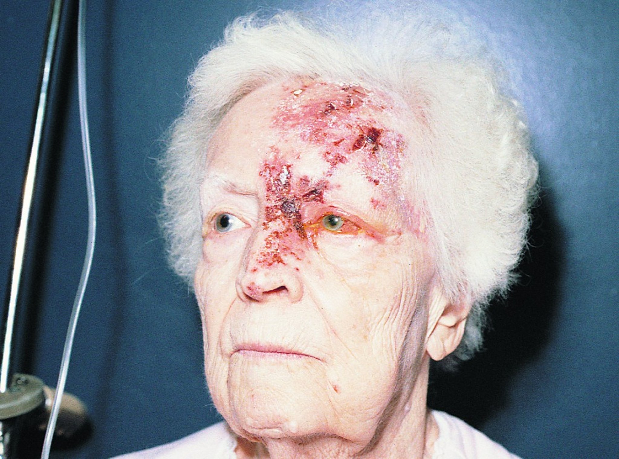
The rash of herpes zoster usually shows up along the path of the nerve that is involved. In this photo, the path of the branches of part of the trigeminal nerve demarcates the rash sharply from surrounding normal skin, that is, between where the vesicles (bumps) appear and where they do not. The appearance of a vesicle on the tip of the nose indicates an increased risk of severe ocular involvement.
© Springer Science+Business Media
Keratitis and/or uveitis may be severe and followed by scarring. Late sequelae—glaucoma, cataract, chronic or recurrent uveitis, corneal scarring, corneal neovascularization, and hypesthesia—are common and may threaten vision. Postherpetic neuralgia may develop later. Patients may develop episcleritis (without increased risk of visual loss) and/or retinitis (with risk of severe visual loss).
Diagnosis of Herpes Zoster Ophthalmicus
Zoster rash on the forehead or eyelid plus eye findings
Diagnosis is based on either a typical acute herpes zoster rash on the forehead, eyelid, and tip of the nose, or on the characteristic pain plus signs of previous zoster rash (eg, atrophic hypopigmented scars). Both skin findings are unilateral (ie, do not cross the midline). Vesicular or bullous lesions in this distribution that do not yet obviously involve the eye should still prompt an ophthalmologic consultation to determine whether the eye is involved. Culture and immunologic or polymerase chain reaction studies of skin at initial evaluation or serial serologic tests are done only when lesions are atypical and the diagnosis uncertain.
Treatment of Herpes Zoster Ophthalmicus
Oral antivirals (eg, acyclovir, famciclovir, valacyclovir)Oral antivirals (eg, acyclovir, famciclovir, valacyclovir)
Sometimes topical corticosteroids
Early treatment with acyclovir, famciclovir, or valacyclovir reduces ocular complications. Patients with uveitis or keratitis require topical corticosteroids (eg, prednisolone acetate). The pupil should be dilated with atropine 1% or scopolamine 0.25% 1 drop 3 times/day. Intraocular pressure must be monitored and treated if it rises significantly above normal values.Early treatment with acyclovir, famciclovir, or valacyclovir reduces ocular complications. Patients with uveitis or keratitis require topical corticosteroids (eg, prednisolone acetate). The pupil should be dilated with atropine 1% or scopolamine 0.25% 1 drop 3 times/day. Intraocular pressure must be monitored and treated if it rises significantly above normal values.
Use of a brief course of high-dose oral corticosteroids to prevent postherpetic neuralgia in patients > 60 years who are in good general health remains controversial.
Medications for neuropathic pain (eg, gabapentin or tricyclic antidepressants) can help relieve symptoms of postherpetic neuralgia.Medications for neuropathic pain (eg, gabapentin or tricyclic antidepressants) can help relieve symptoms of postherpetic neuralgia.
Prevention of Herpes Zoster Ophthalmicus
Recombinant herpes zoster vaccine is recommended for immunocompetent adults ≥ 50 years, regardless of whether they have had herpes zoster or been given the older, live-attenuated vaccine. This recombinant vaccine decreases the chance of getting herpes zoster by approximately 97% for adults 50 to 69 years (1) and 90% for adults ≥ 70 years (2).
Prevention references
1. Lal H, Cunningham AL, Godeaux O, et al: Efficacy of an adjuvanted herpes zoster subunit vaccine in older adults. N Engl J Med 372(22):2087-2096, 2015. doi: 10.1056/NEJMoa1501184
2. Cunningham AL, Lal H, Kovac M, et al: Efficacy of the herpes zoster subunit vaccine in adults 70 years of age or older. N Engl J Med375(11):1019-1032,2016. doi: 10.1056/NEJMoa1603800
Key Points
The eye is affected in about half of cases of V1 varicella-zoster virus reactivation.
Keratitis and/or uveitis can be severe and cause morbidity.
Appearance of the typical herpes zoster rash is usually diagnostic.
Treatment is with oral antivirals and usually topical corticosteroids and pupillary dilation.
Give the recombinant herpes zoster vaccine to all immunocompetent adults Give the recombinant herpes zoster vaccine to all immunocompetent adults≥ 50 years.
Drug Information for the Topic


