


(See also Manometry.)
Impedance planimetry provides real-time assessment of luminal distensibility and geometric changes in response to applied pressure in various parts of the gastrointestinal tract.
A catheter-mounted, balloon-covered probe is placed during a regular endoscopy using sedation and inflated to prespecified pressures. During inflation, pressure sensors along the balloon measure intraluminal pressure and cross-sectional area within a gastrointestinal lumen or sphincter. The measurements are used to calculate a distensibility index (mm2/mm Hg) or compliance of the evaluated area.
The newest generation of probes also produces a real-time graphic display of esophageal contractility patterns.

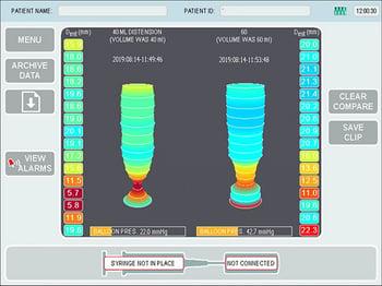
This image shows images of esophagogastric evaluation using impedance planimetry with volumetric tomography comparing esophagogastric junction diameter at various intraballoon pressures.
Impedance planimetry is used in a variety of gastrointestinal conditions, particularly in the esophagus. This technique can be used to evaluate achalasia and can provide useful diagnostic information where high-resolution manometry is nondiagnostic or if the patient cannot tolerate manometry. Recent guidelines recommend its use in the diagnosis and management of achalasia (1, 2). Additionally, in patients undergoing interventions for achalasia (eg, surgery, peroral endoscopic myotomy), measurement of esophagogastric junction distensibility during and after the intervention accurately measures clinical response to interventions and can help guide therapy.
Another potential use of impedance planimetry is in the diagnosis and management of eosinophilic esophagitis. Previous studies have shown decreased esophageal distensibility in patients with eosinophilic esophagitis (3) in whom esophageal fibrosis led to static or minimal changes in cross-sectional area measurements despite increasing balloon pressure and volume. Measurement of esophageal distensibility may show luminal narrowing not seen with conventional endoscopy and is highly suggestive of eosinophilic esophagitis. Further studies are needed to define optimal use of impedance planimetry in this disease.
Impedance planimetry has been studied in the evaluation of gastrointestinal disorders, including gastroesophageal reflux disease (GERD) and hiatus hernia, and in postsurgical settings, including bariatric surgery and fundoplication. Additional studies have assessed its role in disorders of the gastric pylorus (especially in patients with gastroparesis) and in diseases of the anal sphincter. Its utility and specific role in diagnosis and therapeutics require further study.
References
1. Gyawali CP, Carlson DA, Chen JW, et al. ACG clinical guidelines: Clinical use of esophageal physiologic testing. Am J Gastroenterol. 115(9):1412–1428, 2020. doi: 10.14309/ajg.0000000000000734
2. Hirano I, Pandolfino JE, Boeckxstaens GE. Functional lumen imaging probe for the management of esophageal disorders: Expert review from the clinical practice updates committee of the AGA Institute. Clin Gastroenterol Hepatol. 15(3):325–334, 2017. doi: 10.1016/j.cgh.2016.10.022
3. Carlson DA, Lin Z, Hirano I, et al. Evaluation of esophageal distensibility in eosinophilic esophagitis: An update and comparison of functional lumen imaging probe analytic methods. Neurogastroenterol Motil. 28(12):1844–1853, 2016. doi: 10.1111/nmo.12888


