


Doctors can identify many skin disorders simply by looking at the skin. A full skin examination includes examination of the scalp, nails, and mucous membranes. Sometimes the doctor uses a hand-held lens or a dermatoscope (which includes a magnifying lens and a built-in light) to better see the areas of concern.
Revealing characteristics include size, shape, color, and location of the abnormality as well as the presence or absence of other symptoms or signs. To check the distribution of a skin problem, the doctor often asks the person to undress completely, even though the person may have noticed an abnormality on only a small area of skin.
If simply looking at the skin does not provide the doctor with a diagnosis, many tests to identify skin disorders are available.
(See also Structure and Function of the Skin.)
Biopsy
Sometimes a biopsy, in which a small piece of skin is removed for examination under a microscope, must be done.
For this simple procedure, the doctor generally numbs a small area of skin with a local anesthetic and, using a small knife (scalpel), scissors, razor blade (called a shave biopsy), or round cutter (called a punch biopsy), removes a piece of skin. The size of the piece is determined by the type of abnormal growth that is suspected, its location, and the type of tests to be done.
Sometimes the doctor can both diagnose and treat a small tumor by removing the entire tumor along with a small border of normal skin around it (sometimes called an excisional biopsy). The tumor is sent to a laboratory for examination under a microscope.
Scrapings
Culture
If an infection is suspected, a sample of material (such as a skin scraping) can be sent to a laboratory, where the sample is placed in a culture medium (a substance that allows microorganisms to grow). If the sample contains bacteria, fungi, parasites, or viruses, they will often grow in the culture and can then be identified.
Wood Light (Black Light)
A Wood light examination is used when certain skin infections are suspected. The skin is illuminated with an ultraviolet light (also known as a black light) in a dark room. The ultraviolet light makes some fungi or bacteria glow brightly. The light also accentuates the skin's pigment (melanin), making pigmentation abnormalities, such as vitiligo, more visible.

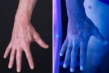
Vitiligo is viewed more easily under a Wood light (right).
Tzanck Testing
A Tzanck test is done to help a doctor diagnose certain diseases caused by viruses, such as herpes simplex and herpes zoster. When these diseases are active, they produce small blisters.
During a Tzanck test, a doctor removes the top of a blister with a sharp blade and then scrapes the blister with a scalpel to obtain fluid. The specimen is examined with a microscope after special stains are applied.
Diascopy
Diascopy is done so that a doctor can observe color changes that occur when pressure is applied to the skin.
During this test, a doctor presses a microscope slide against a lesion to see whether it blanches (becomes less red) or otherwise changes color. Certain kinds of lesions become less red, whereas others do not. Some skin lesions (such as those caused by sarcoidosis) turn a yellowish brown color when this test is done.
Skin Tests
Skin tests, including a "use" test, a patch test, a prick (puncture) test, and an intradermal test, may be done if a doctor suspects an allergic reaction is the cause of a rash.
The use test, in which a suspected substance is applied far from the original area where the rash occurred (usually on the forearm), is useful when perfumes, shampoos, or other substances found in the home may be the cause.
In the patch test, many small samples of common and suspected reaction-causing substances, known as allergens, are applied to the skin (typically on the upper back) under adhesive tape and left on. The skin under the patches is evaluated 48 hours later after the patches are removed and then again at 96 hours. It often takes the skin several days to produce a visible reaction. If a substance produces a characteristic, usually itchy rash, the person is probably allergic to it. In people who have light skin, the color of the rash is usually red. In people who have dark skin, the color of the rash may have less contrast with surrounding skin and thus be more subtle. Sometimes the substances produce an irritation that is not a true allergic reaction.
In the prick test, a drop of an extract of the suspected substance is placed on the skin. Then, the drop is pricked or punctured with a needle to introduce a very tiny amount of the substance into the skin. The skin is then observed for redness (or other color changes), hives, or both, which usually occur within 30 minutes. (See also Skin testing.)
In the intradermal test, tiny amounts of a substance are injected under the skin. The area is then watched for redness (or other color changes) and swelling, which indicate an allergic reaction. (See also Skin testing.)
Although rare, prick and intradermal tests can cause a severe allergic reaction, known as anaphylaxis, which can be life threatening. Therefore, these types of tests should be done only by a trained health care professional.


