


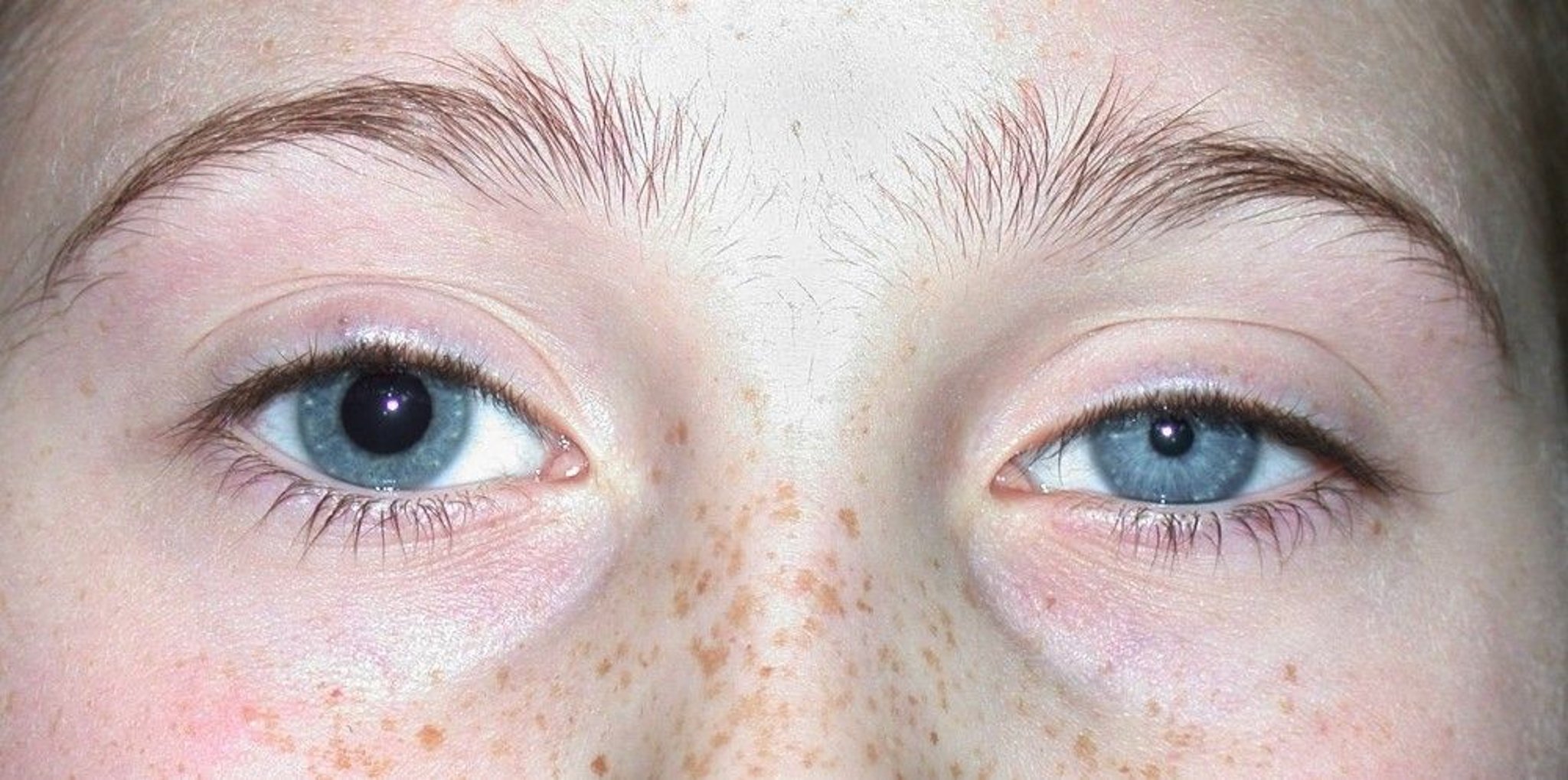
This photo shows anisocoria, which is a visible inequality in pupil diameter. In this case, the anisocoria is due to Horner's syndrome. In physiologic anisocoria, the most common cause of anisocoria, the difference between pupil size is much smaller, typically ≤ approximately 1 mm.
© Springer Science+Business Media
Anisocoria is unequal pupil sizes. Anisocoria itself does not cause symptoms.
Etiology of Anisocoria
The most common cause of anisocoria is:
Physiologic (a benign condition present in up to 20% of people) (1): The difference between pupil sizes in physiologic anisocoria is typically ≤ about 1 mm.
For other causes of anisocoria, see table .
Some Common Causes of Anisocoria
Cause | Suggestive Findings |
|---|---|
Adie tonic pupil (idiopathic impaired constriction) | Pupils that respond more to accommodation than to light; delayed dilation after constriction |
Argyll Robertson pupil (due to syphilis) | Pupils that respond more to accommodation than to light; possibly findings suggesting syphilis |
Congenital iris defects | Associated ocular abnormalities, chromosomal disorder, nonocular congenital defects, chronicity |
Medications and substances (eg, scopolamine patch; cocaine, pilocarpine, animal flea collars or sprays, organophosphates, or aerosolized ipratropium if they contact the eye; cycloplegic, mydriatic, clonidine, or apraclonidine eye drops) Medications and substances (eg, scopolamine patch; cocaine, pilocarpine, animal flea collars or sprays, organophosphates, or aerosolized ipratropium if they contact the eye; cycloplegic, mydriatic, clonidine, or apraclonidine eye drops) | History of use or exposure |
Horner syndrome (eg, congenital, traumatic, postsurgical, due to migraine or lung tumors) | Ptosis, miosis, anhidrosis, delayed dilation after constriction, features of causative disorder |
Iris or other ocular dysfunction after surgery | History of intraocular surgery |
Physiologic anisocoria | Chronicity, absence of symptoms or associated findings, difference of < 1 mm between pupil sizes, normal pupillary light responses |
Third cranial nerve palsy (eg, due to aneurysm or tumor) | Impaired extraocular movements, ptosis |
Traumatic mydriasis | History or evidence of trauma |
Many disorders are accompanied by anisocoria due to iris or neurologic dysfunction (eg, uveitis, stroke, subarachnoid hemorrhage, acute angle-closure glaucoma) but usually manifest with other, clinically significant symptoms (eg, ocular pain, headache, blurred vision, nausea, vomiting, altered mental status, focal neurologic deficits).
Etiology reference
1. Steck RP, Kong M, McCray KL, Quan V, et al. Physiologic anisocoria under various lighting conditions. Clin Ophthalmol. 2018;12:85-89. Published 2018 Jan 4. doi:10.2147/OPTH.S147019
Evaluation of Anisocoria
The goal of evaluation is to elucidate the physiologic mechanism of anisocoria (1). By identifying certain mechanisms (eg, Horner syndrome, third cranial nerve palsy), clinicians can diagnose the occasional serious occult disorder (eg, tumor, aneurysm) manifesting with anisocoria.
History
History of present illness includes the presence, nature, and duration of symptoms. Any history of head or ocular trauma is noted.
Review of systems seeks symptoms that may suggest a cause, such as birth defects or chromosomal abnormalities (congenital defects); droopy eyelid, cough, chest pain, or dyspnea (Horner syndrome); genital lesions, adenopathy, rashes, or fever (syphilis); and headaches or other neurologic symptoms (Horner syndrome or third cranial nerve palsy).
Past medical history includes known ocular disorders and surgeries and exposure to medications.
Physical examination
Pupillary size and light responses should be examined in lighted and dark rooms. Accommodation and extraocular movements should be tested. Ocular structures are inspected by using a slit lamp or other magnification to identify structural abnormalities and ptosis. Other ocular symptoms are evaluated by eye examination as clinically indicated. An old photograph of the patient or the patient’s driver’s license should be examined (under magnification if possible) to see whether anisocoria (and ptosis, if applicable) was present previously.
Red flags
The following findings are of particular concern:
Ptosis
Anhidrosis
Pupils that respond more to accommodation than light
Impaired extraocular movements
Interpretation of findings
If the difference in size is greater in the dark, the smaller pupil is abnormal (because the pupil should dilate in the dark to let in more light). Common causes include Horner syndrome and physiologic anisocoria. The small pupil in Horner syndrome does not dilate after instillation of an ocular dilating drop (eg, 10% cocaine). In physiologic anisocoria, the difference in pupil size may also be equal in light and dark.and physiologic anisocoria. The small pupil in Horner syndrome does not dilate after instillation of an ocular dilating drop (eg, 10% cocaine). In physiologic anisocoria, the difference in pupil size may also be equal in light and dark.
If the difference in pupillary sizes is greater in light, the larger pupil is abnormal (because the pupil should constrict in the light to let in less light). If extraocular movements are impaired, particularly with ptosis, third cranial nerve palsy is likely. If extraocular movements are intact, an ophthalmologist can further differentiate among causes by instilling a drop of a pupillary constrictor (eg, 0.1% pilocarpine). If the large pupil constricts, the cause is probably Adie tonic pupil; if the large pupil does not constrict, the cause is probably pharmacologic (eg, anticholinergic or sympathomimetic medications accidentally instilled in the eye) or structural (eg, traumatic, surgical) damage to the iris. is likely. If extraocular movements are intact, an ophthalmologist can further differentiate among causes by instilling a drop of a pupillary constrictor (eg, 0.1% pilocarpine). If the large pupil constricts, the cause is probably Adie tonic pupil; if the large pupil does not constrict, the cause is probably pharmacologic (eg, anticholinergic or sympathomimetic medications accidentally instilled in the eye) or structural (eg, traumatic, surgical) damage to the iris.
Testing
Additional testing is guided by the symptoms and suspected underlying disorder. Patients with Horner syndrome or third cranial nerve palsy usually require brain MRI or CT and, with Horner syndrome, chest CT. Sexually transmitted infection screening is indicated for Argyll Robertson pupil.
Evaluation reference
1. Gross JR, McClelland CM, Lee MS. An approach to anisocoria. Curr Opin Ophthalmol. 2016;27(6):486-492. doi:10.1097/ICU.0000000000000316
Treatment of Anisocoria
Treatment of anisocoria itself is unnecessary. Underlying disorders (eg, Horner syndrome) should be evaluated and treated as indicated.
Key Points
Physiologic anisocoria is very common and causes < 1 mm of difference between the pupils in size; greater differences require evaluation.
Examining the pupils in light and dark and inspecting an old photograph or the driver’s license of the patient can help identify the abnormal pupil; use of pupillary dilating and constricting drops and further eye examination can provide additional diagnostic information.
Serious disorders should be considered in patients with Horner syndrome or third cranial nerve palsy.
Drug Information for the Topic


