


The brain’s functions are both mysterious and remarkable, relying on billions of nerve cells and the internal communication between them. All thoughts, beliefs, memories, behaviors, and moods arise within the brain. The brain is the site of thought and intelligence, and the control center for the entire body. The brain coordinates the abilities to move, touch, smell, taste, hear, and see. It enables people to form words, speak, and communicate, understand and manipulate numbers, compose and appreciate music, recognize and understand geometric shapes, plan ahead, and even to imagine and fantasize.
The brain reviews all stimuli—from the internal organs, surface of the body, eyes, ears, nose, and mouth. It then reacts to these stimuli by regulating the following:
Position of the body
Movement of limbs
Rate at which the internal organs function
Mood
Viewing the Brain
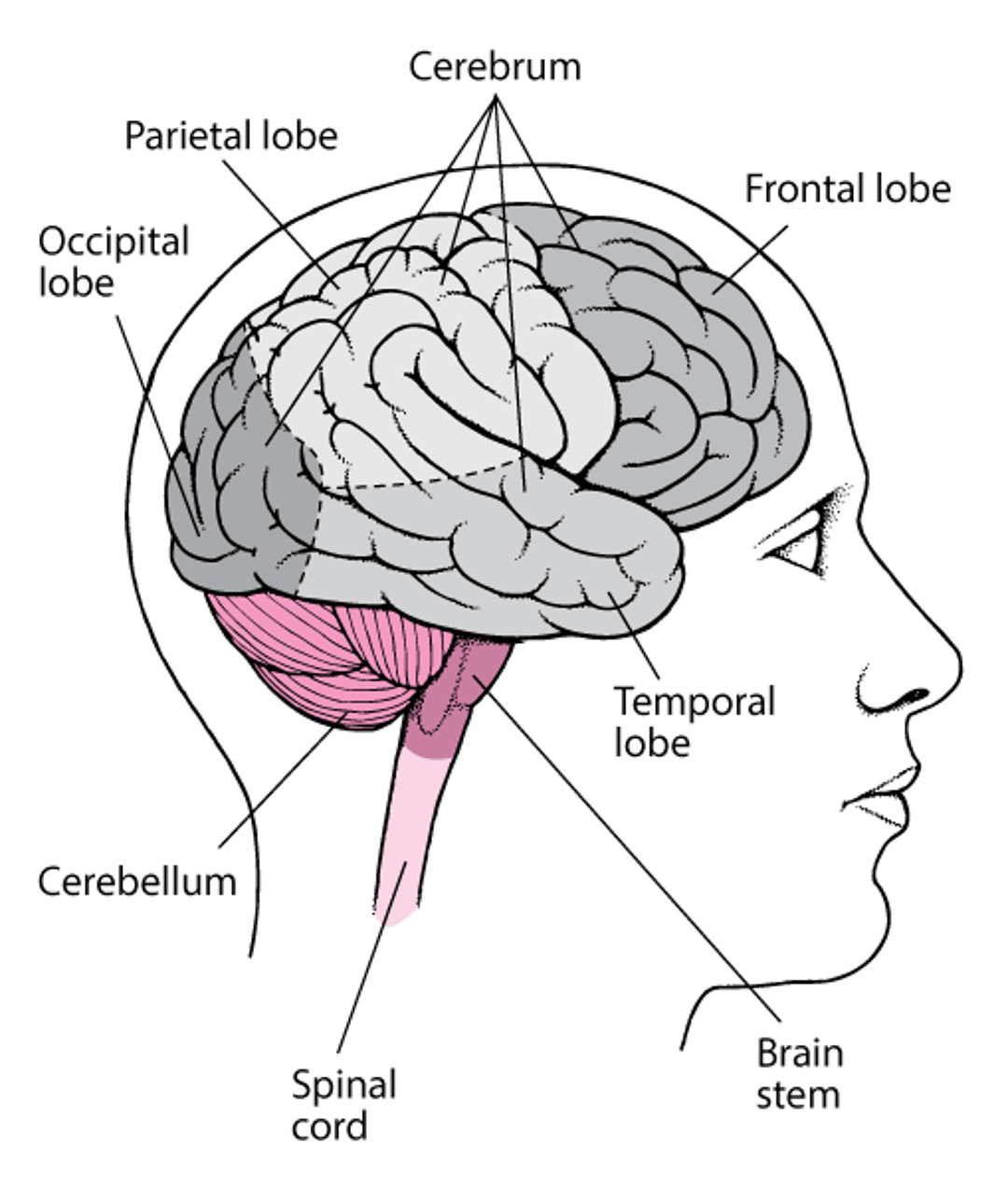
The brain consists of the cerebrum, brain stem, and cerebellum. Each half (hemisphere) of the cerebrum is divided into lobes. |

No computer has yet come close to matching the capabilities of the human brain. However, this sophistication comes with a price. The brain needs constant nourishment. It demands an extremely large amount and continuous flow of blood and oxygen—about 25% of the blood flow from the heart. The overall energy consumption of the brain does not change much over time, but certain areas of the brain use more energy during periods of increased activity (for example, when attempting to learn a new language or learning a new task such as ice skating). A loss of blood flow to the brain for more than about 10 seconds can cause loss of consciousness.
Lack of oxygen or abnormally low sugar (glucose) levels in the blood results in less energy for the brain and can seriously injure the brain within 4 minutes. However, the brain is defended by several mechanisms that can work to prevent these problems. For example, if blood flow to the brain decreases, the brain immediately signals the heart to beat faster and more forcefully, and thus to pump more blood. If the sugar level in the blood becomes too low, the brain signals the adrenal glands to release epinephrine (adrenaline), which stimulates the liver to release stored sugar.
Did You Know...
|
The blood-brain barrier also protects the brain. It is made up of cells that line blood vessels of the brain. These cells allow some substances to reach the brain and block others. The blood-brain barrier is necessary because in the brain, unlike in most of the body, the cells that form the capillary walls are tightly sealed, for example, to protect it from harm caused by toxins and infections. (Capillaries, the smallest of the body’s blood vessels, are where the exchange of nutrients and oxygen between the blood and tissues occurs.) Because the blood-brain barrier controls substances that can enter the brain, penicillin, many chemotherapy agents, some toxic substances, and most proteins cannot pass into the brain. On the other hand, substances such as alcohol, caffeine, and nicotine can pass into the brain. Certain medications, such as antidepressants, are designed so that they can pass through the barrier. Some substances needed by the brain, such as sugar and amino acids, do not readily pass through the barrier. However, the blood-brain barrier has transport systems that move substances the brain needs across the barrier to brain tissue. When the brain is inflamed, as may occur when people have certain infections or tumors, the blood-brain barrier becomes leaky (permeable). When the blood-brain barrier is permeable, some substances (such as certain antibiotics) that normally are unable to pass into the brain are able to do so.
The activity of the brain results from electrical impulses generated by nerve cells (neurons), which process and store information. The impulses pass along the nerve fibers within the brain. How much and what type of brain activity occurs and where in the brain it is initiated depend on a person’s level of consciousness and on the specific activity that the person is doing.
The brain has 3 main parts:
Cerebrum
Brain stem
Cerebellum
Each part (cerebrum, brain stem, and cerebellum) has a number of smaller areas, each with specific functions.
Cerebrum
The cerebrum, the largest part of the brain, contains the following:
The cerebral cortex: This convoluted layer of tissue forms the outer surface of the cerebrum. It consists of a thin layer of gray matter about one-eighth of an inch (2 to 4 millimeters) thick. In adults, the cerebral cortex contains most of the nerve cells in the nervous system.
White matter: White matter consists mainly of nerve fibers (axons) that connect the nerve cells in the cortex with one another, as well as with other parts of the brain and spinal cord. It also contains the support cells (oligodendrocytes) that make the myelin for the nerve cell fibers (to speed the conduction of impulses along nerve fibers). The white matter is located under the cortex.
Subcortical structures: These structures are also located under ("sub-") the cortex—hence, their name. They include the basal ganglia, thalamus, hypothalamus, hippocampus, and the limbic system, which includes the amygdala, olfactory connections (structures that help transmit smell signals), and related structures.
The cerebrum is divided into 2 halves—the left and right cerebral hemispheres. The hemispheres are connected by nerve fibers that form a bridge of white matter (called the corpus callosum) through the middle of the brain. Each hemisphere is further divided into lobes:
Frontal lobe
Parietal lobe
Occipital lobe
Temporal lobe
Each lobe has specific functions, but for most activities, several areas of different lobes in both hemispheres must work together.
The frontal lobes have the following functions:
Initiating many voluntary actions, ranging from looking toward an object of interest, to crossing a street, to relaxing the bladder to urinate
Controlling learned motor skills, such as writing, playing musical instruments, and tying shoelaces
Controlling complex intellectual processes, such as speech, thought, concentration, problem-solving, judgment, and planning for the future
Controlling facial expressions and hand and arm gestures
Coordinating expressions and gestures with mood and feelings
Particular areas of the frontal lobes control specific movements, typically of the opposite side of the body. In most people, the left frontal lobe controls most of the functions involved in using language.
The parietal lobes have the following functions:
Interpreting sensory information from the rest of the body
Controlling body and limb position
Combining impressions of form, texture, and weight into general perceptions
Influencing mathematical skills and language comprehension, as do adjacent areas of the temporal lobes
Storing spatial memories that enable people to orient themselves in space (know where they are) and to maintain a sense of direction (know where they are going)
Processing information that helps people know the position of their body parts
The occipital lobes have the following functions:
Processing and interpreting vision and identifying the shapes of objects
Enabling people to form visual memories
Integrating visual perceptions with the spatial information provided by the adjacent parietal lobes
The temporal lobes have the following functions:
Generating memory and emotions
Processing immediate events into recent and long-term memory
Storing and retrieving long-term memories
Comprehending sounds and images, thus enabling people to recognize other people and objects and to integrate hearing and speech
Subcortical structures include large collections of nerve cells:
The basal ganglia, which coordinate and smooth out movements
The thalamus, which generally organizes sensory messages to and from the highest levels of the brain (cerebral cortex), providing an awareness of such sensations as pain, touch, and temperature
The hypothalamus, which coordinates some of the more automatic functions of the body, such as control of sleep and wakefulness, maintenance of body temperature, regulation of appetite and thirst, and control of hormonal activity of the adjacent pituitary gland
The limbic system, another subcortical structure, consists of structures and nerve fibers located deep within the cerebrum. The parts of the limbic system are the hypothalamus, the amygdala, the thalamus, mammillary bodies, and the hippocampus. This system connects the hypothalamus with other areas of the frontal and temporal lobes. The limbic system controls the experience and expression of emotions, motivation, memory, and learning, as well as some automatic functions of the body. By producing emotions (such as fear, anger, pleasure, and sadness), the limbic system enables people to behave in ways that help them communicate and survive physical and psychological upsets. The hippocampus is also involved in the formation and retrieval of memories, and its connections through the limbic system help connect those memories to the emotions experienced when the memories form. Through the limbic system, memories that are emotionally charged are often easier to recall than those that are not. The limbic system also has input into other areas of the brain, such as the basal ganglia, that control intentional movements of the limbs.
Brain stem
The brain stem connects the cerebrum with the spinal cord. It contains a system of nerve cells and fibers (called the reticular activating system) located deep within the upper part of the brain stem. The reticular activating system controls levels of consciousness and alertness. The brain stem also contains many of the nerve center collections that control movements of the eyes, face, jaw, and tongue, including chewing and swallowing.
The brain stem also automatically regulates critical body functions, such as breathing, blood pressure, and heartbeat, and it helps adjust posture and balance.
If the entire brain stem becomes severely damaged, consciousness is lost, and these automatic body functions cease. That is, all brain activity is lost. This loss is considered brain death. Death soon follows.
However, if the brain stem remains intact, the body may remain alive, even when severe damage to the cerebrum makes awareness, thought, and movement impossible.
Cerebellum
The cerebellum, which lies below the cerebrum just above the brain stem, coordinates the body’s movements. With information it receives from the cerebral cortex and the basal ganglia about the position of the limbs, the cerebellum helps the limbs move smoothly and accurately. It does so by constantly adjusting muscle tone and posture.
The cerebellum interacts with areas in the brain stem called vestibular nuclei, which are connected with the organs of balance (semicircular canals) in the inner ear. Together, these structures provide a sense of balance, making walking upright possible.
The cerebellum also stores memories of practiced movements, enabling highly coordinated movements, such as a ballet dancer’s pirouette, to be done with speed and balance. The cerebellum contributes to thought functions such as attention, language, and emotion.
Meninges
Both the brain and spinal cord are covered by 3 layers of tissue (meninges) that protect them:
Tissues Covering the Brain
Within the skull, the brain is covered by 3 layers of tissue called the meninges:
Between the arachnoid membrane and pia mater is the subarachnoid space. This space contains cerebrospinal fluid, which flows through the meninges, fills the spaces within the brain, and helps cushion the brain and spinal cord. |

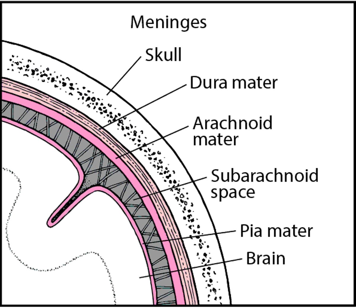
The leathery dura mater is the outermost and toughest layer.
The delicate, spider web–like arachnoid mater is the middle layer.
The thin pia mater is the innermost layer, which adheres to the brain and spinal cord.
The space between the arachnoid mater and the pia mater (the subarachnoid space) is a channel for cerebrospinal fluid, which helps protect the brain and spinal cord.
Cerebrospinal fluid helps to cushion the brain against sudden jarring and minor injury and also to remove waste products from the brain. The cerebrospinal fluid is contained in a network of spaces within the brain called ventricles. Cerebrospinal fluid, formed by specialized cells that line the ventricles, enters the brain along the outside of blood vessels and flows over the surface of the brain between the meninges. The fluid is taken up by support cells (glial cells) and distributed throughout the brain, filling internal spaces within the brain (the 4 cerebral ventricles). Eventually, the fluid leaves the brain to enter the body's blood vessels. As the cerebrospinal fluid flows through the brain, it removes discarded proteins and other waste products from the brain tissue. Lymphatic vessels in the brain also help in waste removal. This removal process occurs mainly when people are sleeping, which highlights the importance of sleep.
The brain and its meninges are contained in a tough, bony protective structure, the skull. The spinal cord connects to the brain at the base of the brain stem.


