

Eyelid injuries consist of contusions and lacerations and are usually caused by physical trauma.
(See also Overview of Eye Trauma.)
Eyelid injuries
Eyelid contusions (which result in black eyes that usually affect both the upper and lower eyelids simultaneously) are more significant cosmetically than clinically, although more serious injuries may sometimes accompany them and should not be overlooked. Uncomplicated contusions are treated with ice packs to inhibit swelling during the first 24 to 48 hours.
Severe blunt trauma to the periocular region can result in orbital hemorrhage and orbital compartment syndrome with the potential for loss of vision. It is imperative to check visual function and examine the globe in all eyelid injuries. If eyelid swelling is too extensive to allow examination of the globe, an ophthalmologist should be consulted emergently (1).

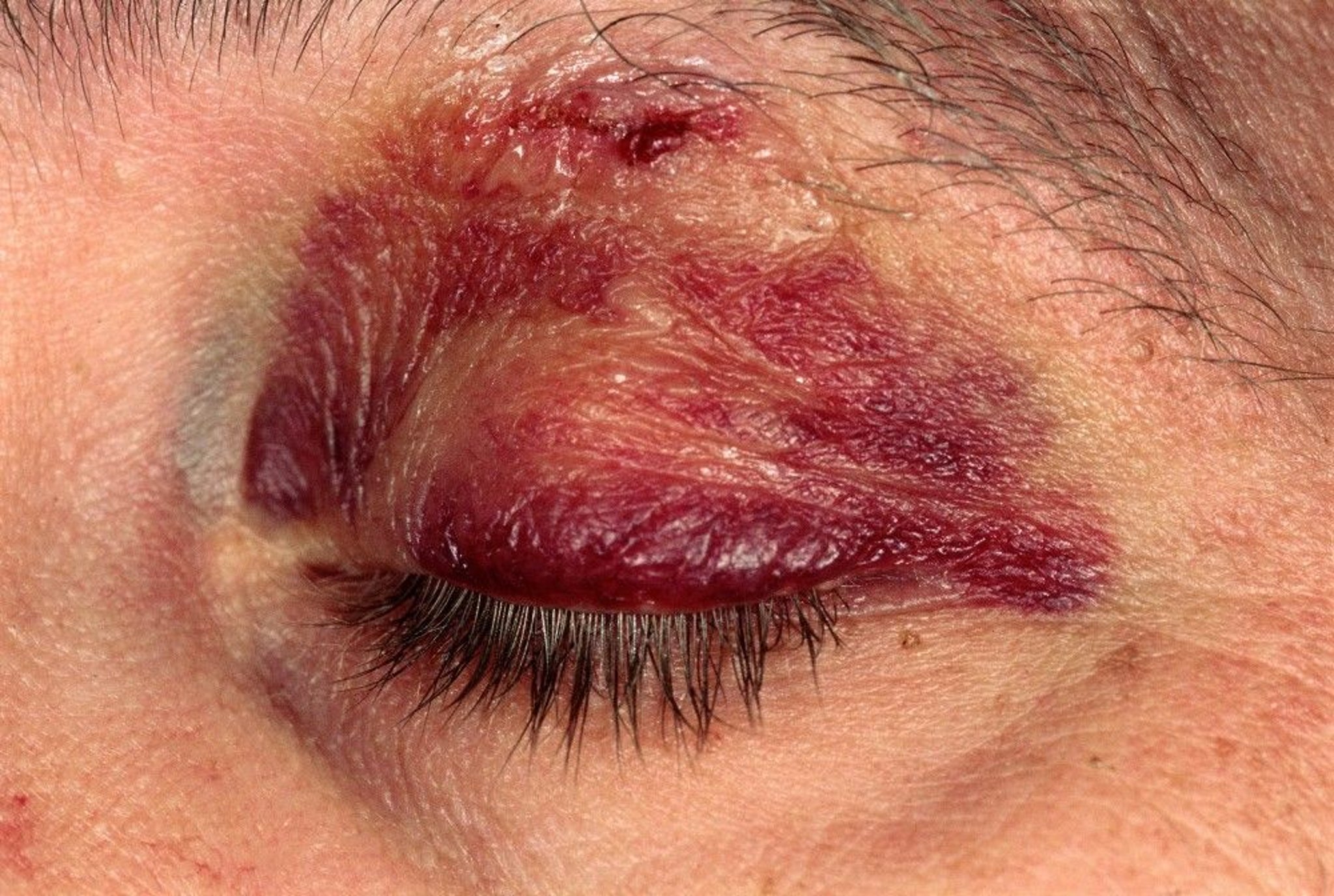
This close-up shows a black eye (periorbital bruising or ecchymosis).
SCOTT CAMAZINE/SCIENCE PHOTO LIBRARY
Eyelid lacerations
Minor eyelid lacerations that do not involve the lid margin or tarsal plate may be repaired with nylon or polypropylene (or, in some populations such as children, with absorbable suture such as plain gut) 6-0 or 7-0 sutures. Superficial lacerations may not require suture repair if the wound edges are not under tension and are well approximated. Sometimes wound-closure strips or tissue adhesives may be used. If tissue adhesives are used, extra care must be taken to ensure that the upper and lower eyelids are not accidentally adhered together.
Lacerations of the lid margin are best repaired by an ophthalmic surgeon to ensure accurate apposition and to avoid a notch in the eyelid contour. Complicated lid lacerations, which include those of the medial portion of the lower or upper eyelid (possibly involving the lacrimal canaliculus), through-and-through lacerations, those in which the patient has ptosis, and those that expose orbital fat or involve the tarsal plate, should also be repaired by an ophthalmic surgeon (2).
References
1. Scoville NM, Ding L, Stacey AW: Success rates of lateral canthotomy and cantholysis for treatment of orbital compartment syndrome. Am J Emerg Med 70:140-143, 2023. doi: 10.1016/j.ajem.2023.05.037
2. Chang EL, Rubin PA: Management of complex eyelid lacerations. Int Ophthalmol Clin 2002;42(3):187-201. doi:10.1097/00004397-200207000-00020
Key Points
Consult an ophthalmologist if an eyelid laceration is complicated (eg, through the margin, tarsal plate, or canaliculus, causing ptosis, or exposing orbital fat).
Globe trauma may cause globe laceration, cataract, lens dislocation, glaucoma, vitreous hemorrhage, or retinal damage (hemorrhage, detachment, or edema).
Suspect globe rupture if trauma results in a visible corneal or scleral laceration, leaking aqueous humor, an unusually shallow or deep anterior chamber, or an irregular pupil.


