Inflammatory breast carcinoma is a rare form of breast cancer that is highly aggressive and often fatal. It presents with erythema and edema, causing a thickened, dimpled appearance of the breast skin (called peau d'orange) and often with diffuse enlargement of the breast but no palpable breast lump. Diagnosis is by biopsy combined with clinical manifestations. Treatment is with chemotherapy, mastectomy, and radiation.
Inflammatory breast cancer is a rare type of breast cancer. In the United States, it had an incidence of 3 per 100,000 people in the United States from 1973 to 2015 (1), and it comprises approximately 1 to 5% of all breast cancers (2). It has a poor prognosis, although advances in treatment have improved survival rates.
Inflammatory breast cancer is a clinical-pathologic entity. It is not a distinct histologic type; most cases are ductal, but some are lobular. The characteristic clinical findings of erythema and edema are caused by lymphedema, and histology reveals tumor emboli within the dermal lymphatics.
General references
1. Abraham HG, Xia Y, Mukherjee B, Merajver SD. Incidence and survival of inflammatory breast cancer between 1973 and 2015 in the SEER database. Breast Cancer Res Treat. 2021;185(1):229-238. doi:10.1007/s10549-020-05938-2
2. Tadros A, Diskin B, Sevilimedu V, et al. Trends in Guideline-Concordant Care for Inflammatory Breast Cancer. JAMA Netw Open. 2025;8(2):e2454506. Published 2025 Feb 3. doi:10.1001/jamanetworkopen.2024.54506
Diagnosis of Inflammatory Breast Cancer
Breast examination
Breast and lymph node imaging
Core breast biopsy and breast skin biopsy
The diagnosis of inflammatory breast cancer is primarily clinical, based on a characteristic history and physical examination in a patient with invasive breast carcinoma. Diagnostic criteria are (1)
Rapid onset of breast erythema, edema and/or peau d’orange (thickened, dimpled appearance similar to that of an orange peel), and/or warm breast, with or without an underlying palpable mass
Duration of findings of no more than 6 months
Erythema occupying at least one-third of the breast
Pathologic confirmation of invasive carcinoma
Mammography should be performed in patients with suspected inflammatory breast cancer. Findings may include breast skin thickening and trabecular distortion, and a mass may be present. Lymph node metastases are often present at diagnosis, and ultrasound is performed to evaluate the breast and lymph nodes. MRI is performed if mammogram or ultrasound images are ambiguous.
Breast core biopsy is performed to confirm invasive carcinoma and evaluate hormone receptor and HER2 status. If clinical criteria are met, punch biopsies can be performed of affected breast skin to possibly detect dermal lymphovascular tumor emboli. However, the finding of tumor in the dermal lymphatics is not sufficient to make the diagnosis in the absence of clinical findings, and the absence of tumor emboli does not exclude inflammatory cancer.
For breast cancer staging, the tumor in inflammatory breast cancer is classified as T4d. The definition of T stage 4 includes tumors with direct extension to the skin, and "d" is a classification specifically for inflammatory breast cancer (a, b, and c refer to extension to the chest wall and/or skin, in tumors that do not meet criteria for inflammatory breast cancer).

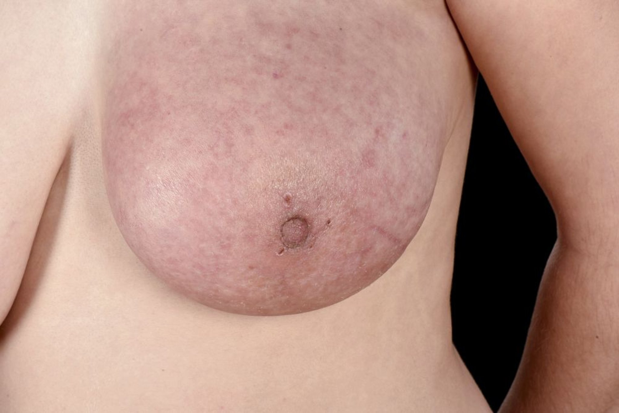
This photo shows diffuse erythema and edema in the left breast of patient with inflammatory breast cancer.
MID ESSEX HOSPITAL SERVICES NHS TRUST/SCIENCE PHOTO LIBRARY

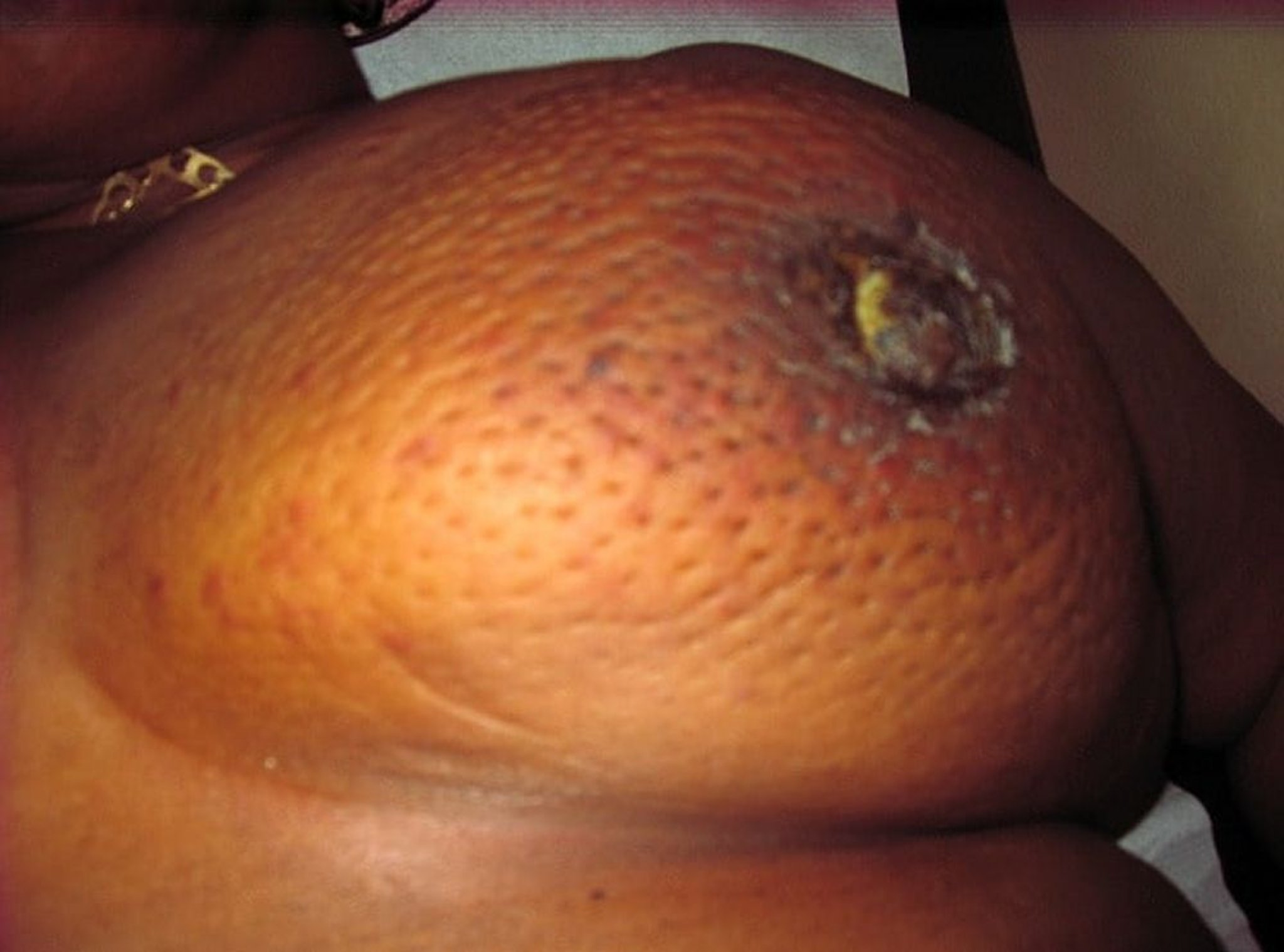
Sometimes in inflammatory breast cancer and very advanced cancers, the breast appears inflamed, and the skin appears thickened, resembling orange peel (peau d’orange).
"Creative Commons Patient with inflammatory breast cancer" by Epidemiology and surgical management of breast cancer in gynecological department of Douala General Hospital (Scientific Figure on ResearchGate) is licensed under Creative Commons 2.0. Available from: https://www.researchgate.net/figure/Patient-with-inflammatory-breast-cancer_fig2_234162338 [accessed 3 Oct, 2019].
Diagnosis reference
1. Dawood S, Merajver SD, Viens P, et al. International expert panel on inflammatory breast cancer: consensus statement for standardized diagnosis and treatment. Ann Oncol. 2011;22(3):515-523. doi:10.1093/annonc/mdq345
Treatment of Inflammatory Breast Cancer
Neoadjuvant chemotherapy
Mastectomy and lymph node dissection
Treatment of underlying tumor
Inflammatory breast cancer is typically treated with neoadjuvant chemotherapy, with anti-HER2 monoclonal antibodies for HER2-positive tumors.
If the patient responds to the neoadjuvant chemotherapy, this is followed by mastectomy and chest wall radiation and then postoperative chemotherapy.
Tumors that do not respond to initial chemotherapy are treated with additional chemotherapy and/or radiation therapy.