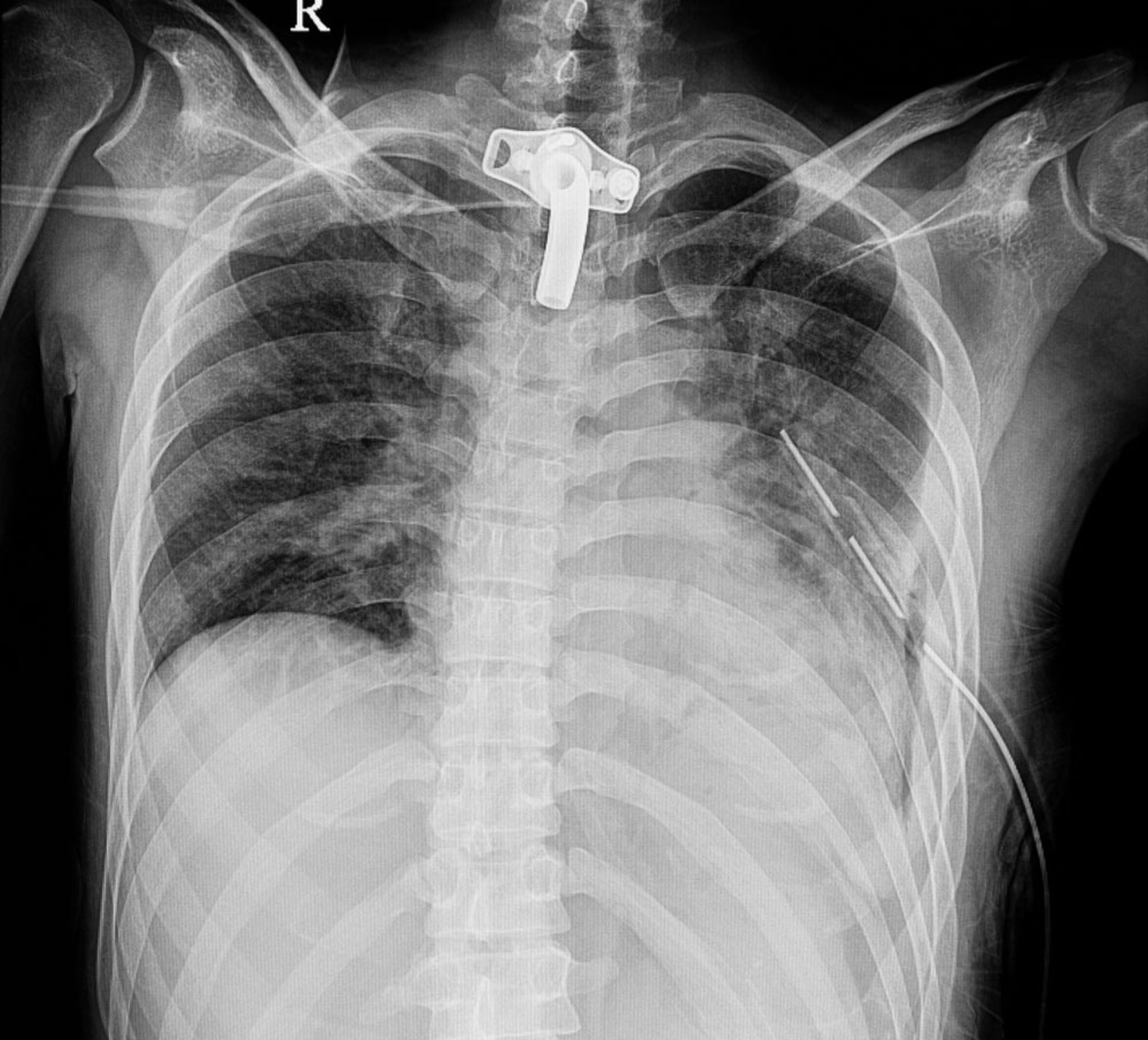
Pulmonary Contusion (Radiograph)
This chest radiograph shows pulmonary contusion with typical findings of patchy, nonsegmental air-space opacities in the left mid-to-lower lung. Hemothorax and an implantable cardioverter-defibrillator (ICD) are also present.
SOPONE/stock.adobe.com
In these topics