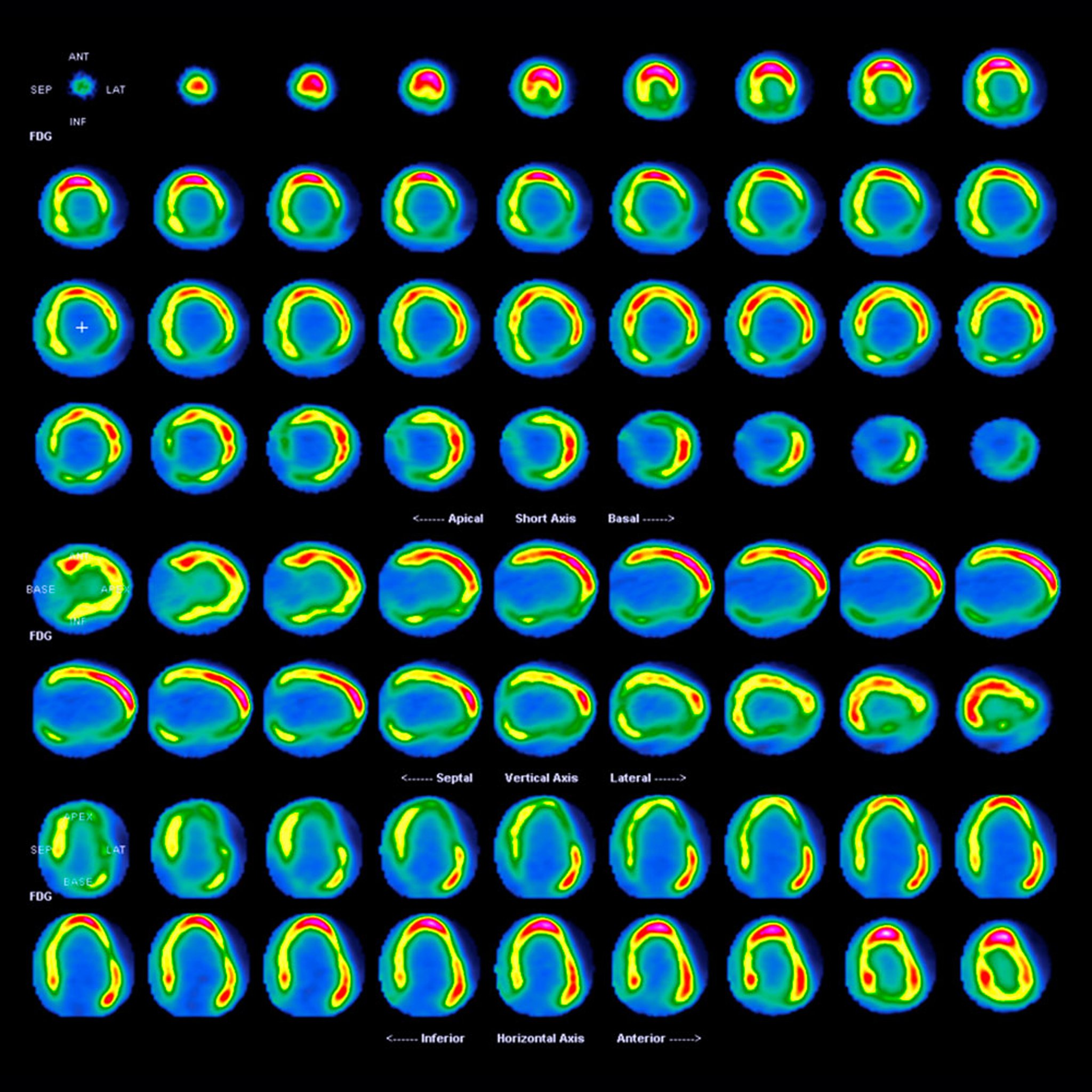
FDG PET-CT Scan in Dilated Cardiomyopathy
This scan shows sequences of combined computed tomography (CT) and positron emission tomography (PET) scans of a heart with dilated cardiomyopathy.
The sequences are short-axis from apex to base (upper), vertical long-axis from septum to lateral wall (middle), and horizontal long-axis from inferior to anterior (lower) views. The order of each sequence runs across from upper left to lower right. The PET scan used here uses F-18 FDG. Here, there is reduced FDG uptake in portions of the myocardium.
CENTRE JEAN-PERRIN/SCIENCE PHOTO LIBRARY
In these topics