

Achilles tendon tears (ruptures) most often result from ankle dorsiflexion, particularly when the tendon is taut. Diagnosis is by examination and sometimes MRI. Treatment is splinting in plantar flexion and immediate referral to an orthopedic surgeon; surgical repair may be necessary.
(See also Overview of Sprains and Other Soft-Tissue Injuries.)
Achilles tendon tears are common. They typically occur during running or jumping and are most common among middle-aged men and athletes. Very rarely, spontaneous Achilles tendon tears have occurred in people who take fluoroquinolone antibiotics or glucocorticoids.
Achilles tendon tears may be partial or complete.
Symptoms and Signs of Achilles Tendon Tears
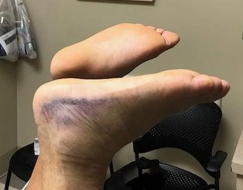
Pain in the distal calf makes walking difficult, particularly when the tear is complete. The calf may be swollen and bruised.
Complete tears may result in a palpable defect and usually occur 2 to 6 cm proximal to the tendon's insertion.
Diagnosis of Achilles Tendon Tears
Primarily history and physical examination
Sometimes MRI or ultrasound
Diagnosis of Achilles tendon tears is clinical (1). The patient's ability to flex the ankle does not exclude a tear.
If clinicians suspect an Achilles tendon tear, 3 main tests can be performed to help confirm the diagnosis.
For the Thompson test (calf squeeze test), the patient is prone, and the calf is squeezed to elicit plantar flexion. Results include the following:
If the tear is complete, ankle plantar flexion is absent or decreased.
If the tear is partial, results are sometimes normal, so these tears are often missed.
The Thompson test is 96% sensitive and 93% specific for Achilles tendon tears (1).

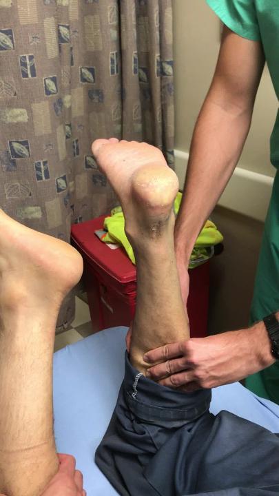
The Matles test is used to assess resting tension. The patient is prone with the knee bent at 90° to shorten the gastrocnemius. The patient's feet are compared. Results include the following:
If the Achilles tendon is intact, plantar flexion of the ankle to 20 to 30° occurs.
If the Achilles tendon is torn, the foot falls to a neutral position.
The Matles test is 88% sensitive and 85% specific for Achilles tendon tears (1).

The purpose of the Matles test is to have the examiner look for natural plantar flexion in the foot with an intact Achilles and for the absence of plantar flexion (indicating a positive Matles test) in the injured ankle. In the Matles test, the patient lies prone with the knees flexed, and the examiner assesses how the foot lies. In this image, the foot in front shows absence of plantar flexion (ie, the foot falls completely flat (to the neutral position with 0° plantar flexion). This result is a positive Matles test. The foot in the background shows 20 to 30° plantar flexion, indicating that the Achilles tendon is not ruptured (ie, the Achilles tendon is intact).
For palpation of the tendon gap, the patient is asked to stand on the affected leg (if possible). Then the clinician gently palpates the course of Achilles tendon and feels for a gap; a gap indicates that the tendon is torn.
Physical examination is more sensitive than MRI for detecting an Achilles tendon tear (2). A study of patients with an Achilles tendon tear (2012) found that if all 3 tests (Thompson, Matles, palpation of the gap) are positive, sensitivity for an Achilles tear is 100%. According to the American Academy of Orthopedic Surgeons guidelines (2010), diagnosis of a tear requires only one of the following (3):
2 of these 3 tests are positive.
1 of the tests is positive and ankle plantar flexion is weakened.
Ultrasound is increasingly being used to confirm tendon tears or to differentiate between partial and complete tears when imaging is required. Diagnostic accuracy appears to be good when performed by experienced operators.
Diagnosis references
1. Maffulli N: The clinical diagnosis of subcutaneous tear of the Achilles tendon: A prospective study in 174 patients. Am J Sports Med 26 (2):266–270, 1998. doi:10.1177/03635465980260021801
2. Garras DN, Raikin SM, Bhat SB, et al: MRI is unnecessary for diagnosing acute Achilles tendon ruptures: Clinical diagnostic criteria. Clin Orthop Relat Res 470 (8):2268–2273, 2012. doi: 10.1007/s11999-012-2355-y
3. Chiodo CP, Glazebrook M, Bluman EM, et al: Diagnosis and treatment of acute Achilles tendon rupture: Practice guideline. J Am Acad Orthop Surg 18 (8):503–510, 2010. doi: 10.5435/00124635-201008000-00007
Treatment of Achilles Tendon Tears
Splinting in plantar flexion
Immediate orthopedic referral
Sometimes surgical repair
Initial treatment of Achilles tendon tears consists of splinting with the ankle in plantar flexion and immediate referral to an orthopedic surgeon.
Achilles tendon tears can be treated with nonoperative management (splinting and progressive range of motion rehabilitation) or with surgical repair.
Nonoperative management includes a posterior ankle splint with the ankle in plantar flexion initially and then progressive plantar flexion to neutral positioning range of motion exercises.
Surgical repair of the tendon can be performed with an open technique as well as a minimally invasive/percutaneous approach.
Nonoperative management has a slightly higher re-rupture rate but otherwise operative and nonoperative approaches have similar long term outcomes (1).
Treatment reference
1. Dold AP. Acute Achilles Tendon Ruptures: An Update on Current Management Strategies. J Am Acad Orthop Surg. 2024;33(16):881-889. Published 2024 Oct 8. doi:10.5435/JAAOS-D-24-00275


