


Trauma to the external ear (pinna or auricle) may result in hematoma, laceration, avulsion, or fracture.
Most pinna wounds may undergo primary closure if they are clean and have viable tissue edges and if repair is performed within approximately 24 hours of injury (1). Wounds that are contaminated or are at high risk of infection (eg, bite wounds, cartilage with a penetrating injury or piercing) are typically managed with delayed primary closure; secondary reconstruction is performed if needed for cosmetic deformities.
Chondritis is a potential complication following external ear injuries. However, data are limited and practice varies regarding routine antibiotic prophylaxis; if prescribed, antibiotics should cover pseudomonas and staphylococci (2, 3).
Subperichondrial hematoma (cauliflower ear)
The external ear (pinna or auricle) is a single piece of cartilage covered by the perichondrium (dense, vascularized connective tissue that covers the surface of cartilage) and skin. The perichondrium supplies blood to the auricular cartilage.
Blunt trauma to the pinna may cause a subperichondrial hematoma; the accumulation of large amounts of blood between the perichondrium and cartilage can interrupt the blood supply to the cartilage and render all or part of the pinna a shapeless, reddish purple mass. Avascular necrosis of the cartilage then occurs and fibrocartilaginous deposition may follow, which causes the characteristic "cauliflower ear" of wrestlers, boxers, and rugby players (4).
Treatment consists of promptly evacuating the accumulated blood through an incision (2, 5). Reaccumulation of the hematoma is prevented by placing a pressure dressing with through-and-through ear sutures over dental gauze rolls or petroleum (plain or antimicrobial) gauze; some clinicians also insert a Penrose drain to allow continued drainage. Pressure dressings are left in place for approximately 1 week.

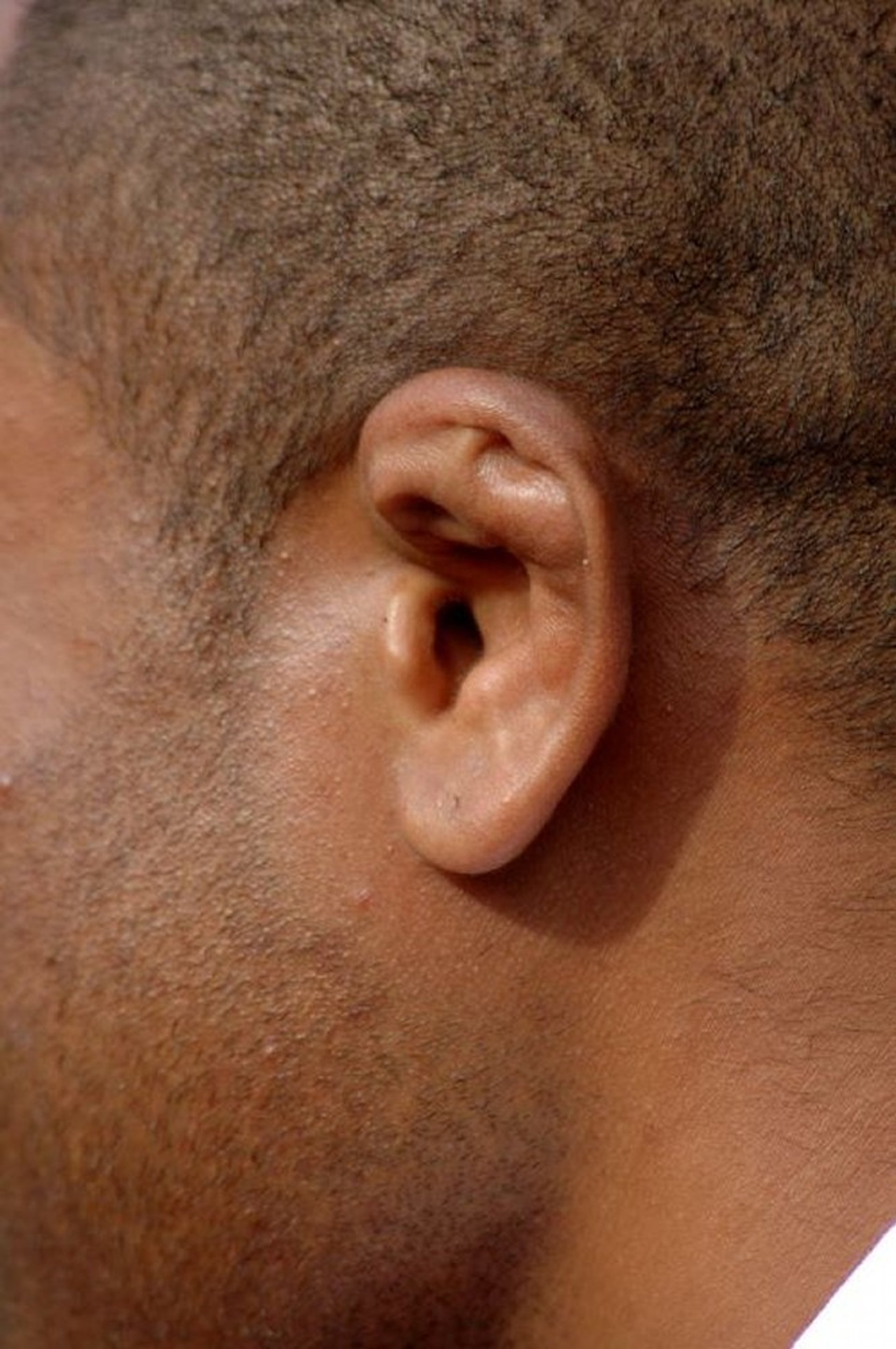
This photo shows deformed cartilage in the superior aspect of the pinna.
RICHARD WAREHAM FOTOGRAFIE/SCIENCE PHOTO LIBRARY
Pearls & Pitfalls
|
Lacerations of the pinna
In lacerations of the pinna, the skin margins are reapproximated and sutured whenever possible. If the cartilage is damaged (eg, punctured or lacerated), it is repaired; secondary reconstruction may be required after the tissue has healed. Damaged cartilage, whether repaired or not, is splinted externally with a cotton (eg, dental roll) or antibiotic-impregnated gauze pressure dressing to prevent development of a subperichondrial hematoma.
Lacerations of the pinna caused by a human or animal bite are at high risk of infection, including infection of the cartilage, a potentially severe complication. Treatment begins with meticulous debridement of devitalized tissue. Prophylactic antibiotics should be given, (6) and possibly antivirals if the source of the bite has a known or suspected infection for which there is post-exposure prophylaxis (eg, HIV, hepatitis B, hepatitis C, rabies) (see table Antimicrobials for Bite Wounds) (7). Tetanus toxoid should be administered to patients who have not had toxoid vaccination within 10 years.
Avulsions
Complete or partial avulsions are repaired by an otolaryngologist, facial plastic surgeon, or plastic surgeon.
Trauma secondary to mandibular fractures
Forceful blows to the mandible may be transmitted to the anterior wall of the ear canal (posterior wall of the glenoid fossa). Displaced fragments from a fractured anterior wall may cause stenosis of the canal and must be reduced or removed surgically.
General references
1. Capellan O, Hollander JE. Management of lacerations in the emergency department. Emerg Med Clin North Am. 2003;21(1):205-231. doi:10.1016/s0733-8627(02)00087-1
2. Long B, Mason J, Bridwell RE, et al. Managing Auricular Hematoma: An Emergency Medicine Narrative Review. J Emerg Med. 2025;69:62-75. doi:10.1016/j.jemermed.2024.08.021
3. Appelbaum RD, Farrell MS, Gelbard RB, et al. Antibiotic prophylaxis in injury: an American Association for the Surgery of Trauma Critical Care Committee clinical consensus document. Trauma Surg Acute Care Open. 2024;9(1):e001304. Published 2024 Jun 3. doi:10.1136/tsaco-2023-001304
4. Giffin CS. Wrestler's ear: pathophysiology and treatment. Ann Plast Surg. 1992;28(2):131-139. doi:10.1097/00000637-199202000-00002
5. Prasad HK, Sreedharan S, Prasad HS, et al. Perichondritis of the auricle and its management. J Laryngol Otol. 2007;121(6):530-534. doi:10.1017/S0022215107005877
6. Stevens DL, Bisno AL, Chambers HF, et al. Practice guidelines for the diagnosis and management of skin and soft tissue infections: 2014 update by the Infectious Diseases Society of America. Clin Infect Dis. 2014;59(2):e10-e52. doi:10.1093/cid/ciu444
7. Kennedy SA, Stoll LE, Lauder AS. Human and other mammalian bite injuries of the hand: evaluation and management. J Am Acad Orthop Surg. 2015;23(1):47-57. doi:10.5435/JAAOS-23-01-47


